. 2014 Mar 13:5:28.
doi: 10.3389/fneur.2014.00028. eCollection 2014.

Magnetic vestibular stimulation in subjects with unilateral labyrinthine disorders

Affiliations

Magnetic vestibular stimulation in subjects with unilateral labyrinthine disorders

Bryan K Ward et al. Front Neurol. .

Abstract

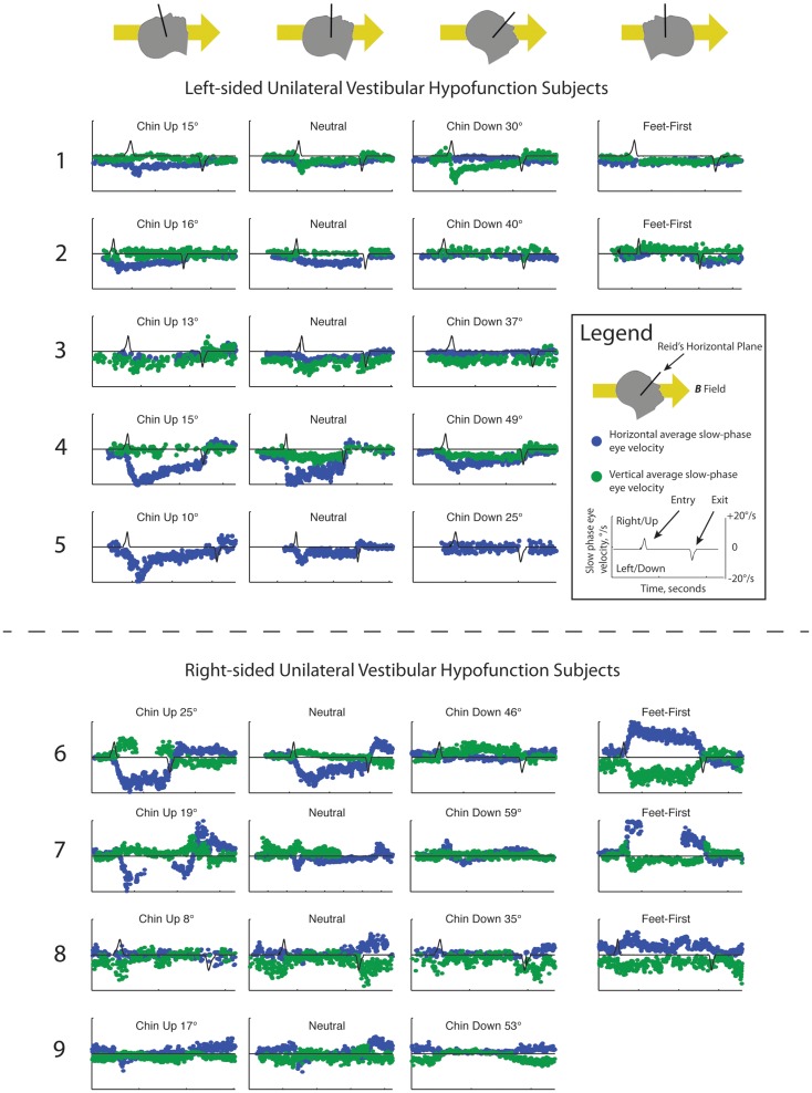
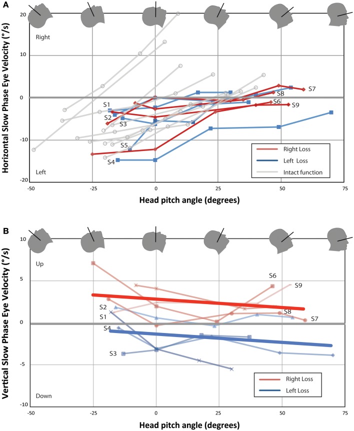
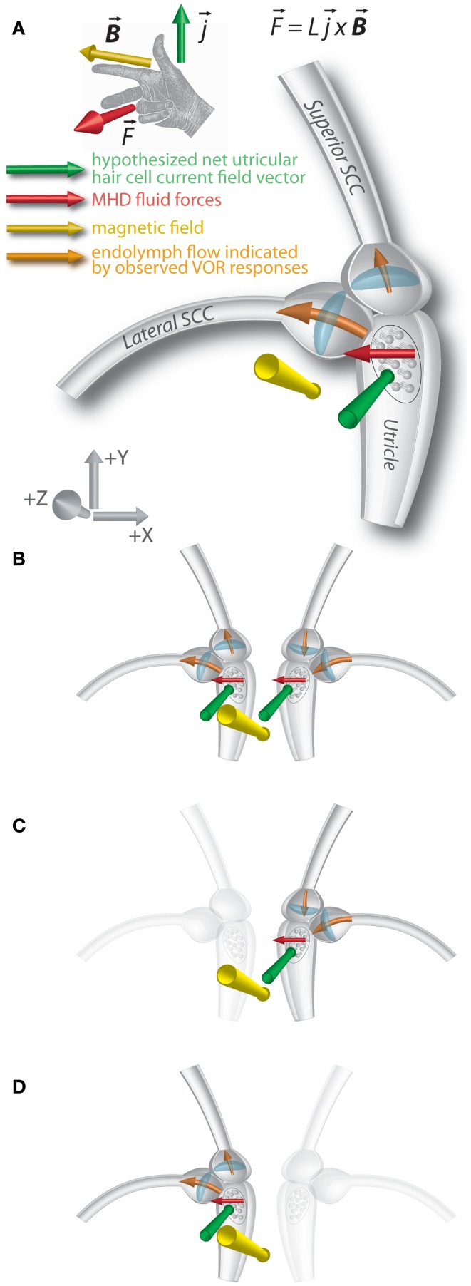
We recently discovered that static magnetic fields from high-strength MRI machines induce nystagmus in all normal humans, and that a magneto-hydrodynamic Lorentz force, derived from ionic currents in the endolymph and pushing on the cupula, best explains this effect. Individuals with no labyrinthine function have no nystagmus. The influence of magnetic vestibular stimulation (MVS) in individuals with unilateral deficits in labyrinthine function is unknown and may provide insight into the mechanism of MVS. These individuals should experience MVS, but with a different pattern of nystagmus consistent with their unilateral deficit in labyrinthine function. We recorded eye movements in the static magnetic field of a 7 T MRI machine in nine individuals with unilateral labyrinthine hypofunction, as determined by head impulse testing and vestibular-evoked myogenic potentials (VEMP). Eye movements were recorded using infrared video-oculography. Static head positions were varied in pitch with the body supine, and slow-phase eye velocity (SPV) was assessed. All subjects exhibited predominantly horizontal nystagmus after entering the magnet head-first, lying supine. The SPV direction reversed when entering feet-first. Pitching chin-to-chest caused subjects to reach a null point for horizontal SPV. Right unilateral vestibular hypofunction (UVH) subjects developed slow-phase-up nystagmus and left UVH subjects, slow-phase-down nystagmus. Vertical and torsional components were consistent with superior semicircular canal excitation or inhibition, respectively, of the intact ear. These findings provide compelling support for the hypothesis that MVS is a result of a Lorentz force and suggest that the function of individual structures within the labyrinth can be assessed with MVS. As a novel method of comfortable and sustained labyrinthine stimulation, MVS can provide new insights into vestibular physiology and pathophysiology.

Keywords: Lorentz; magnetic; magneto-hydrodynamics; semicircular canals; vestibular.


Figures

Figure 1
Figure 1
Slow-phase nystagmus eye velocity over time for all subjects. Each column represents a separate trial with the subject’s head pitched either chin up or chin down from a head-neutral supine position. The last column represents feet-first in subjects in which this data was available. Horizontal slow-phase velocity is shown with blue dots and vertical slow-phase velocity with green dots. Solid black line represents electromagnetic induction (dB/dt), measured using a wire coil. Electromagnetic induction peak and trough indicate, respectively, entry and exit from the bore of the magnet.
Figure 2
Figure 2
Change in nystagmus slow-phase horizontal (A) and vertical (B) eye velocity from baseline eye velocity for all UVH subjects. Each point represents the difference in average SPV between a 40-s time interval once the subject has entered the bore and baseline average SPV in darkness prior to entry. Right-sided UVH subjects are labeled to the right and left-sided UVH subjects to the left of each line. Ten subjects with bilateral intact vestibular function from Roberts et al. are plotted in (A) for comparison (3). Lines of best-fit (linear least-squares method) are shown in (B) for right-sided UVH and left-sided UVH. (A) All subjects developed a slow-phase left nystagmus that decreased in velocity with chin down head pitch, with four subjects demonstrating reversal of horizontal nystagmus direction with head pitch beyond a null position. (B) Right-sided loss subjects developed slow-phase-up nystagmus inside the magnetic field and left-sided loss subjects developed slow-phase-down nystagmus.
Figure 3
Figure 3
Lorentz force vector diagram of magnetic vestibular stimulation (MVS). (A) An ionic current in a magnetic field results in a magneto-hydrodynamic (MHD) force (F), represented by the cross product of the current (j) and magnetic field vectors (B). L represents the scalar length over which the current flows. A right-hand rule demonstrates this relationship. The MHD force induces endolymph flow, which deflects the horizontal and superior canal cupulae. Axis represents RAS radiological coordinate system [+X/right, +Y/anterior, +Z/superior]. The images show the magnetic field vector in the −Z orientation. (B) In individuals with intact vestibular function on both sides, effects of MHD stimulation in the right and left lateral canal cupulae sum, and horizontal nystagmus is observed. The forces on the superior canal cupulae are inhibitory on the right and excitatory on left, so no vertical eye movements are observed. (C) In those with left-sided loss, the force on the right superior canal cupula is inhibitory, and downward slow-phases are observed in the magnetic field. (D) In those with right-sided loss, the force on the left superior canal cupula is excitatory and upward slow phases are observed in the magnetic field.

References

    1. Cason AM, Kwon B, Smith JC, Houpt TA. Labyrinthectomy abolishes the behavioral and neural response of rats to a high-strength static magnetic field. Physiol Behav (2009) 97:36–4310.1016/j.physbeh.2009.01.018 - DOI - PubMed
    1. Houpt TA, Houpt CE. Circular swimming in mice after exposure to a high magnetic field. Physiol Behav (2010) 100:284–9010.1016/j.physbeh.2010.02.021 - DOI - PMC - PubMed
    1. Roberts DC, Marcelli V, Gillen JS, Carey JP, Della Santina CC, Zee DS. MRI magnetic field stimulates rotational sensors of the brain. Curr Biol (2011) 21(19):1635–4010.1016/j.cub.2011.08.029 - DOI - PMC - PubMed
    1. Della Santina CC, Potyagaylo V, Migliaccio AA, Minor LB, Carey JP. Orientation of human semicircular canals measured by three-dimensional multiplanar CT reconstruction. J Assoc Res Otolaryngol (2005) 6:191–20610.1007/s10162-005-0003-x - DOI - PMC - PubMed
    1. Nguyen KD, Welgampola MS, Carey JP. Test-retest reliability and age-related characteristics of the ocular and cervical vestibular evoked myogenic potential tests. Otol Neurotol (2010) 31:793–80210.1097/MAO.0b013e3181e3d60e - DOI - PMC - PubMed