

Structural changes in hair follicles and sebaceous glands of hairless mice following exposure to sulfur mustard
- PMID: 24662110
- PMCID: PMC4120277
- DOI: 10.1016/j.yexmp.2014.03.002
Structural changes in hair follicles and sebaceous glands of hairless mice following exposure to sulfur mustard
Abstract
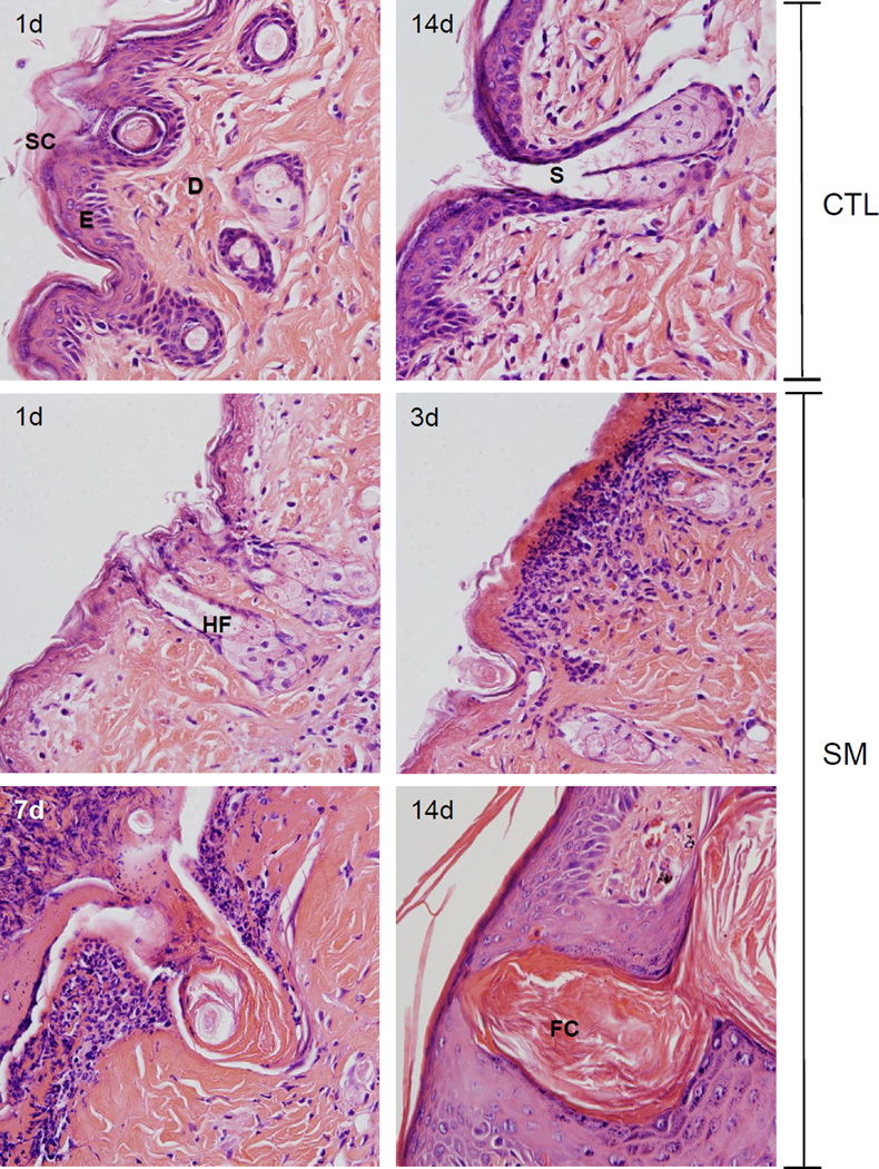
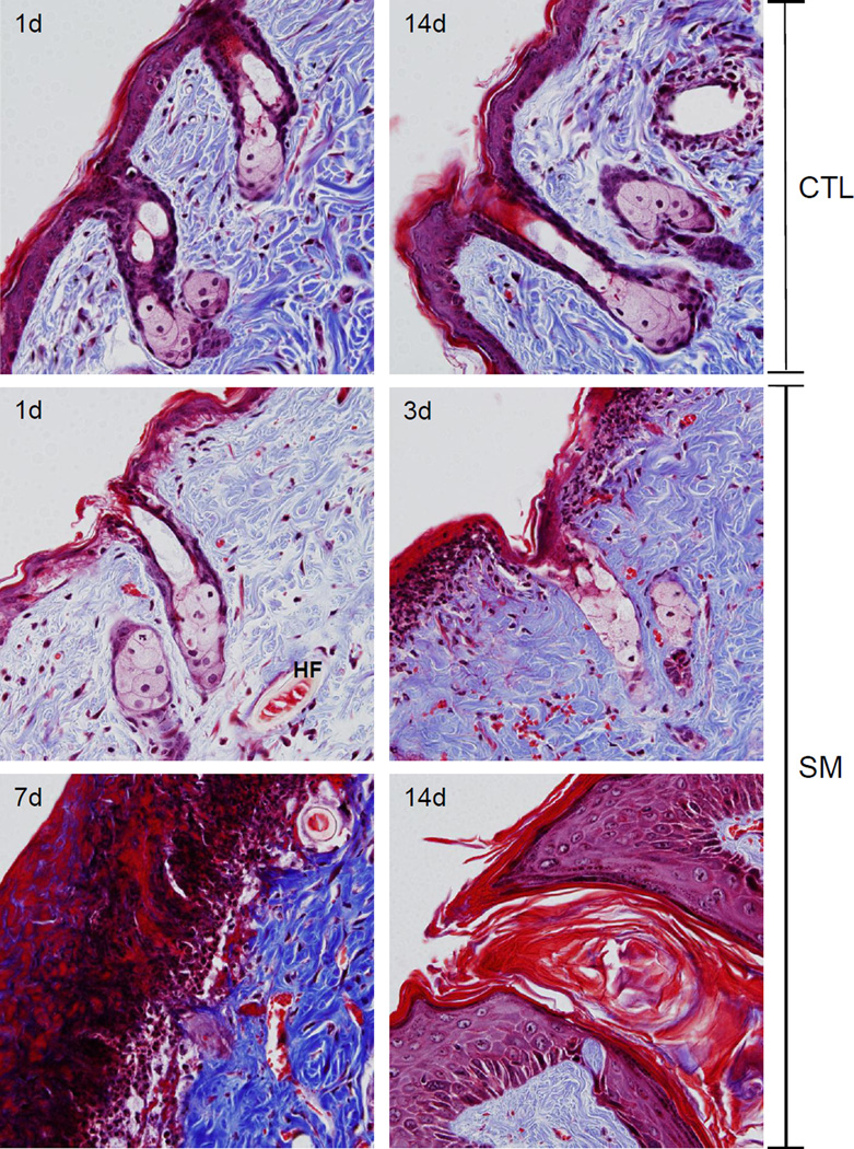
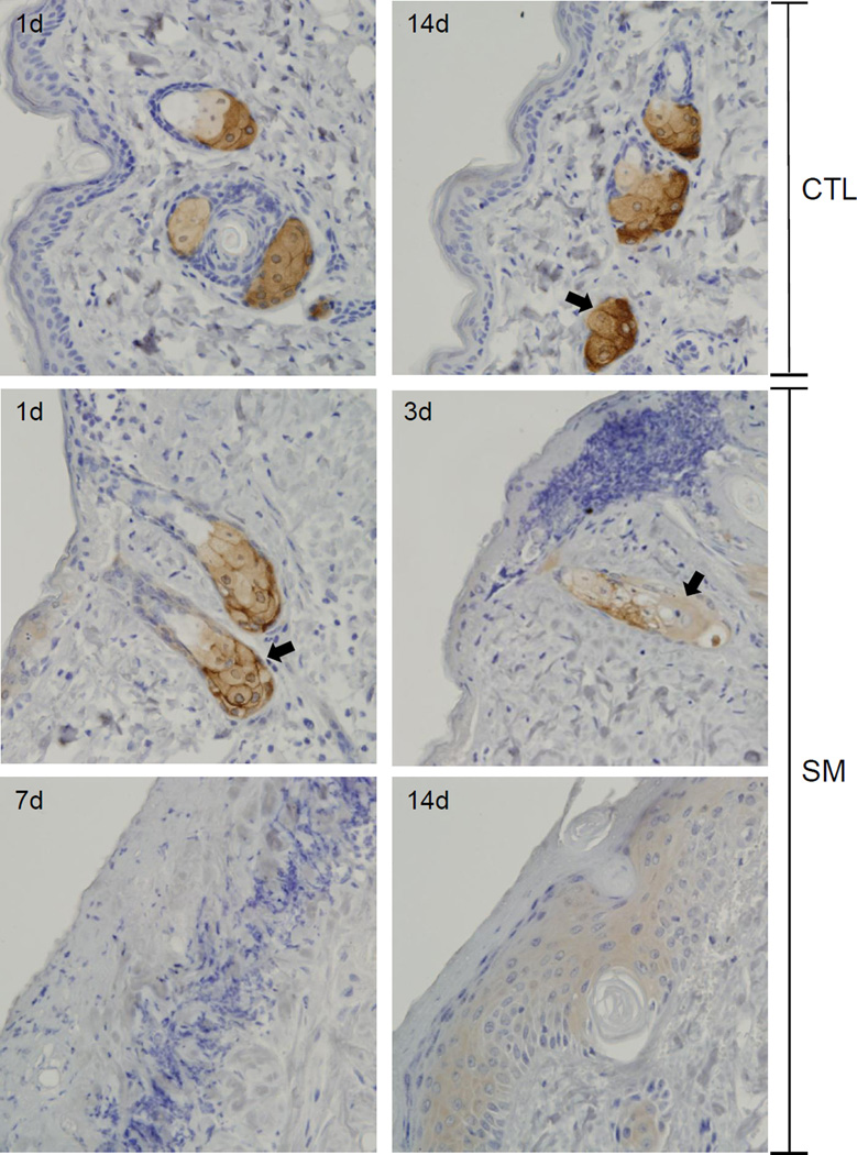
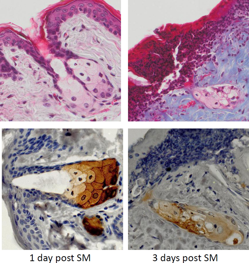
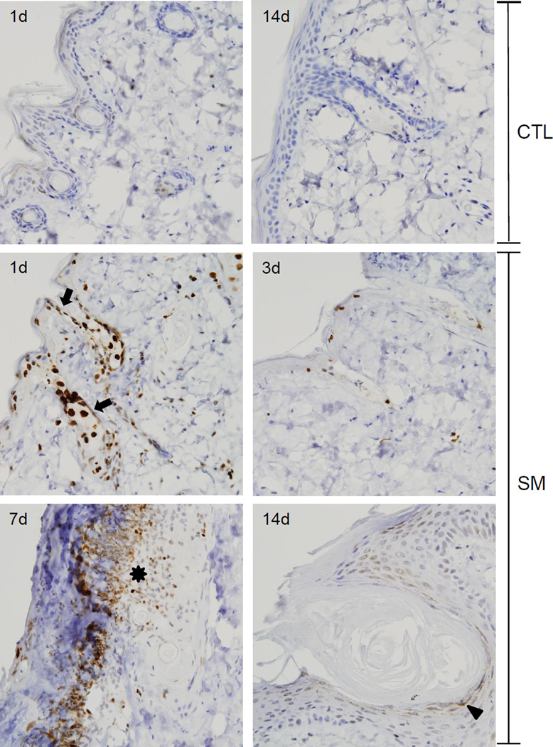
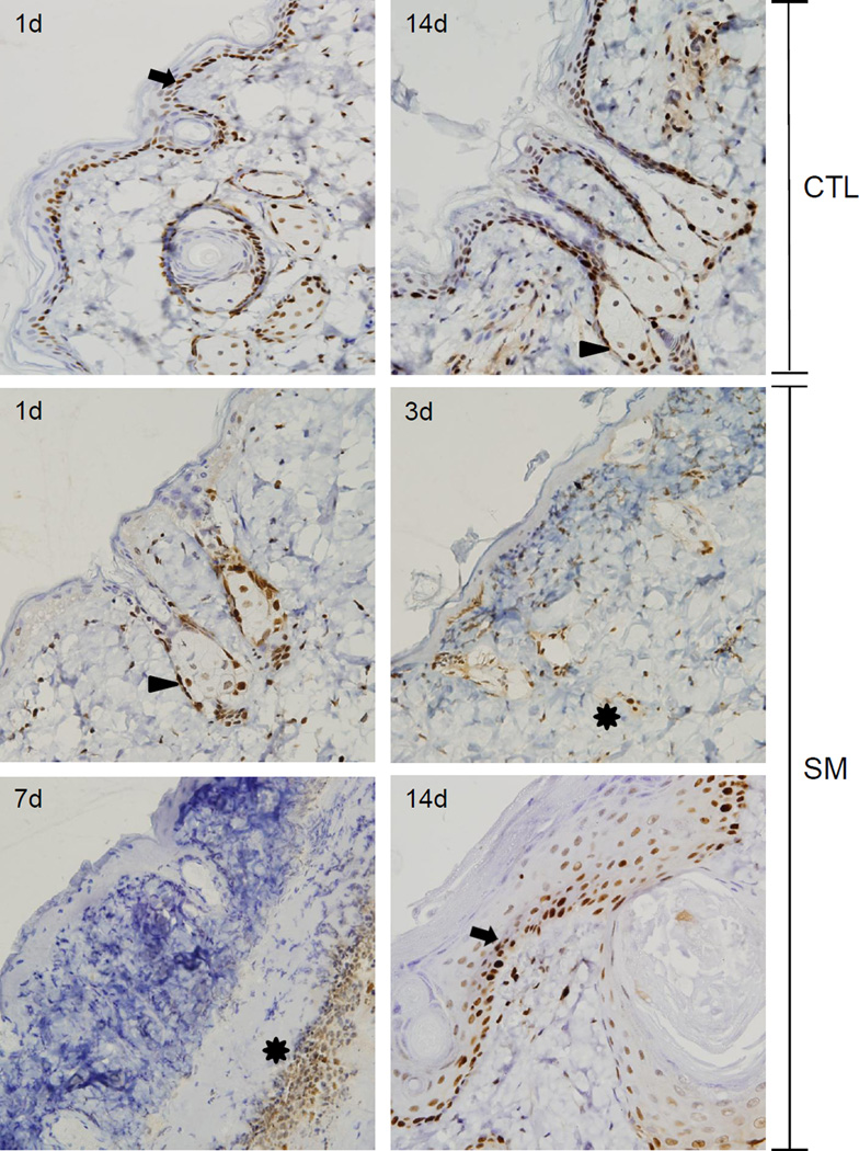
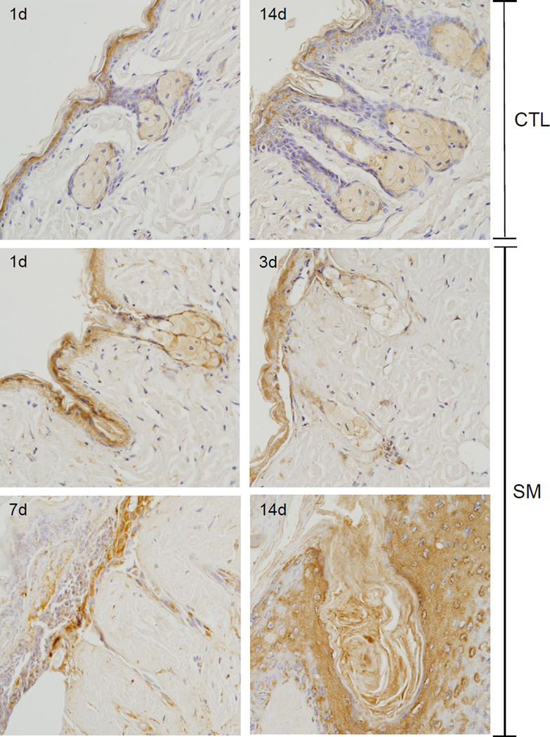
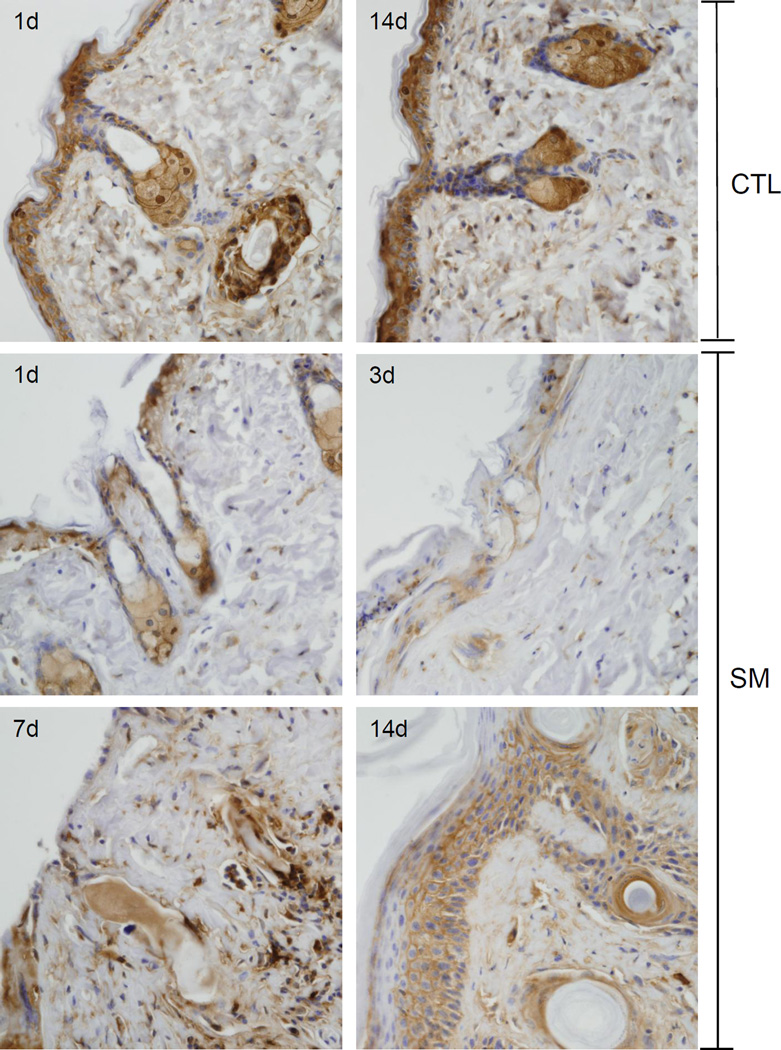
Sulfur mustard (SM) is a bifunctional alkylating agent causing skin inflammation, edema and blistering. A hallmark of SM-induced toxicity is follicular and interfollicular epithelial damage. In the present studies we determined if SM-induced structural alterations in hair follicles and sebaceous glands were correlated with cell damage, inflammation and wound healing. The dorsal skin of hairless mice was treated with saturated SM vapor. One to seven days later, epithelial cell karyolysis within the hair root sheath, infundibulum and isthmus was apparent, along with reduced numbers of sebocytes. Increased numbers of utriculi, some with connections to the skin surface, and engorged dermal cysts were also evident. This was associated with marked changes in expression of markers of DNA damage (phospho-H2A.X), apoptosis (cleaved caspase-3), and wound healing (FGFR2 and galectin-3) throughout pilosebaceous units. Conversely, fatty acid synthase and galectin-3 were down-regulated in sebocytes after SM. Decreased numbers of hair follicles and increased numbers of inflammatory cells surrounding the utriculi and follicular cysts were noted within the wound 3-7 days post-SM exposure. Expression of phospho-H2A.X, cleaved caspase-3, FGFR2 and galectin-3 was decreased in dysplastic follicular epidermis. Fourteen days after SM, engorged follicular cysts which expressed galectin-3 were noted within hyperplastic epidermis. Galectin-3 was also expressed in basal keratinocytes and in the first few layers of suprabasal keratinocytes in neoepidermis formed during wound healing indicating that this lectin is important in the early stages of keratinocyte differentiation. These data indicate that hair follicles and sebaceous glands are targets for SM in the skin.
Keywords: Hair follicles; Phosphorylated histone H2A.X; Sebaceous glands; Sulfur mustard.
Copyright © 2014 Elsevier Inc. All rights reserved.
Figures

Similar articles
-
Skin remodeling and wound healing in the Gottingen minipig following exposure to sulfur mustard.Exp Mol Pathol. 2020 Aug;115:104470. doi: 10.1016/j.yexmp.2020.104470. Epub 2020 May 21. Exp Mol Pathol. 2020. PMID: 32445752 Free PMC article.
-
Tissue injury and repair following cutaneous exposure of mice to sulfur mustard.Ann N Y Acad Sci. 2016 Aug;1378(1):118-123. doi: 10.1111/nyas.13125. Epub 2016 Jul 2. Ann N Y Acad Sci. 2016. PMID: 27371823 Free PMC article. Review.
-
Structural changes in the skin of hairless mice following exposure to sulfur mustard correlate with inflammation and DNA damage.Exp Mol Pathol. 2011 Oct;91(2):515-27. doi: 10.1016/j.yexmp.2011.05.010. Epub 2011 Jun 13. Exp Mol Pathol. 2011. PMID: 21672537 Free PMC article.
-
Therapeutic potential of a non-steroidal bifunctional anti-inflammatory and anti-cholinergic agent against skin injury induced by sulfur mustard.Toxicol Appl Pharmacol. 2014 Oct 15;280(2):236-44. doi: 10.1016/j.taap.2014.07.016. Epub 2014 Aug 13. Toxicol Appl Pharmacol. 2014. PMID: 25127551 Free PMC article.
-
Pathophysiological Effects of Sulfur Mustard on Skin and its Current Treatments: Possible Application of Phytochemicals.Comb Chem High Throughput Screen. 2021;24(1):3-19. doi: 10.2174/1386207323666200717150414. Comb Chem High Throughput Screen. 2021. PMID: 32679016 Review.
Cited by
-
DNA damage signaling in the cellular responses to mustard vesicants.Toxicol Lett. 2020 Jun 15;326:78-82. doi: 10.1016/j.toxlet.2020.03.008. Epub 2020 Mar 12. Toxicol Lett. 2020. PMID: 32173488 Free PMC article.
-
Skin remodeling and wound healing in the Gottingen minipig following exposure to sulfur mustard.Exp Mol Pathol. 2020 Aug;115:104470. doi: 10.1016/j.yexmp.2020.104470. Epub 2020 May 21. Exp Mol Pathol. 2020. PMID: 32445752 Free PMC article.
-
Skin Models Used to Define Mechanisms of Action of Sulfur Mustard.Disaster Med Public Health Prep. 2023 Oct 18;17:e551. doi: 10.1017/dmp.2023.177. Disaster Med Public Health Prep. 2023. PMID: 37849329 Free PMC article. Review.
-
Mustard vesicants alter expression of the endocannabinoid system in mouse skin.Toxicol Appl Pharmacol. 2016 Jul 15;303:30-44. doi: 10.1016/j.taap.2016.04.014. Epub 2016 Apr 26. Toxicol Appl Pharmacol. 2016. PMID: 27125198 Free PMC article.
-
Tissue injury and repair following cutaneous exposure of mice to sulfur mustard.Ann N Y Acad Sci. 2016 Aug;1378(1):118-123. doi: 10.1111/nyas.13125. Epub 2016 Jul 2. Ann N Y Acad Sci. 2016. PMID: 27371823 Free PMC article. Review.
References
-
- Arck P, Paus R. From the brain-skin connection: the neuroendocrine-immune misalliance of stress and itch. Neuroimmunomodulation. 2006;13:347–356. - PubMed
-
- Bacchi CE, Gown AM. Detection of cell proliferation in tissue sections. Braz J Med Biol Res. 1993;26:677–687. - PubMed
-
- Black AT, Hayden PJ, Casillas RP, Heck DE, Gerecke DR, Sinko PJ, Laskin DL, Laskin JD. Expression of proliferative and inflammatory markers in a fullthickness human skin equivalent following exposure to the model sulfur mustard vesicant, 2-chloroethyl ethyl sulfide. Toxicol Appl Pharmacol. 2010;249:178–187. - PMC - PubMed
-
- Cao Z, Said N, Amin S, Wu HK, Bruce A, Garate M, Hsu DK, Kuwabara I, Liu FT, Panjwani N. Galectins-3 and-7, but not galectin-1, play a role in reepithelialization of wounds. J Biol Chem. 2002;277:42299–42305. - PubMed
Publication types
MeSH terms
Substances
Grants and funding
- R01 CA132624/CA/NCI NIH HHS/United States
- CA132624/CA/NCI NIH HHS/United States
- U01 NS079249/NS/NINDS NIH HHS/United States
- T32 ES007148/ES/NIEHS NIH HHS/United States
- NS079249/NS/NINDS NIH HHS/United States
- P30 ES005022/ES/NIEHS NIH HHS/United States
- CA093798/CA/NCI NIH HHS/United States
- R01 CA093798/CA/NCI NIH HHS/United States
- U54 AR055073/AR/NIAMS NIH HHS/United States
- ES005022/ES/NIEHS NIH HHS/United States
- ES004738/ES/NIEHS NIH HHS/United States
- U54AR055073/AR/NIAMS NIH HHS/United States
- R01 ES004738/ES/NIEHS NIH HHS/United States
- R55 CA093798/CA/NCI NIH HHS/United States
LinkOut - more resources
Full Text Sources
Other Literature Sources
Research Materials
Miscellaneous
