. 2014 May 9;289(19):13308-22.
doi: 10.1074/jbc.M114.559088. Epub 2014 Mar 24.

ADAM10 is the major sheddase responsible for the release of membrane-associated meprin A

Affiliations

ADAM10 is the major sheddase responsible for the release of membrane-associated meprin A

Christian Herzog et al. J Biol Chem. .

Abstract

Meprin A, composed of α and β subunits, is a membrane-bound metalloproteinase in renal proximal tubules. Meprin A plays an important role in tubular epithelial cell injury during acute kidney injury (AKI). The present study demonstrated that during ischemia-reperfusion-induced AKI, meprin A was shed from proximal tubule membranes, as evident from its redistribution toward the basolateral side, proteolytic processing in the membranes, and excretion in the urine. To identify the proteolytic enzyme responsible for shedding of meprin A, we generated stable HEK cell lines expressing meprin β alone and both meprin α and meprin β for the expression of meprin A. Phorbol 12-myristate 13-acetate and ionomycin stimulated ectodomain shedding of meprin β and meprin A. Among the inhibitors of various proteases, the broad spectrum inhibitor of the ADAM family of proteases, tumor necrosis factor-α protease inhibitor (TAPI-1), was most effective in preventing constitutive, phorbol 12-myristate 13-acetate-, and ionomycin-stimulated shedding of meprin β and meprin A in the medium of both transfectants. The use of differential inhibitors for ADAM10 and ADAM17 indicated that ADAM10 inhibition is sufficient to block shedding. In agreement with these results, small interfering RNA to ADAM10 but not to ADAM9 or ADAM17 inhibited meprin β and meprin A shedding. Furthermore, overexpression of ADAM10 resulted in enhanced shedding of meprin β from both transfectants. Our studies demonstrate that ADAM10 is the major ADAM metalloproteinase responsible for the constitutive and stimulated shedding of meprin β and meprin A. These studies further suggest that inhibiting ADAM 10 activity could be of therapeutic benefit in AKI.

Keywords: ADAM ADAMTS; ADAM10; ADAM17; Kidney; Meprin; Metalloprotease; Mouse; Shedding; siRNA.


Figures
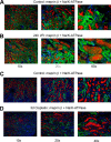
FIGURE 1.
FIGURE 1.
Meprin A distribution in kidneys of mice subjected to ischemia-reperfusion or cisplatin injury. A–D, double-immunofluorescence of meprin A (red) and Na/K-ATPase (green) in kidney sections of control mice (A and C) and mice subjected to IR (24 h of reperfusion after 45 min of ischemia) (B) or cisplatin injury (D). Kidney tissue was fixed in phosphate-buffered 4% formalin and paraffin-embedded. Tissue sections (8 μm) were incubated with goat polyclonal anti-meprin-β and rabbit polyclonal anti-Na+/K+-ATPase antibodies overnight. After washing with PBS, slices were incubated with secondary antibodies from Molecular Probes (donkey anti-goat Alexa Fluor 594 (red) and donkey anti-rabbit Alexa Fluor 488 (green). Nuclei were stained with DAPI. Epifluorescence pictures were recorded on an Olympus (IX51, IX71, or BX51) microscope.
FIGURE 2.
FIGURE 2.
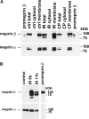
Shedding of meprin β after IR and cisplatin (CP) injury. A, total homogenates, cytosolic and membrane fractions were prepared from kidneys of control mice and mice subjected to 24 h of IR injury and probed for meprin β by Western blot. ctrl total, control total. B, urine samples (normalized for protein content) from control mice and mice subjected to IR injury (6 and 11 h of reperfusion injury after 30 min of ischemia, as indicated) were probed for meprin β and meprin α by Western blot. Recombinant human promeprin β lacking its membrane anchor purified from HEK293 cells described earlier (21) is shown as a reference control in last lane. The individual lanes as shown were obtained from the same Western blot.
FIGURE 3.
FIGURE 3.
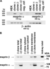
Characterization of meprin-expressing HEK293 cell lines. A, RT-PCRs for meprin β mRNA expression in stable meprin β HEK293 transfectants and for both meprin α and meprin β mRNA expression in stable meprin αβ co-transfectants were performed. Negative control lane (neg. ctrl) represents RT-PCR in the absence of the cDNA template. PCR products were separated on a 1% agarose gel stained with ethidium bromide. β-Actin is shown as a loading control. B, total cell lysates, membrane, and cytosolic fractions of HEK cells stably transfected with meprin β alone or co-transfected with meprin α and β expression plasmids were assayed for protein expression of meprin β and meprin α by Western blot. Total lysate of an untreated C57BL/6n mouse kidney is shown for comparison. Aliquots containing equal protein amounts were probed with a meprin β- or meprin α-specific antibody, and signals were detected with ECL reagent.
FIGURE 4.
FIGURE 4.
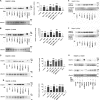
Effect of various protease inhibitors on meprin β shedding in meprin β and meprin αβ clones in the presence and absence of PMA and IM. A, effect of protease inhibitors on shedding of meprin β in HEK cells stably transfected with meprin β in the presence of PMA. The indicated cells were treated with 50 nm PMA for 30 min prior to the addition of 25 μm TAPI-1, 25 μm DAPT, 10 μm GM6001, 100 nm d-VFK-CMK, or 44 μm MMP2/9-1 inhibitors. Medium supernatants were centrifuged for 30 min at 100,000 × g to remove cellular debris, and aliquots of equal protein content were analyzed by Western blot for the presence of meprin β (proMeprin β lane). Recombinant human promeprin β lacking its membrane anchor purified from HEK293 cells described earlier (21) is shown as a reference (left panel). Albumin detected by Ponceau S stain is shown as a loading control. Western blot band intensities were evaluated by densitometry normalized to those observed in medium supernatant of the corresponding PMA-treated clones (right panel). Error bars represent S.E., n = 4. **, p < 0.01, ***, p < 0.001. B, effect of protease inhibitors on secretion of meprin β in HEK cells stably co-transfected with meprin α and meprin β (meprin A) in the presence of PMA. Cells were treated with inhibitors in the presence and absence of PMA and processed for shedding of meprin β in the medium (left panel). Western blot band intensities were quantified by densitometry (right panel). Error bars represent S.E., n = 4. *, p < 0.05, ***, p < 0.001. C, effect of protease inhibitors on shedding of meprin β in HEK cells stably transfected with meprin β in the presence of IM. The indicated cells were treated with 2.5 μm IM for 30 min prior to the addition of 25 μm TAPI-1, 25 μm DAPT, 10 μm GM6001, 100 nm d-VFK-CMK, or 44 μm MMP2/9-1 inhibitors. Medium supernatants were centrifuged for 30 min at 100,000 × g to remove cellular debris and aliquots of equal protein content analyzed by Western blot for the presence of meprin β. Recombinant human promeprin β (proMeprin β lane) lacking its membrane anchor purified from HEK293 cells described earlier (21) is shown as a reference (left panel). Albumin detected by Ponceau S stain is shown as a loading control. Western blot band intensities were evaluated by densitometry normalized to those observed in medium supernatant of the corresponding IM treated clones (right panel). Error bars represent S.E., n = 4. *, p < 0.05, **, p < 0.01, ****, p < 0.0001. D, effect of protease inhibitors on secretion of meprin β in HEK cells stably co-transfected with meprin α and meprin β (meprin A) in the presence of IM. Cells were treated with inhibitors in the presence and absence of IM and processed for shedding of meprin β in the medium (left panel). Western blot band intensities were quantified by densitometry (right panel). Error bars represent S.E., n = 4. **, p < 0.01. E and F, detection of promeprin β shed from HEK cells. Western blots shown in panels C and D, respectively, were reprobed with a meprin β prodomain-specific antibody. G, detection of meprin β and meprin α shed from HEK cells stably co-transfected with meprin β and meprin α treated with IM or PMA in the presence or absence of TAPI-1.
FIGURE 5.
FIGURE 5.
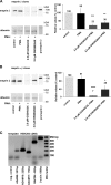
Effect of specific inhibitors of the ADAM family of proteases on meprin β shedding in meprin β and meprin αβ clones. A, HEK293 cells stably transfected with meprin β were treated with highly specific inhibitors of ADAM10 and ADAM17 in the presence or absence of PMA (30 ng/ml) for 4 h. The inhibitors used were GI254023X (2 μm) and GW280264X (2 μm). Medium supernatants were centrifuged for 30 min at 100,000 × g to remove cellular debris, and aliquots of equal protein content were analyzed by Western blot for the presence of meprin β (left panel). The individual lanes as shown were obtained from the same Western blot. Recombinant human promeprin β lacking its membrane anchor purified from HEK293 cells described earlier (21) is shown as a reference. Western band intensities were evaluated by densitometry normalized to those observed in medium supernatant of the corresponding untreated clones (right panel). Error bars represent S.E., n = 3. **, p < 0.01, ***, p < 0.001. B, HEK293 cells stably co-transfected with meprin αβ (meprin A) were treated with highly specific inhibitors of ADAM10 and ADAM17 in the presence and absence of PMA (30 ng/ml) for 4 h as described for panel A. Medium supernatants were analyzed by Western blot for the presence of meprin β (left panel). The individual lanes as shown were obtained from the same Western blot. Western band intensities were evaluated by densitometry normalized to those observed in medium supernatant of the corresponding untreated clones (right panel). Error bars represent S.E., n = 3, *, p < 0.05, ****, p < 0.0001. C, untransfected HEK293 cells were tested for the presence of ADAM9, ADAM10, and ADAM17 mRNA by RT-PCR using human ADAM9-, ADAM10-, and ADAM17-specific primers as described under “Experimental Procedures.” neg. control, negative control.
FIGURE 6.
FIGURE 6.
Effect of siRNAs to ADAM9, ADAM10, and ADAM17 on meprin β shedding in meprin β and meprin αβ-expressing clones in the absence and presence of PMA or IM. A, ADAM10 expression was knocked down with siRNA pools to human ADAM10 in HEK293 or HEK cells stably transfected with meprin β or co-transfected with meprin β and meprin α. Scrambled siRNA was used as a negative control. Forty-eight hours after transfection, cells were incubated with PMA (30 ng/ml), IM (2.5 μm), or vehicle for 4 h. Cell lysates were analyzed for expression of ADAM10 by Western blot. α-Actinin served as a loading control. B, ADAM17 expression was knocked down with siRNA pools to human ADAM17 in HEK293 or HEK cells stably transfected with meprin β or co-transfected with meprin β and meprin α. Scrambled siRNA was used as a negative control. Forty-eight hours after transfection, cells were incubated with PMA (30 ng/ml), IM (2.5 μm), or vehicle for 4 h. Cell lysates were analyzed for expression of ADAM10 by Western blot; α-actinin served as a loading control. C, ADAM9, ADAM10, or ADAM17 expression was knocked down with siRNA pools to the indicated ADAMs in HEK293 cells stably transfected with meprin β. Scrambled siRNA was used as a negative control. Cells (48 h after transfection) were incubated with PMA (30 ng/ml) or vehicle for 4 h. Medium supernatants were harvested, and aliquots of equal protein amount were analyzed by Western blot for the presence of meprin β (left panels). Albumin detected by Ponceau S stain is shown as a loading control. This panel shows a representative Western blot from three independent experiments. Western band intensities were evaluated by densitometry normalized to those observed in medium supernatant of the corresponding clone treated with scrambled siRNA (right panel). Numbers in the diagrams represent the average band intensities, and bars represent the S.E. obtained from 3 independent experiments. *, p < 0.05 was considered statistically significant. D, ADAM9, ADAM10, or ADAM17 expression was knocked down with siRNA pools to the indicated ADAMs in HEK293 cells stably co-transfected with meprin α and β. Cells (48 h after transfection) were incubated with PMA (30 ng/ml) or vehicle for 4 h. Medium supernatants were analyzed by Western blot for the presence of meprin β as described for panel B (left panel). Albumin detected by Ponceau S stain is shown as a loading control. This panel shows a representative Western blot from three independent experiments. Western band intensities were evaluated by densitometry normalized to those observed in medium supernatant of the corresponding clone treated with scrambled siRNA (right panel). Numbers in the diagrams represent the average band intensities, and bars represent the S.E. obtained from 3 independent experiments. **, p < 0.01, ***, p < 0.001.
FIGURE 7.
FIGURE 7.
Overexpression of ADAM10 leads to increased shedding of meprin β shedding in meprin β- and meprin αβ-expressing clones. A, HEK293 cells stably transfected with meprin β or co-transfected with meprin β and meprin α were transiently transfected with bovine ADAM10-HA in pcDNA3, empty vector as control, or Lipofectamine 2000 alone (LF only). Cell lysates were prepared 48 h after transfection, and aliquots of equal protein amount were analyzed by Western blot for ADAM10. α-Actinin was used as a loading control. This panel shows a representative Western blot from five independent experiments. B, HEK293 cells stably transfected with meprin β or co-transfected with meprin α and β were transiently transfected with bovine ADAM10-HA in pcDNA3, empty vector as control, or Lipofectamine 2000 alone (LF only). Medium supernatants were harvested 48 h after transfection and analyzed by Western blot for meprin β shedding (left panel). This panel shows a representative Western blot from five independent experiments. Western band intensities were evaluated by densitometry (right panel). Band intensities of clones transfected with empty vector were used as controls for normalization. Numbers in the diagrams represent the average band intensities, and bars represent the S.E. of the mean obtained from 4–5 independent experiments as indicated. **, p < 0.01, ***, p < 0.001.
FIGURE 8.
FIGURE 8.
Meprin β shedding in primary renal tubular epithelial cells (RTEC) treated with inhibitors TAPI-1 or GI254023X in the presence or absence of PMA and ionomycin. Primary renal tubular epithelial cells were treated with 50 nm PMA or 2.5 μm ionomycin or vehicle (DMSO) for 30 min prior to the addition of 25 μm TAPI-1, 2 μm GI254023X, or vehicle. After 2 h, medium supernatants were collected and centrifuged at 500 × g for 5 min to remove cells, and the supernatants were centrifuged at 100,000 × g for 30 min to remove cellular debris. Aliquots of equal protein content were analyzed by Western blots for the presence of meprin β. Recombinant human promeprin β lacking its membrane anchor purified from HEK293 cells described earlier (21) is shown as a reference (left panel). Albumin detected by Ponceau S stain is shown as a loading control. Western blot intensities were evaluated by densitometry normalized to those observed in medium supernatants of the corresponding control (right panel). Numbers represent the average of 5 independent experiments. Error bars represent S.E., n = 5. * p < 0.05, ** p < 0.01, *** p < 0.001.
FIGURE 9.
FIGURE 9.
Illustration of meprin A ectodomain shedding with putative cleavage sites by ADAM10. The picture represents meprin A in the hetero-tetrameric form β2α2. The two β subunits provide the membrane anchor, and the α subunits are covalently linked to the β subunits by disulfide bridges. Putative cleavage sites by ADAM10 are located at the junction of the AM and the EGF-like domain. The panel on the right shows the individual domains of a meprin β subunit.

Similar articles

Cited by

References

    1. Sterchi E. E., Stöcker W., Bond J. S. (2008) Meprins, membrane-bound and secreted astacin metalloproteinases. Mol. Aspects Med. 29, 309–328 - PMC - PubMed
    1. Becker-Pauly C., Höwel M., Walker T., Vlad A., Aufenvenne K., Oji V., Lottaz D., Sterchi E. E., Debela M., Magdolen V., Traupe H., Stöcker W. (2007) The α and β subunits of the metalloprotease meprin are expressed in separate layers of human epidermis, revealing different functions in keratinocyte proliferation and differentiation. J. Invest. Dermatol. 127, 1115–1125 - PubMed
    1. Lottaz D., Hahn D., Müller S., Müller C., Sterchi E. E. (1999) Secretion of human meprin from intestinal epithelial cells depends on differential expression of the α and β subunits. Eur. J. Biochem. 259, 496–504 - PubMed
    1. Lottaz D., Maurer C. A., Hahn D., Büchler M. W., Sterchi E. E. (1999) Nonpolarized secretion of human meprin α in colorectal cancer generates an increased proteolytic potential in the stroma. Cancer Res. 59, 1127–1133 - PubMed
    1. Crisman J. M., Zhang B., Norman L. P., Bond J. S. (2004) Deletion of the mouse meprin β metalloprotease gene diminishes the ability of leukocytes to disseminate through extracellular matrix. J. Immunol. 172, 4510–4519 - PubMed

Publication types

MeSH terms

LinkOut - more resources