. 2014 Mar 24:5:3501.
doi: 10.1038/ncomms4501.

Molecular basis for erythromycin-dependent ribosome stalling during translation of the ErmBL leader peptide

Affiliations

Molecular basis for erythromycin-dependent ribosome stalling during translation of the ErmBL leader peptide

Stefan Arenz et al. Nat Commun. .

Abstract

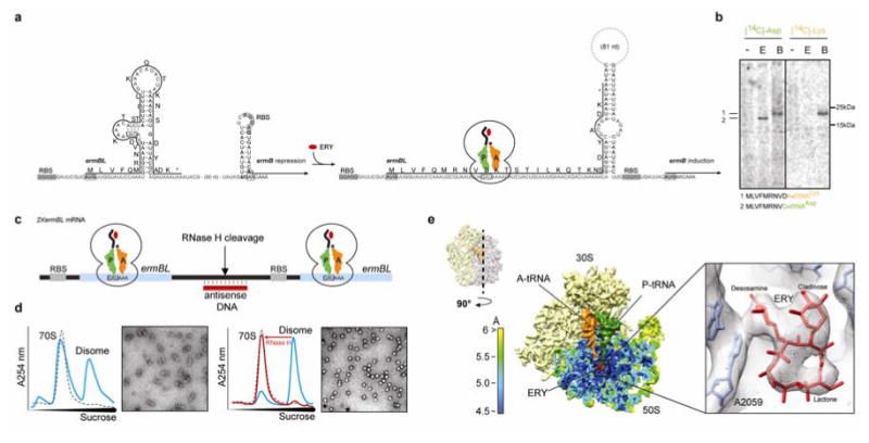
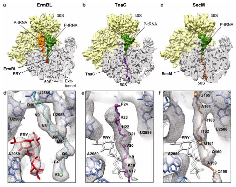
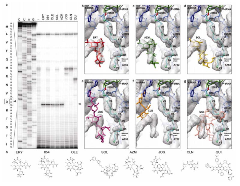
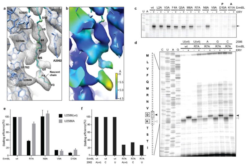
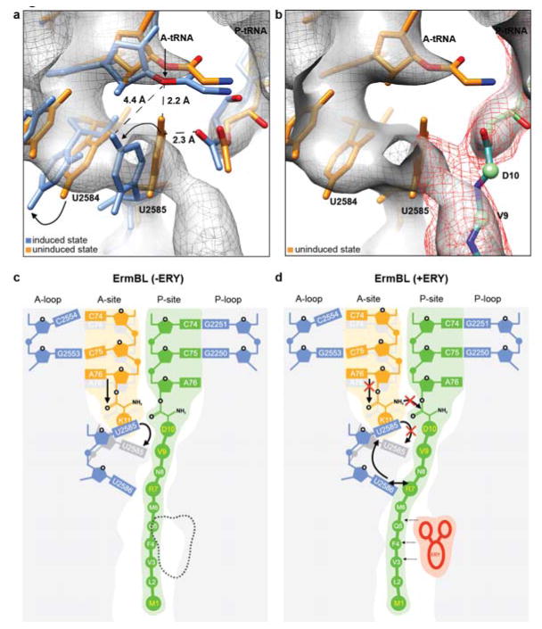
In bacteria, ribosome stalling during translation of ErmBL leader peptide occurs in the presence of the antibiotic erythromycin and leads to induction of expression of the downstream macrolide resistance methyltransferase ErmB. The lack of structures of drug-dependent stalled ribosome complexes (SRCs) has limited our mechanistic understanding of this regulatory process. Here we present a cryo-electron microscopy structure of the erythromycin-dependent ErmBL-SRC. The structure reveals that the antibiotic does not interact directly with ErmBL, but rather redirects the path of the peptide within the tunnel. Furthermore, we identify a key peptide-ribosome interaction that defines an important relay pathway from the ribosomal tunnel to the peptidyltransferase centre (PTC). The PTC of the ErmBL-SRC appears to adopt an uninduced state that prevents accommodation of Lys-tRNA at the A-site, thus providing structural basis for understanding how the drug and the nascent peptide cooperate to inhibit peptide bond formation and induce translation arrest.


Figures

Figure 1
Figure 1. Cryo-EM structure of an ErmBL-stalled ribosome complex
a, Schematic for ermBL-dependent regulation of ermB translation in the presence of erythromycin (ERY). b, In vitro translation of ErmBL using either 14C-Asp or 14C-Lys in the absence (-) or presence (E) of 50 μM ERY. In the control samples (B), where translation was carried-out in the absence of ERY, but in presence of borrelidin (a Thr-tRNA synthetase inhibitor) translation arrest occurred after an 11 amino acid nascent chain was (i.e. with codon 12 in the A-site i.e. one codon further compared to the natural stall site) polymerized thereby providing a mobility marker and confirming 14C-Lys incorporation in the absence of ERY. c, The bicistronic 2XermBL mRNA was translated in vitro in the presence of 10 μM ERY in order to generate ErmBL-SRC disomes. Complementary DNA oligo and RNase H cleavage converts disomes to monosomes, as shown by (d) sucrose density centrifugation and negative stain EM. e, Surface and cross-section of the ErmBL-SRC, containing 30S (yellow), 50S (colored according to local resolution), A-tRNA (orange), P-tRNA (green) and erythromycin (red). Inset shows electron density for erythromycin (grey mesh) with fitted crystal structure (PDB3OFR).
Figure 2
Figure 2. Path of the ErmBL compared to TnaC and SecM nascent chains
a-c, Comparison of cryo-EM structures of ErmBL-SRC, TnaC-SRC and SecM-SRC, with a transverse section of the 50S subunit (grey) displaying the path of the respective nascent chains through the ribosomal tunnel. d-f, Paths of the (d) ErmBL (teal), (e) TnaC (purple) and (f) SecM (brown) nascent chains. Erythromycin (ERY) is colored red in (d) ErmBL-SRC, whereas the superimposed position of ERY is colored white in (e) TnaC-SRC and (f) SecM-SRC.
Figure 3
Figure 3. Superimposition of various antibiotics relative to ErmBL
a, Detection of ribosome stalling by toeprinting during translation of ermBL, in the presence of the antibiotics erythromycin (ERY), ITR-054 (054), oleandomycin (OLE), solithromycin (SOL), azithromycin (AZM), josamycin (JOS), clindamycin (CLN) and quinupristin (QUI). The arrowed toeprint band indicates that ErmBL arrests translation with the Asp (D10) codon (boxed) located in the P-site. b, Chemical structures of erythromycin (ERY), macrolides ITR-054 (054), oleandomycin (OLE), the ketolide solithromycin (SOL) and the azalide azithromycin (AZM), compared with the macrolide josamycin (JOS), the lincosamide clindamycin (CLN) and the streptogramin B quinupristin (QUI). Differences between erythromycin and respective drugs are highlighted. c, ErmBL-SRC map (grey mesh) with molecular model for ErmBL-tRNA (teal), 23S rRNA (blue) and erythromycin (red, PDB3OFR). d-h, as (c), but with relative binding positions of (d) azithromycin (AZM, green, PDB1M1K), (e) SOL (yellow, PDB3ORB), (f) josamycin (JOS, purple, PDB2O44), (g) clindamycin (CLN, orange, PDB3OFZ) and (h) quinupristin (QUI, pink, PDB1SM1).
Figure 4
Figure 4. Interactions of the ErmBL nascent chain with the ribosome
a, Interactions of ErmBL (teal) with 23S rRNA (blue) within the exit tunnel of the ribosome. b, Local resolution map of view shown in (a). c, Alanine-scanning mutagenesis of ErmBL and effect of mutant peptides on ribosome stalling (arrowed) in the presence (+) and absence (-) of erythromycin (ERY) as determined by toe-printing. d, Toe-printing of wildtype (wt) and mutant (R7A) ErmBL on E. coli ribosomes with wild type (wt) or mutated 23S rRNA nucleotide 2586, in the presence (+) or absence (-) of erythromycin. All the reactions contained borrelidin, a Thr-tRNA synthetase inhibitor, which, in the absence of erythromycin-dependent arrest at the Asp (D10) codon, causes translation arrest at the Lys (K11) codon. e, Influence of the U2586A mutation on stalling efficiency with the wild type (wt) and mutant ErmBL peptides (an average of two independent experiments; error bars represent the standard deviation of the mean). f, The mutations of A2062 do not affect ErmBL-stalling, nor rescue stalling impaired by the R7A mutation.
Figure 5
Figure 5. The uninduced state of the PTC and an unaccommodated A-tRNA in the ErmBL-SRC prevent peptide bond formation
a-b, During accommodation of the A-tRNA, U2585 and U2584 undergo conformational changes (arrowed) necessary for peptide bond formation-. In the ErmBL-SRC, the electron density suggests that U2585 retains an unaccommodated state (orange, PDB1VQ6)- thus preventing proper placement of the A-tRNA in the PTC (blue, PDB1VQN)-. b, Electron density between the ErmBL nascent chain (dark mesh) and U2585 indicates that the C-terminal amino acids of ErmBL directly interact with U2585, likely restricting its movement. c, d, Model for the translation of ErmBL (c) in the absence of erythromycin (canonical translation), and (d) translation arrest in the presence of erythromycin (stalling). In (d), the drug restricts the conformational space available for the ErmBL nascent chain such that interactions with U2586 and U2585 are established. We propose that this prevents movement of U2585 from the uninduced to induced state and thus, hinders accommodation of the A-tRNA and peptide bond formation.

References

    1. Ito K, Chiba S. Arrest peptides: cis-acting modulators of translation. Annu Rev Biochem. 2013;82:171–202. - PubMed
    1. Ramu H, Mankin A, Vazquez-Laslop N. Programmed drug-dependent ribosome stalling. Mol Microbiol. 2009;71:811–824. - PubMed
    1. Vázquez-Laslop N, Ramu H, Mankin AS. In: Ribosomes. Structure, function, evolution. Rodnina MV, Wintermeyer W, Green R, editors. Springer-Verlag; 2011. pp. 377–392.
    1. Vazquez-Laslop N, Ramu H, Klepacki D, Kannan K, Mankin AS. The key function of a conserved and modified rRNA residue in the ribosomal response to the nascent peptide. EMBO J. 2010;29:3108–3117. - PMC - PubMed
    1. Mayford M, Weisblum B. The ermC leader peptide: amino acid alterations leading to differential efficiency of induction by macrolide-lincosamide-streptogramin B antibiotics. J Bacteriol. 1990;172:3772–3779. - PMC - PubMed

Publication types

MeSH terms

Associated data