. 2013:49:112-24.
doi: 10.1540/jsmr.49.112.

Chloride channel blockers promote relaxation of TEA-induced contraction in airway smooth muscle

Affiliations

Chloride channel blockers promote relaxation of TEA-induced contraction in airway smooth muscle

Peter D Yim et al. J Smooth Muscle Res. 2013.

Abstract

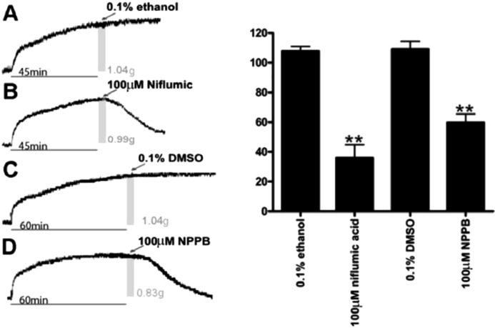
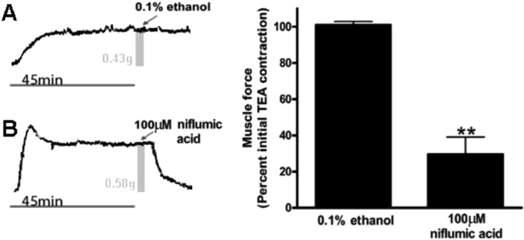
Enhanced airway smooth muscle (ASM) contraction is an important component in the pathophysiology of asthma. We have shown that ligand gated chloride channels modulate ASM contractile tone during the maintenance phase of an induced contraction, however the role of chloride flux in depolarization-induced contraction remains incompletely understood. To better understand the role of chloride flux under these conditions, muscle force (human ASM, guinea pig ASM), peripheral small airway luminal area (rat ASM) and airway smooth muscle plasma membrane electrical potentials (human cultured ASM) were measured. We found ex vivo guinea pig airway rings, human ASM strips and small peripheral airways in rat lungs slices relaxed in response to niflumic acid following depolarization-induced contraction induced by K(+) channel blockade with tetraethylammonium chloride (TEA). In isolated human airway smooth muscle cells TEA induce depolarization as measured by a fluorescent indicator or whole cell patch clamp and this depolarization was reversed by niflumic acid. These findings demonstrate that ASM depolarization induced contraction is dependent on chloride channel activity. Targeting of chloride channels may be a novel approach to relax hypercontractile airway smooth muscle in bronchoconstrictive disorders.


Figures

Fig. 1.
Fig. 1.
Plasma membrane potential in human airway smooth muscle. Left: Representative tracings of real-time FLIPR potentiometric dye fluorescence emissions after cultured human airway smooth muscle cells were treated with either buffer, 100 μM niflumic acid, 40 mM KCl, 10 mM TEA or 10 μM NS1619. These tracings display directionality of the fluorescence change in relation to depolarization (upward deflection: TEA, KCl) and hyperpolarization (downward deflection: niflumic acid, NS1619). Right: FLIPR potentiometric dye fluorescence emissions (RFU), measures at maximum change values, after cultured airway smooth muscle cells were treated with either buffer (control), 40 mM KCl, 10 μM NS1619, 100 μM niflumic acid or 10 mM TEA. * P<0.01 for niflumic (–195 ± 22 maximum ΔRFU) (n=11); ** P<0.001 for NS1619 (–341 ± 37.7 maximum ΔRFU), TEA (152 ± 16.6 maximum ΔRFU) or KCl (195 ± 20.5 maximum ΔRFU) compared to control (n=4).
Fig. 2.
Fig. 2.
Airway smooth muscle whole cell electrical recording of membrane potential. A representative tracing of voltage recordings in a cultured human airway smooth muscle cell under current clamp in a single whole cell recording. Resting membrane potential was –56.4 ± 6.5 mV (n=3). 10 mM TEA depolarized cultured human airway smooth muscle cells and 100 μM niflumic acid treatment after 10 mM TEA returned membrane potential to near baseline resting values (n=3).
Fig. 3.
Fig. 3.
TEA induced contractions of guinea pig airway smooth muscle are relaxed by chloride channel blockers NPPB and niflumic acid. Left: Representative tracings of guinea pig tracheal rings contracted with 10 mM TEA with the addition of either vehicle controls (0.1% ethanol or 0.1% DMSO) or chloride channel antagonists. A and B, the gray bars represent the 45 min time point when treatments were applied. The associated muscle force at the time of treatment is also noted as 1.04 and 0.99 grams, respectively. C and D, the gray bars represent the 60 min time point when treatments were applied. The associated muscle force at the time of treatment is also noted as 1.04 and 0.88 grams respectively. Right: Muscle force expressed as a percent of the plateau TEA-induced contraction. Airway smooth muscle demonstrated a significant reduction in muscle force 30 mins after treatment with 100 μM niflumic acid (n=6) (36.1 ± 8.8% of the initial TEA-induced force) (**P< 0.001 compared to ethanol control). Airway smooth muscle demonstrated a significant reduction in muscle force 30 mins after treatment with 100 μM NPPB (n=6) (59.8 ± 5.8% of the initial TEA-induced force at 30 min) (**P< 0.001 compared to DMSO control). Thirty minutes after treatment with ethanol (n=6) or DMSO (n=4) vehicles the resulting muscle force was not significantly different from the initial TEA-induced force (108 ± 3.2% and 109 ± 5.1%, respectively).
Fig. 4.
Fig. 4.
TEA induced contractions in human airway smooth muscle are relaxed by the chloride channel blocker niflumic acid. Left: Representative tracings of human tracheal strips muscle force with the addition of TEA and subsequently treated with either ethanol vehicle control or 100 μM niflumic acid. A and B, the gray bars represent the 45 min time point when treatments were applied. The associated muscle force at the time of treatment is also noted as 0.43 and 0.58 grams, respectively. Right: Muscle force expressed as a percent of plateau TEA-induced contraction. Human airway smooth muscle demonstrated a significant reduction in TEA-induced muscle force at 30 min after the addition of 100 μM niflumic acid (n=6) (29.6 ± 9.7% of the initial TEA-induced force) (** P<0.001 compared to ethanol control). Thirty minutes after treatment with ethanol vehicle (n=6) the resulting muscle force was not significantly different from the initial TEA-induced force (101 ± 1.9%).
Fig. 5.
Fig. 5.
Rat lung slice luminal area measurements. A) Representative images of video recorded rat lung slices. Images (1)–(4) correlate to graphically represented luminal area tracing demarcated by arrows ()–(4) in panel B. Photograph (1) is a rat bronchiole without treatment. Photograph (2) is the same rat bronchiole treated with 100 nM ACh. Photograph (3) is the same rat bronchiole after a buffer wash and treated with 50 mM KCl. Photograph (4) is the same rat bronchiole after a buffer wash and treated with 10 mM TEA. B) Left: A representative tracing of bronchiolar luminal area extracted from video recorded images expressed as a percent of baseline luminal area (denoted by (1)). Right: Airway luminal area was measured as a percentage of baseline luminal areas at points (2), (3) and (4). (n=5 for all groups). Mean intraluminal area values of TEA was not significantly different between ACh and KCl groups (n=5, P>0.05). C) Left: A representative tracing of bronchiolar luminal area measurements showing niflumic acid (NFA) direct relaxation of TEA contraction. Right: Airway luminal area as a percentage of baseline luminal area at points before and after niflumic acid were significantly different (n=4, *P<0.01).

References

    1. Sondik EJ, Madans JH, Gentleman JF. Summary Health Statistics for U.S. Adults: National Health Interview Survey 2011, U.S. Department of Health and Human Services. Vital and Health Statistics ed. Center for Disease Prevention and Control, 2012: 1–218. - PubMed
    1. Wijesinghe M, Weatherall M, Perrin K, Harwood M, Beasley R. Risk of mortality associated with formoterol: a systematic review and meta-analysis. Eur Respir J. 2009; 34(4): 803–11. doi: 10.1183/09031936.00159708 - DOI - PubMed
    1. Hirota S, Helli P, Janssen LJ. Ionic mechanisms and Ca2+ handling in airway smooth muscle. Eur Respir J. 2007; 30(1): 114–33. doi: 10.1183/09031936.00147706 - DOI - PubMed
    1. Janssen LJ, Sims SM. Spontaneous transient inward currents and rhythmicity in canine and guinea-pig tracheal smooth muscle cells. Pflugers Arch. 1994; 427(5-6): 473–80. doi: 10.1007/BF00374263 - DOI - PubMed
    1. Hirota S, Trimble N, Pertens E, Janssen LJ. Intracellular Cl- fluxes play a novel role in Ca2+ handling in airway smooth muscle. Am J Physiol Lung Cell Mol Physiol. 2006; 290(6): L1146–53. doi: 10.1152/ajplung.00393.2005 - DOI - PubMed

Publication types

MeSH terms