. 2014 Sep 11;33(37):4613-22.
doi: 10.1038/onc.2014.66. Epub 2014 Mar 24.

Exosomal HIF1α supports invasive potential of nasopharyngeal carcinoma-associated LMP1-positive exosomes

Affiliations
Free PMC article

Exosomal HIF1α supports invasive potential of nasopharyngeal carcinoma-associated LMP1-positive exosomes

M Aga et al. Oncogene. .
Free PMC article

Abstract

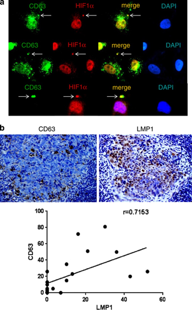
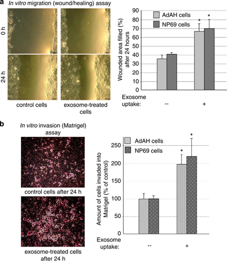
It has emerged recently that exosomes are potential carriers of pro-tumorigenic factors that participate in oncogenesis. However, whether oncogenic transcription factors are transduced by exosomes is unknown. Hypoxia-inducible factor-1α (HIF1α) transcriptionally regulates numerous key aspects of tumor development and progression by promoting a more aggressive tumor phenotype, characterized by increased proliferation and invasiveness coupled with neoangiogenesis. It has been shown that the principal oncoprotein of Epstein-Barr virus (EBV), latent membrane protein 1 (LMP1), drives oncogenic processes and tumor progression of the highly invasive EBV malignancy, nasopharyngeal carcinoma (NPC). We now demonstrate that endogenous HIF1α is detectable in exosomes and that LMP1 significantly increases levels of HIF1α in exosomes. HIF1 recovered from exosomes retains DNA-binding activity and is transcriptionally active in recipient cells after exosome uptake. We also show that treatment of EBV-negative cells with LMP1-exosomes increases migration and invasiveness of NP cell lines in functional assays, which correlates with the phenotype associated with epithelial-mesenchymal transition (EMT). In addition, we provide evidence that HIF1α itself participates in exosome-mediated pro-metastatic effects in recipient cells, as exosome-mediated delivery of active and inactive forms of HIF1α results in reciprocal changes in the expression of E- and N-cadherins associated with EMT. Further, immunohistochemical analysis of NPC tumor tissues revealed direct correlation between protein levels of LMP1 and of the endosome/exosome marker tetraspanin, CD63, which suggests an increase in exosome formation in this EBV-positive malignancy. We hypothesize that exosome-mediated transfer of functional pro-metastatic factors by LMP1-positive NPC cells to surrounding tumor cells promotes cancer progression.


Figures

Figure 1
Figure 1
The EBV oncogene LMP1 increases endogenous levels of the transcription factor HIF1α in exosomes secreted by NP cells. (a) Purified exosomal fractions from NP69 cells stably expressing LMP1 or the control and AdAH nasopharyngeal cells transiently transfected with LMP1 or a control-expression vector were analyzed by immunoblotting. Total protein lysates of exosome pellets, isolated from supernatant fluids, were normalized to exosome-specific markers HSC70 and flotillin2. (b) Purified exosomes from NP69-LMP1 and 293T cells transiently transfected with HIF1α expression plasmids were subjected to ultrastructural examination and immunogold labeling for HIF1α. Exosomes were stained with HIF1α primary and colloidal gold-labeled secondary antibodies. (c) LMP1-negative AdAH cells were treated with graduated amounts of exosome fractions isolated from LMP1-expressing AdAH cells. After 24 h, western blotting analyses were used to detect LMP1, HIF1α and GAPDH (glyceraldehyde 3-phosphate dehydrogenase) expression in recipient AdAH cells.
Figure 2
Figure 2
Exosome-mediated transfer of functionally active HIF1α to recipient cells. (a) 293T cells (LMP1-negative) were transfected with pcDNA3 control and HA-tagged WT or HA-tagged DN HIF1α expression vectors along with HRE-luciferase reporter plasmid (see Materials and methods). Luciferase assays for HIF1α activity and western blotting for determination of HA-tagged proteins were performed 24 h post transfection. (b) Exosomes from the same transfected cells were purified, as described in Materials and methods. The presence of HIF1α WT and DN mutant in exosomes was determined by western blotting with HA-tag antibodies. Exosomal fractions were normalized to HSC70 and flotillin2 (exosomal markers). (c) Recipient 293T cells were transfected with HRE-luciferase reporter and exposed to purified control, WT or DN HIF1α-positive exosomes as described in panel (b). Twenty-four hours later, luciferase assays were performed and normalized to β-galactosidase activity. The mean s.ds. were determined for experiments performed in triplicate. (d) The DNA-binding capacity of exosomal HIF1α was determined by in vitro electrophoretic mobility shift assay (EMSA): exosome protein fractions were incubated with HRE probe (see Materials and methods) according to the BD manufacturer's protocol and separated by non-denaturating 6% PAGE. To detect shifts of HRE–HIF1 complexes, the gel was stained with SYBR green EMSA to visualize DNA (left), and after transfer, the membrane was probed with HIF1α antibodies for specific identification of exosomal HIF1 in complex with the HRE promoter (top right). Coomassie staining was used for total protein levels (bottom right).
Figure 3
Figure 3
Physiological correlation between MVB marker and LMP1/HIF1α levels. (a) LMP1-positive NP69 cells were fixed in 4% paraformaldehyde and co-stained with the late endosome (MVB) marker CD63 (green) and HIF1α (red). Shown are representative images. (b) Direct correlation between MVB marker and LMP1 expression levels in NPC tissue samples was determined. Representative images of immunohistochemical analysis of paraffin-embedded NPC specimens with CD63 (top, left) and LMP1 (top, right) antibodies are presented. Dark-brown staining indicates CD63 and LMP1, respectively. In addition, expression scores for CD63 and LMP1 in 20 NPC tissue specimens (see Materials and methods) were determined. Average percentages of tumor cells staining for LMP1 and CD63 are plotted. Linear regression analyses were used to ascertain the correlation between CD63 and LMP1 expression levels (bottom).
Figure 4
Figure 4
LMP1-positive exosomes and exosomal HIF1α change expression of EMT markers in recipient cells. (a) LMP1-negative NP cells were exposed to purified exosomes secreted by LMP1-positive and -negative cells. Twenty-four hours later, expression levels of endogenous HIF1α, E-cadherin and N-cadherin were determined in recipient cell lysates by western blotting analyses. GAPDH (glyceraldehyde 3-phosphate dehydrogenase) levels served as loading control. (b) AdAH cells were incubated with LMP1-expressing exosomes from Flag-LMP1-transfected cells for 24 h, and then the cells were fixed and stained with Flag and N-cadherin antibodies ( × 20 (top) and × 40 (bottom) magnification, respectively). The Flag staining (red) indicates the delivery of the exosomal cargo to the recipient cells. The absence of the Flag staining served as a negative control for exosomal uptake. Anti-N-cadherin (green) staining shows the increase in N-cadherin endogenous levels in the cells that received the exosomes. (c) Recipient 293T cells (LMP1-negative) were exposed to purified exosomes secreted by cells transfected with control, WT HIF1α and DN HIF1α vectors. Exosome-mediated delivery of HIF1α proteins to recipient cells was evaluated by staining with HA-tag antibodies. Changes in endogenous E- and N-cadherin protein levels in recipient cells after exosome uptake were analyzed with the corresponding antibodies.
Figure 5
Figure 5
LMP1-positive exosomes increase motility and invasiveness of NP cells in functional assays. (a) Sub-confluent LMP1-negative AdAH and NP69 cells that had been incubated with or without LMP1-expressing exosomes were scratched and cultured for 24 h. The widths of the ‘wound' (scratched areas) were measured by ImageJ software (http://rsb.info.nih.gov/ij/), and the percentage of the wound healed was calculated by the following formula: ‘wounded area filled (%)'=100%−(width after 24 h/width at beginning) × 100% as shown in the histogram. At left is a representative image of AdAH control and LMP1-exosome-treated cells 0 and 24 h after the scratch was applied (*P<0.01, T-test). (b) The suspension of NP69 and AdAH cells incubated with or without LMP1-expressing exosomes was added to the insert of a transwell cell-culture chamber containing Matrigel and incubated for 24 h. Medium containing 10% fetal bovine serum was added to the bottom of the chamber. Cells that attached to the underside of the membrane were fixed and counted, and the average number of cells per field of view was determined (mean±s.d.; n=3; *P<0.01, T-test). At left, representative images of control and LMP1-exosome-treated AdAH cells that invaded into the Matrigel after 24 h, stained with crystal violet.

References

    1. Daker M, Ahmad M, Khoo AS. Quercetin-induced inhibition and synergistic activity with cisplatin - a chemotherapeutic strategy for nasopharyngeal carcinoma cells. Cancer Cell Int. 2012;12:34. - PMC - PubMed
    1. Raab-Traub N. Novel mechanisms of EBV-induced oncogenesis. Curr Opin Virol. 2012;2:453–458. - PMC - PubMed
    1. Yoshizaki T, Wakisaka N, Pagano JS.Epstein-Barr virus invasion and metastasis. In: Robertson Erle S (ed).Epstein-Barr Virus Caister Academic Press: Philadelphia, PA, USA; 2005171–196.
    1. Thiery JP, Sleeman JP. Complex networks orchestrate epithelial-mesenchymal transitions. Nat Rev Mol Cell Biol. 2006;7:131–142. - PubMed
    1. Yoshizaki T, Kondo S, Wakisaka N, Murono S, Endo K, Sugimoto H, et al. Pathogenic role of Epstein-Barr virus latent membrane protein-1 in the development of nasopharyngeal carcinoma. Cancer Lett. 2013;337:1–7. - PubMed

Publication types

MeSH terms