

Assessment of optic nerve head drusen using enhanced depth imaging and swept source optical coherence tomography
- PMID: 24662838
- PMCID: PMC4523639
- DOI: 10.1097/WNO.0000000000000115
Assessment of optic nerve head drusen using enhanced depth imaging and swept source optical coherence tomography
Abstract
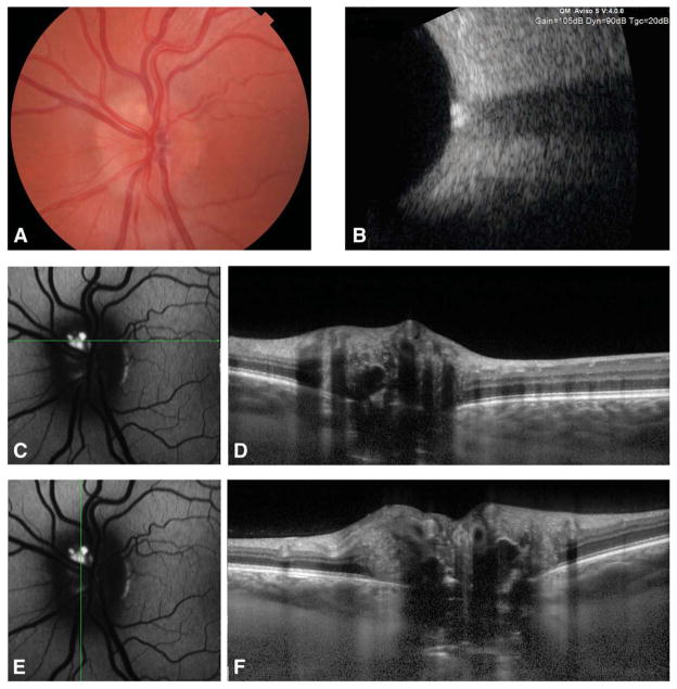
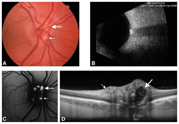
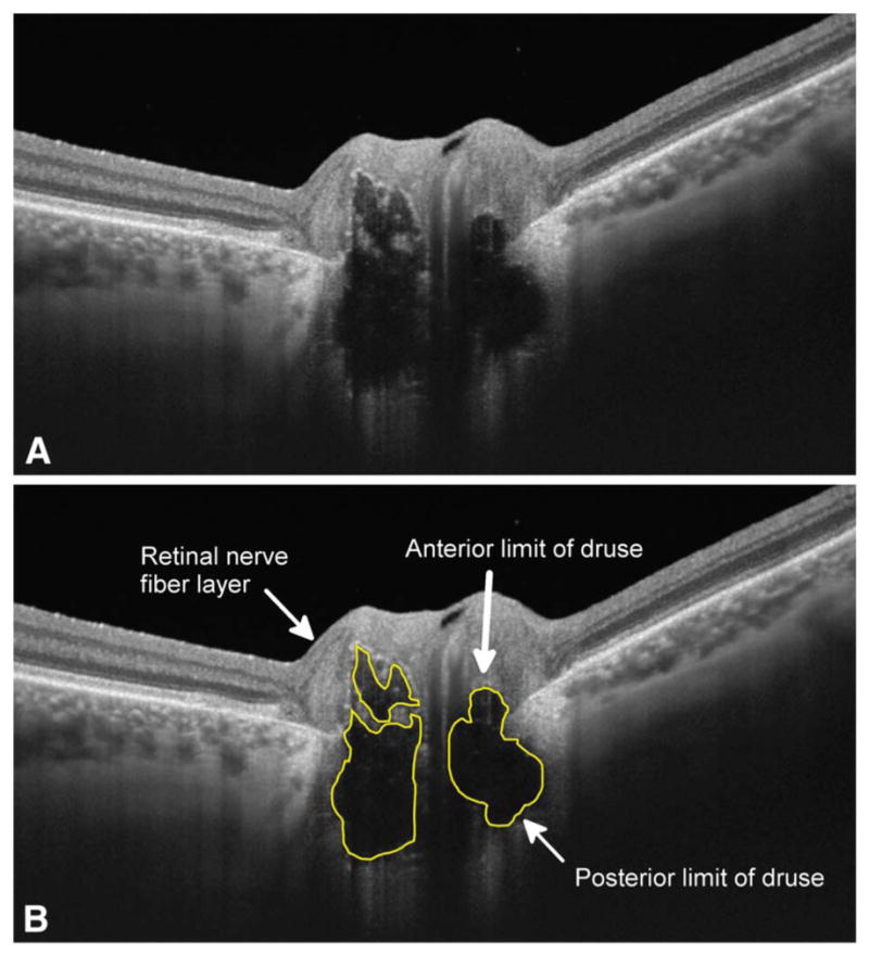
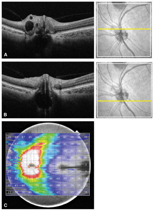
Background: Optic nerve head drusen (ONHD) are calcific deposits buried or at the surface of the optic disc. Although ONHD may be associated with progressive visual field defects, the mechanism of drusen-related field loss is poorly understood. Methods for detecting and imaging disc drusen include B-scan ultrasonography, fundus autofluorescence, and optical coherence tomography (OCT). These modalities are useful for drusen detection but are limited by low resolution or poor penetration of deep structures. This review was designed to assess the potential role of new OCT technologies in imaging ONHD.
Evidence acquisition: Critical appraisal of published literature and comparison of new imaging devices to established technology.
Results: The new imaging modalities of enhanced depth imaging optical coherence tomography (EDI-OCT) and swept source optical coherence tomography (SS-OCT) are able to provide unprecedented in vivo detail of ONHD. Using these devices it is now possible to quantify optic disc drusen dimensions and assess integrity of neighboring retinal structures, including the retinal nerve fiber layer.
Conclusions: EDI-OCT and SS-OCT have the potential to allow better detection of longitudinal changes in drusen and neural retina and improve our understanding of drusen-related visual field loss.
Figures
References
-
- Lam BL, Morais CG, Jr, Pasol J. Drusen of the optic disc. Curr Neurol Neurosci Rep. 2008;8:404–408. - PubMed
-
- Grippo TA. Optic disc drusen. Glaucoma Today. 2012;10:19–23.
-
- Auw-Haedrich C, Staubach F, Witschel H. Optic disk drusen. Surv Ophthalmol. 2002;47:515–532. - PubMed
-
- Sato T, Mrejen S, Spaide RF. Multimodal imaging of optic disc drusen. Am J Ophthalmol. 2013;156:275–282. e1. - PubMed
Publication types
MeSH terms
Grants and funding
LinkOut - more resources
Full Text Sources
Other Literature Sources
Medical
