Review
. 2014 Mar 21;15(3):5094-114.
doi: 10.3390/ijms15035094.

No stress! Relax! Mechanisms governing growth and shape in plant cells

Affiliations
Review

No stress! Relax! Mechanisms governing growth and shape in plant cells

Gea Guerriero et al. Int J Mol Sci. .

Abstract

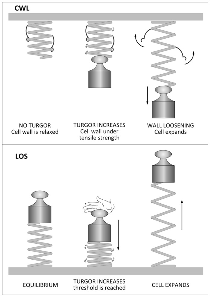
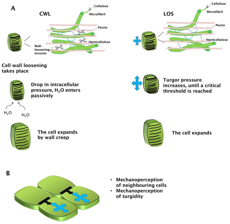
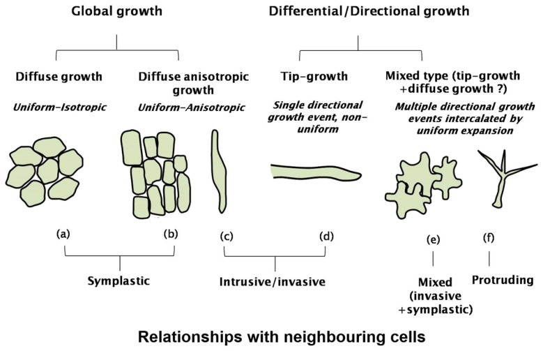
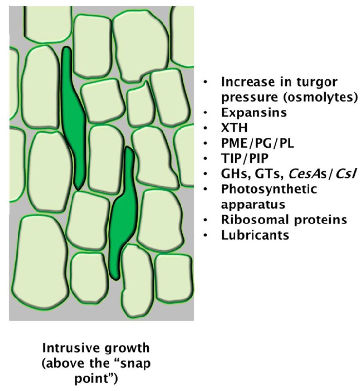
The mechanisms through which plant cells control growth and shape are the result of the coordinated action of many events, notably cell wall stress relaxation and turgor-driven expansion. The scalar nature of turgor pressure would drive plant cells to assume spherical shapes; however, this is not the case, as plant cells show an amazing variety of morphologies. Plant cell walls are dynamic structures that can display alterations in matrix polysaccharide composition and concentration, which ultimately affect the wall deformation rate. The wide varieties of plant cell shapes, spanning from elongated cylinders (as pollen tubes) and jigsaw puzzle-like epidermal cells, to very long fibres and branched stellate leaf trichomes, can be understood if the underlying mechanisms regulating wall biosynthesis and cytoskeletal dynamics are addressed. This review aims at gathering the available knowledge on the fundamental mechanisms regulating expansion, growth and shape in plant cells by putting a special emphasis on the cell wall-cytoskeleton system continuum. In particular, we discuss from a molecular point of view the growth mechanisms characterizing cell types with strikingly different geometries and describe their relationship with primary walls. The purpose, here, is to provide the reader with a comprehensive overview of the multitude of events through which plant cells manage to expand and control their final shapes.


Figures

Figure 1.
Figure 1.
Schematic representation of the two principles (turgor pressure and wall tensile stress) regulating plant cell growth. The top part of the figure illustrates the CWL (Chemical Wall Loosening) mechanism by using a spring, hooks and a weight, while the lower part schematizes the LOS model. In the CWL model, the spring (which represents the cell wall), under the action of the weight (the internal turgor pressure), is in tensile stress and the potential energy of the system increases; when the transducer (wall loosening enzymes) releases the weight by unfastening the hooks holding the spring (which represent the inter/intra-chain bonds of wall polysaccharides), the energy accumulated is transformed into kinetic energy (growth). Therefore, the action of wall loosening enzymes is required for growth, according to the CWL model. According to the LOS model, the compression of the spring (i.e., the cell wall) by an increase in turgor pressure (here represented as a hand pushing the weight), proceeds until a threshold is reached (wall yield threshold). Beyond this threshold, the wall yields and expansion occurs. Therefore, according to the LOS model, growth depends on the intrinsic properties of the wall.
Figure 2.
Figure 2.
Models of plant cell expansion and mechanosensory signals. (A) Cartoons depicting the steps marking growth according to the CWL and LOS models; the dotted line represents the critical yield threshold of the wall; (B) Mechanosensory signals coming from neighbouring cells (black arrows) and from the turgidity of the expanding cells (blue arrows).
Figure 3.
Figure 3.
Cartoon depicting the two main plant cell growth events: global and differential/directional growth. Details concerning the cell types (a) fruit parenchyma cells; (b) shoot/root epidermal cells; (c) bast fibre; (d) pollen tube; (e) leaf pavement cells; (f) trichome and the relationship with the neighbouring cells are added to show the different expansion modalities displayed by plant cells. The question mark indicating the mixed-type growth refers to a hypothetical mechanism involving both tip- and diffuse growth.
Figure 4.
Figure 4.
Cartoon representing the diffuse (intrusive) growth of bast fibres (dark green cell grows showing intrusive extremities, as in [20]). The box shows important factors/genes involved [20,47]. Abbreviations: XTH xyloglucan endotransglycosylase/hydrolase, PME pectin methylesterase, PG polygalacturonase, PL pectin lyase, TIP tonoplast intrinsic protein, PIP plasma membrane intrinsic protein, GHs glycosylhydrolases, GTs, glycosytransferases, CesAs cellulose synthases, Csl cellulose synthase-like.
Figure 5.
Figure 5.
Schematic representation of the cytoskeletal architecture in a jigsaw puzzle-like pavement cell (as described in [36,53]). On the right, schematic molecular details on the relationship between Rho proteins and cytoskeleton are reported for the molecular control of interdigitated domains (invagination/lobe).
Figure 6.
Figure 6.
Schematic diagram of the secretion process in pollen tubes. Only the key elements involved in cell wall synthesis are shown (PME, PMEI, CESA, CALS). The gradient of gray in the cell wall indicates the status of methyl-esterification of pectins (light gray: fully methyl-esterified pectins; dark gray: acid pectins). Cellulose is shown as random black lines in the tube apex and as more organized lines in distal parts. Callose is shown as red bands in the pollen tube shanks. TGN, Trans-Golgi-Network. Actin filaments (red lines) are shown as interacting with the Golgi, indicating that they are involved in the transport of vesicles to the apex. Microtubules (green lines) are shown as positioned in the cortical region where they presumably could interact with CALS and CESA. Secretion is indicated to occur not exactly at the apex but in a region immediately close. Endocytotic processes are hypothesized to occur both at the extreme apex and in more distal regions [67,99].

References

    1. Geitmann A., Ortega J.K. Mechanics and modeling of plant cell growth. Trends Plant Sci. 2009;1:467–478. - PubMed
    1. Cosgrove D.J. Growth of the plant cell wall. Nat. Rev. Mol. Cell Biol. 2005;6:850–861. - PubMed
    1. Humphrey T.V., Bonetta D.T., Goring D.R. Sentinels at the wall: Cell wall receptors and sensors. New Phytol. 2007;176:7–21. - PubMed
    1. Hamann T., Bennett M., Mansfield J., Somerville C. Identification of cell-wall stress as a hexose-dependent and osmosensitive regulator of plant responses. Plant J. 2009;57:1015–1026. - PubMed
    1. Seifert G.J., Blaukopf C. Irritable walls: The plant extracellular matrix and signaling. Plant Physiol. 2010;153:467–478. - PMC - PubMed

LinkOut - more resources