A recombinant novirhabdovirus presenting at the surface the E Glycoprotein from West Nile Virus (WNV) is immunogenic and provides partial protection against lethal WNV challenge in BALB/c mice
- PMID: 24663075
- PMCID: PMC3963854
- DOI: 10.1371/journal.pone.0091766
A recombinant novirhabdovirus presenting at the surface the E Glycoprotein from West Nile Virus (WNV) is immunogenic and provides partial protection against lethal WNV challenge in BALB/c mice
Abstract
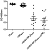
West Nile Virus (WNV) is a zoonotic mosquito-transmitted flavivirus that can infect and cause disease in mammals including humans. Our study aimed at developing a WNV vectored vaccine based on a fish Novirhabdovirus, the Viral Hemorrhagic Septicemia virus (VHSV). VHSV replicates at temperatures lower than 20°C and is naturally inactivated at higher temperatures. A reverse genetics system has recently been developed in our laboratory for VHSV allowing the addition of genes in the viral genome and the recovery of the respective recombinant viruses (rVHSV). In this study, we have generated rVHSV vectors bearing the complete WNV envelope gene (EWNV) (rVHSV-EWNV) or fragments encoding E subdomains (either domain III alone or domain III fused to domain II) (rVHSV-DIIIWNV and rVHSV-DII-DIIIWNV, respectively) in the VHSV genome between the N and P cistrons. With the objective to enhance the targeting of the EWNV protein or EWNV-derived domains to the surface of VHSV virions, Novirhadovirus G-derived signal peptide and transmembrane domain (SPG and TMG) were fused to EWNV at its amino and carboxy termini, respectively. By Western-blot analysis, electron microscopy observations or inoculation experiments in mice, we demonstrated that both the EWNV and the DIIIWNV could be expressed at the viral surface of rVHSV upon addition of SPG. Every constructs expressing EWNV fused to SPG protected 40 to 50% of BALB/cJ mice against WNV lethal challenge and specifically rVHSV-SPGEWNV induced a neutralizing antibody response that correlated with protection. Surprisingly, rVHSV expressing EWNV-derived domain III or II and III were unable to protect mice against WNV challenge, although these domains were highly incorporated in the virion and expressed at the viral surface. In this study we demonstrated that a heterologous glycoprotein and non membrane-anchored protein, can be efficiently expressed at the surface of rVHSV making this approach attractive to develop new vaccines against various pathogens.
Conflict of interest statement
Figures


Similar articles
-
Complete Protection against Influenza Virus H1N1 Strain A/PR/8/34 Challenge in Mice Immunized with Non-Adjuvanted Novirhabdovirus Vaccines.PLoS One. 2016 Oct 6;11(10):e0164245. doi: 10.1371/journal.pone.0164245. eCollection 2016. PLoS One. 2016. PMID: 27711176 Free PMC article.
-
Recombinant viral hemorrhagic septicemia virus with rearranged genomes as vaccine vectors to protect against lethal betanodavirus infection.Front Immunol. 2023 Mar 14;14:1138961. doi: 10.3389/fimmu.2023.1138961. eCollection 2023. Front Immunol. 2023. PMID: 36999033 Free PMC article.
-
Effect of exchange of viral hemorrhagic septicemia virus (VHSV) G protein's signal peptide with piscidin signal peptide on virus replication and immunogenicity.Fish Shellfish Immunol. 2025 Sep;164:110455. doi: 10.1016/j.fsi.2025.110455. Epub 2025 May 26. Fish Shellfish Immunol. 2025. PMID: 40436152
-
Immunization of flavivirus West Nile recombinant envelope domain III protein induced specific immune response and protection against West Nile virus infection.J Immunol. 2007 Mar 1;178(5):2699-705. doi: 10.4049/jimmunol.178.5.2699. J Immunol. 2007. PMID: 17312111
-
A reverse genetics system for the Great Lakes strain of viral hemorrhagic septicemia virus: the NV gene is required for pathogenicity.Mar Biotechnol (NY). 2011 Aug;13(4):672-83. doi: 10.1007/s10126-010-9329-4. Epub 2010 Oct 9. Mar Biotechnol (NY). 2011. PMID: 20936318
Cited by
-
Complete Protection against Influenza Virus H1N1 Strain A/PR/8/34 Challenge in Mice Immunized with Non-Adjuvanted Novirhabdovirus Vaccines.PLoS One. 2016 Oct 6;11(10):e0164245. doi: 10.1371/journal.pone.0164245. eCollection 2016. PLoS One. 2016. PMID: 27711176 Free PMC article.
-
Recombinant viral hemorrhagic septicemia virus with rearranged genomes as vaccine vectors to protect against lethal betanodavirus infection.Front Immunol. 2023 Mar 14;14:1138961. doi: 10.3389/fimmu.2023.1138961. eCollection 2023. Front Immunol. 2023. PMID: 36999033 Free PMC article.
-
Generation of Recombinant Viral Hemorrhagic Septicemia Virus (rVHSV) Expressing Two Foreign Proteins and Effect of Lengthened Viral Genome on Viral Growth and In Vivo Virulence.Mol Biotechnol. 2016 Apr;58(4):280-6. doi: 10.1007/s12033-016-9926-1. Mol Biotechnol. 2016. PMID: 26921191
References
-
- Kramer LD (2007) West Nile Virus. . Lancet Neurol. 6: 171–181. - PubMed
-
- McLean R, Ubico S (2007) In: Infectious Diseases of Wild Birds. Thomas N, Hunter D, Atkinson C, editor. Iowa: Blacwell Publishing;. Arboviruses in Birds; pp. 17–62.
-
- Mostashari F, Bunning ML, Kitsutani PT, Singer DA, Nash D, et al. (2001) Epidemic West Nile encephalitis, New York, 1999: results of a household-based seroepidemiological survey. Lancet 358: 261–264. - PubMed
Publication types
MeSH terms
Substances
LinkOut - more resources
Full Text Sources
Other Literature Sources

