Review
. 2014 May;11(5):255-65.
doi: 10.1038/nrcardio.2014.28. Epub 2014 Mar 25.

The inflammatory response in myocardial injury, repair, and remodelling

Affiliations
Review

The inflammatory response in myocardial injury, repair, and remodelling

Nikolaos G Frangogiannis. Nat Rev Cardiol. 2014 May.

Abstract

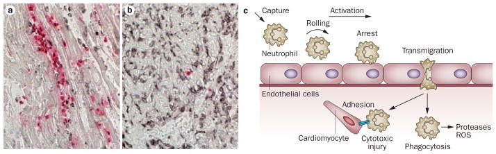
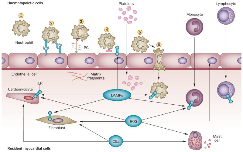
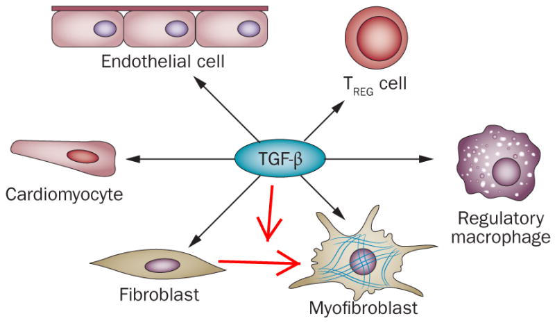
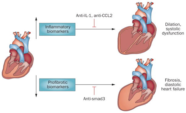
Myocardial infarction triggers an intense inflammatory response that is essential for cardiac repair, but which is also implicated in the pathogenesis of postinfarction remodelling and heart failure. Signals in the infarcted myocardium activate toll-like receptor signalling, while complement activation and generation of reactive oxygen species induce cytokine and chemokine upregulation. Leukocytes recruited to the infarcted area, remove dead cells and matrix debris by phagocytosis, while preparing the area for scar formation. Timely repression of the inflammatory response is critical for effective healing, and is followed by activation of myofibroblasts that secrete matrix proteins in the infarcted area. Members of the transforming growth factor β family are critically involved in suppression of inflammation and activation of a profibrotic programme. Translation of these concepts to the clinic requires an understanding of the pathophysiological complexity and heterogeneity of postinfarction remodelling in patients with myocardial infarction. Individuals with an overactive and prolonged postinfarction inflammatory response might exhibit left ventricular dilatation and systolic dysfunction and might benefit from targeted anti-IL-1 or anti-chemokine therapies, whereas patients with an exaggerated fibrogenic reaction can develop heart failure with preserved ejection fraction and might require inhibition of the Smad3 (mothers against decapentaplegic homolog 3) cascade. Biomarker-based approaches are needed to identify patients with distinct pathophysiologic responses and to rationally implement inflammation-modulating strategies.


Conflict of interest statement

Competing interests

The author declares no competing interests.

Figures

Figure 1
Figure 1. Cytotoxic inflammatory injury following myocardial infarction
Myocardial infarction is associated with an intense inflammatory reaction and infiltration of the infarct with abundant leukocytes. a. canine infarct (1h coronary occlusion/24 h reperfusion) stained with MAC387 (red), a marker for newly recruited myeloid cells (neutrophils and monocytes), and an anti-macrophage antibody (black). Abundant newly recruited leukocytes closely associated with viable cardiomyocytes. b. The same canine infarct after 7 days of reperfusion. The density of MAC387 positive cells is markedly reduced; however, mature macrophages are still abundant (black) reflecting repression of the acute inflammatory reaction. c. The close spatial association of leukocytes and viable cardiomyocytes in the border zone and the injurious potential of subsets of blood-derived cells generated the concept of leukocyte-mediated cardiomyocyte injury. Neutrophils interact with endothelial cells, roll along the endothelial surface, decelerate to a firm arrest, transmigrate across the vascular wall, infiltrate the infarct, and adhere to viable cardiomyocytes exerting cytotoxic effects and extending ischemic injury. Infiltrating leukocytes are also important role for infarct repair by releasing proteases and ROS, thereby clearing the wound from dead cells and debris. Abbreviations: ROS, reactive oxygen species.
Figure 2
Figure 2. The post-infarction inflammatory response
In the infarcted myocardium, dying cardiomyocytes and damaged matrix release DAMPs that activate TLR signaling in myocardial cells, triggering an inflammatory reaction. Activation of the complement cascade and ROS generation also help initiate the inflammatory reaction. Dying and surviving cardiomyocytes, endothelial cells, resident cardiac fibroblasts, resident mast cells and newly recruited neutrophils monocytes and platelets participate in the post-infarction inflammatory response. However, their relative contributions remain unclear. Leukocytes are recruited through activation of a multistep adhesion cascade. . Capture (1) of circulating leukocytes by activated endothelial cells is followed by rolling (2), mediated through interactions involving the selectins. Rolling leukocytes are activated (3) by chemokines bound to proteoglycans (PG) on the endothelial surface. Activated leukocytes express integrins and adhere to endothelial cells (4). Strengthening of the adhesive interaction (5) between leukocytes and endothelial cells is followed by transmigration of the cells into the infarcted area (6). Abbreviations: DAMPs, danger-associated molecular patterns; TLR, Toll-like receptor; ROS, reactive oxygen species
Figure 3
Figure 3. TGF-β is a key mediator in post-infarction remodeling
TGF-β exerts anti-inflammatory actions, inducing a regulatory macrophage phenotype, promoting regulatory Treg cell activation and reducing adhesion molecule synthesis by endothelial cells. TGF-β is also critically involved in fibroblast to myofibroblast conversion by activating a pro-fibrotic program.
Figure 4
Figure 4. Biomarker-based approaches to target the inflammatory response in patients with acute myocardial infarction
Patients surviving a myocardial infarction exhibit pathophysiologically heterogeneous responses, which is in part independent on the size of the infarct. Distinct pathophysiologic responses might be due to differences in genetic background and to the presence of conditions (such as diabetes mellitus or hypertension) that affect inflammatory and fibrogenic pathways. After myocardial infarction, some patients develop progressive dilation and systolic dysfunction, whereas others develop diastolic heart failure. Dilation might reflect excessive inflammatory activity causing matrix degradation; conversely, diastolic heart failure might indicate overactive pro-fibrotic signaling. We propose the use of inflammatory biomarkers (such as serum cytokine and chemokine levels) and of profibrotic markers (including indicators of matrix synthesis and remodeling) to stratify patients into subpopulations based on the predominant pathophysiology. Patients with overactive inflammation may benefit from targeted inhibition of inflammatory signals (anti-IL1 or anti-MCP1 strategies), whereas patients with profibrotic responses might benefit from inhibition of the TGF-β/smad cascade.

References

    1. Mallory GK, White PD, Salcedo-Salgar J. The speed of healing of myocardial infarction. A study of the pathologic anatomy in seventy-two cases. Am Heart J. 1939;18:647.
    1. Entman ML, et al. Neutrophil induced oxidative injury of cardiac myocytes. A compartmented system requiring CD11b/CD18-ICAM-1 adherence. J Clin Invest. 1992;90:1335–1345. - PMC - PubMed
    1. Yamazaki T, et al. Expression of intercellular adhesion molecule-1 in rat heart with ischemia/reperfusion and limitation of infarct size by treatment with antibodies against cell adhesion molecules. Am J Pathol. 1993;143:410–418. - PMC - PubMed
    1. Simpson PJ, et al. Reduction of experimental canine myocardial reperfusion injury by a monoclonal antibody (anti-Mo1, anti-CD11b) that inhibits leukocyte adhesion. J Clin Invest. 1988;81:624–629. - PMC - PubMed
    1. Tojo SJ, et al. Reduction of rat myocardial ischemia and reperfusion injury by sialyl Lewis x oligosaccharide and anti-rat P-selectin antibodies. Glycobiology. 1996;6:463–469. - PubMed

Publication types

MeSH terms