. 2014 Mar 24;9(3):e92257.
doi: 10.1371/journal.pone.0092257. eCollection 2014.

Esterase LpEst1 from Lactobacillus plantarum: a novel and atypical member of the αβ hydrolase superfamily of enzymes

Affiliations

Esterase LpEst1 from Lactobacillus plantarum: a novel and atypical member of the αβ hydrolase superfamily of enzymes

Yanaisis Alvarez et al. PLoS One. .

Abstract

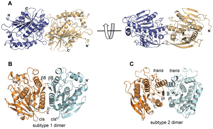
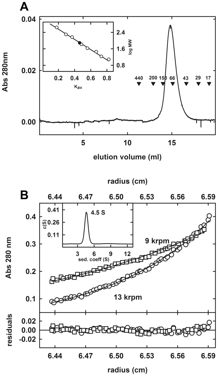
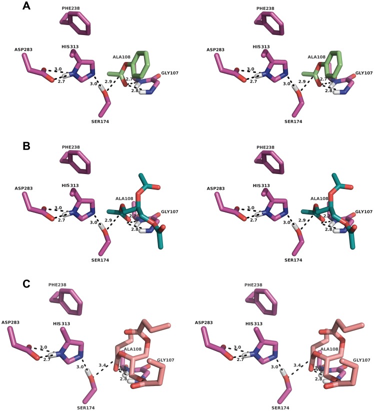
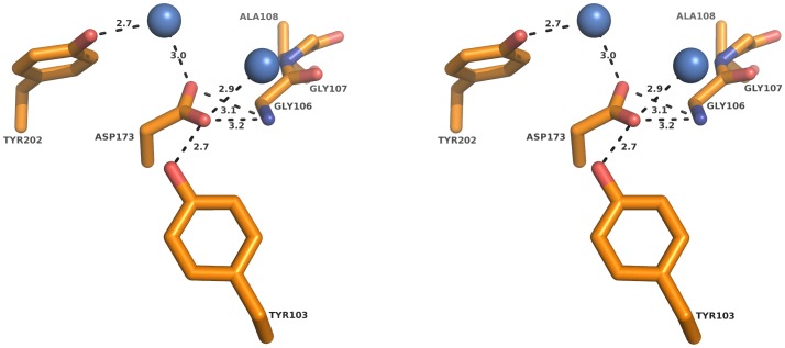
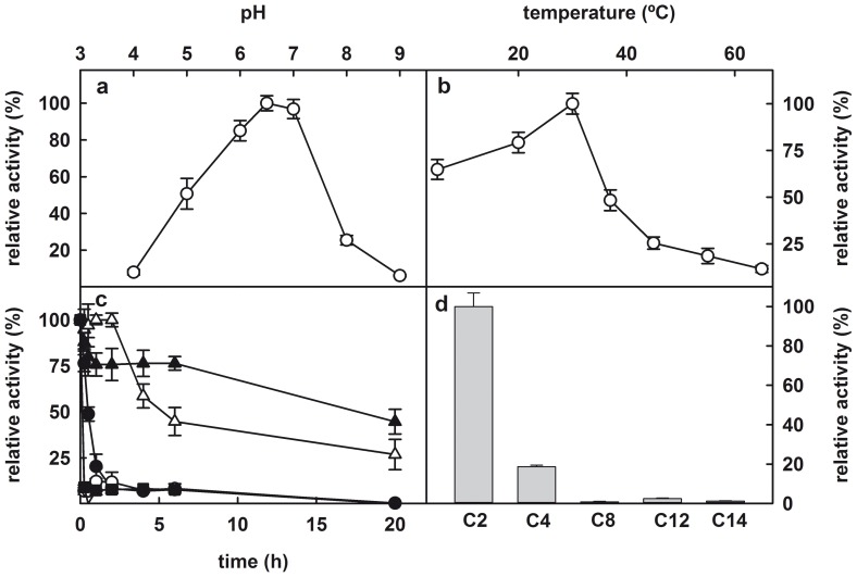
The genome of the lactic acid bacterium Lactobacillus plantarum WCFS1 reveals the presence of a rich repertoire of esterases and lipases highlighting their important role in cellular metabolism. Among them is the carboxylesterase LpEst1 a bacterial enzyme related to the mammalian hormone-sensitive lipase, which is known to play a central role in energy homeostasis. In this study, the crystal structure of LpEst1 has been determined at 2.05 Å resolution; it exhibits an αβ-hydrolase fold, consisting of a central β-sheet surrounded by α-helices, endowed with novel topological features. The structure reveals a dimeric assembly not comparable with any other enzyme from the bacterial hormone-sensitive lipase family, probably echoing the specific structural features of the participating subunits. Biophysical studies including analytical gel filtration and ultracentrifugation support the dimeric nature of LpEst1. Structural and mutational analyses of the substrate-binding pocket and active site together with biochemical studies provided insights for understanding the substrate profile of LpEst1 and suggested for the first time the conserved Asp173, which is adjacent to the nucleophile, as a key element in the stabilization of the loop where the oxyanion hole resides.


Conflict of interest statement

Competing Interests: The authors have declared that no competing interests exist.

Figures

Figure 1
Figure 1. Protein sequence alignment between LpEst1 and homologs from the hormone-sensitive lipase (HSL) family.
PestE, carboxylesterase PestE from Pyrobaculum calidifontis; AFEST, carboxylesterase AFEST from Archaeoglobus fulgidus; Sto-Est, thermophilic esterase Sto-Est from Sulfolobus tokodaii; EstE7, esterase EstE7 from environmental samples; EstE5, esterase EstE5 from environmental samples; EstE2, thermophilic esterases EstE2 from Alicyclobacillus acidocaldarius; HEst, heroin esterase from Rhodococcus sp.; BFAE, Brefeldin A esterase from Bacillus subtilis. Residues from LpEst1 in Residues from LpEst1 in sterase fblue cylinders and orange arrows, respectively. Residues forming the catalytic triad are marked with an asterisk. Colour code for boxes is as follows: red, conserved residues in all proteins; yellow, highly conserved positions; cyan, residues that coincide with the expected canonical sequence motif characteristic of enzymes from the HSL family.
Figure 2
Figure 2. Crystal structure of the LpEst1 subunit.
(A) Ribbon representation of the LpEst1 subunit; two different views are depicted. Canonical β-strands forming the core β-sheet are shown in yellow, whereas the two N-terminal, non-canonical ones are shown in orange. α-Helices are in blue, except the first two previous to the first β-strand from the core β-sheet (β3), which are shown in green. (B) Topology diagram of the LpEst1 fold. Colour code for the secondary structure elements are as in (A). The positions of the residues forming the catalytic triad Ser174, Asp283 and His313 are indicated as S, D and H, respectively, and those forming the oxyanion hole, Gly107 and Ala108, are indicated as GA. (C) Representative 2FobsFcalc density map contoured at 1σ.
Figure 3
Figure 3. Three-dimensional comparisons of cap regions from bacterial HSL enzymes.
(A) Superposition of the cap regions of LpEst1 (yellow) and PestE from P. calidifontis (green), which has been chosen arbitrarily as a representative model of the predominant pattern (see the text). (B) Superposition of the cap region from BFAE (dark green) and that from PestE. (C) Superposition of cap regions from PestE (green), AFEST (cyan), EstE2 (grey), EstE5 (light brown), EstE7 (magenta), heroin esterase (pale green) and Sto-Est from (blue). All structures are represented as ribbon models.
Figure 4
Figure 4. Dimeric assembly of LpEst1 and classification of the dimers of the HSL family members.
(A) Two orthogonal views of a dimer of LpEst1. Each subunit is shown as ribbon model with different colour. (B) Subtype 1 of dimers (PDB entry: 3aik), characterized by a cis-cis association of subunits. Cis is arbitrarily defined as the side of the protein with respect to the plane of the core β-sheet where the α-helix downstream the canonical strand β8 is situated (see the text for details). (C) Subtype 2 of dimers (PDB entry: 1lzl), characterized by a trans-trans association of subunits. Trans is arbitrarily defined as the side of the protein with respect to the plane of the core β-sheet where the α-helix upstream the canonical strand β8 is situated. The orientation of is this dimer is as in (B).
Figure 5
Figure 5. Analysis of the oligomeric state of LpEst1 in solution.
(A) Analytical gel-filtration of LpEst1 on Superdex 200 10/300 GL Tricorn column. The elution profile of LpEst1 is shown together with the elution positions for some standard proteins (molecular mass in kDa). Inset, semilog plot of the molecular mass of all standards used versus their Kav values (open circles). The closed circle indicates the position of the Kav value of LpEst1 interpolated in the regression line (solid line) (B) Analytical ultracentrifugation analysis of LpEst1. Sedimentation equilibrium analysis of LpEst1 (10 μM) in Tris buffer (20 mM Tris-HCl, pH 8.0, and 0.1 M NaCl) at 9,000 rpm (open squares) and 13,000 (open circles). Absorbance at 280 nm is plotted against the radial position from the center of the rotor. The fit to the data set (solid line curves) corresponds to an ideal species with a molecular mass of 77.2±4.2 kDa (n = 3). Residuals from this fit are shown in the panel at the bottom. Calculations were done with the program Heteroanalysis . Inset, sedimentation coefficient c(s) distributions for LpEst1 (10 μM) in Tris buffer (20 mM Tris-HCl, pH 8.0 with 0.1 M NaCl). Raw sedimentation velocity profiles for this analysis were acquired using absorbance at 280 nm, 45,000 rpm, 20 °C, and different times (not shown). Calculations were done with the program Sedfit .
Figure 6
Figure 6. Intersubunit binding energy decomposition for dimer and tetramer formation.
Colour code is as follows: black, electrostatic term; orange, van der Waals term; blue, hydrogen bond term; yellow, total contribution, including desolvation energies (not shown).
Figure 7
Figure 7. Distribution of the energetically relevant residues within the LpEst1 contacting interface.
These residues are basically hydrophobic in agreement with the relevance of this type of interactions in the stabilization of the dimer. The bidentate hydrogen bonding interaction between the side-chain carboxamide groups of Gln189 (close up view) is situated at the core of the interface. The 2FobsFcalc density map is contoured at 1σ.
Figure 8
Figure 8. Stereoview of the network of interactions present around the catalytic triad of LpEst1.
Residues forming the catalytic triad (Ser174, His313 and Asp283) and those coordinating a well-ordered water molecule that is also identified in the HSL family members (blue sphere) are shown as sticks. The 2FobsFcalc density map is contoured at 1σ (green: amino acid side chains; blue: water molecule) Distances are in Å.
Figure 9
Figure 9. Stereoviews of three putative LpEst1:substrate complexes resulting from docking studies with CRDOCK.
The three complexes correspond to phenyl acetate (a), triacetin (b) and tributyrin (c). In all cases, interactions are observed between the nucleophile (Ser174) and also reveal the stabilizing effect of the oxyanion hole formed by the backbone nitrogen atoms of Gly107 and Ala108. Residues are shown as stick models. Distances are in Å.
Figure 10
Figure 10. Stereoview of the environment around the residue Asp173.
The side chain of the Asp173 residue is highly oriented towards the loop where the oxyanion hole resides probably contributing to its stabilization. The network of hydrogen bonds is shown as dashed lines. Participating residues are shown as sticks and water molecules as blue spheres. Distances are in Å.
Figure 11
Figure 11. Biochemical characterization of LpEst1.
(A) Dependence on pH of hydrolytic activity of LpEst1 against pNPA. (B) Dependence on temperature of hydrolytic activity of LpEst1 against pNPA. The optimum temperature for esterase activity was ∼30 °C. (C) Analysis of the temperature stability of LpEst1. The enzyme was incubated in 50 mM sodium phosphate buffer pH 7.0 at 22 °C (closed triangles), 30 °C (open triangles), 37 °C (closed circles), 45 °C (open circles) and 55 °C (closed squares) for 15 min, 30 min, and 1, 2, 4, 6 and 20 h. The values correspond to the mean of three independent experiments. (D) Dependence of the esterase activity of LpEst1 on the chain length of p-nitrophenyl (p-NP): p-NP acetate (C2), p-NP butyrate (C4); p-NP caprylate (C8); p-NP laureate (C12); and p-NP myristate (C14).

References

    1. Arpigny JL, Jaeger KE (1999) Bacterial lipolytic enzymes: classification and properties. Biochem J 343: 177–183. - PMC - PubMed
    1. Fojan P, Jonson PH, Petersen MTN, Petersen SB (2000) What distinguishes an esterase from a lipase: A novel structural approach. Biochimie 82: 1033–1041. - PubMed
    1. Reis P, Holmberg K, Watke H, Leser ME, Miller R (2009) Lipases at interfaces: A review. Adv Colloid Interf Sci 147–148: 237–250. - PubMed
    1. Sussman JL, Harel M, Frolow F, Oefner C, Goldman A, et al. (1991) Atomic structure of acetylcholinesterase from Torpedo californica: a prototypic acetylcholine-binding protein. Science 253: 872–879. - PubMed
    1. Zhu X, Larsen NA, Basran A, Bruce NC, Wilson IA (2003) Observation of an arsenic adduct in an acetyl esterase crystal structure. J Biol Chem 278: 2008–2014. - PubMed

Publication types

LinkOut - more resources