. 2014 Mar 24;9(3):e92648.
doi: 10.1371/journal.pone.0092648. eCollection 2014.

Evidence of trem2 variant associated with triple risk of Alzheimer's disease

Affiliations

Evidence of trem2 variant associated with triple risk of Alzheimer's disease

Zainularifeen Abduljaleel et al. PLoS One. .

Abstract

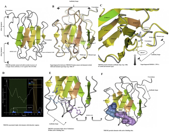
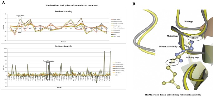
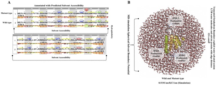
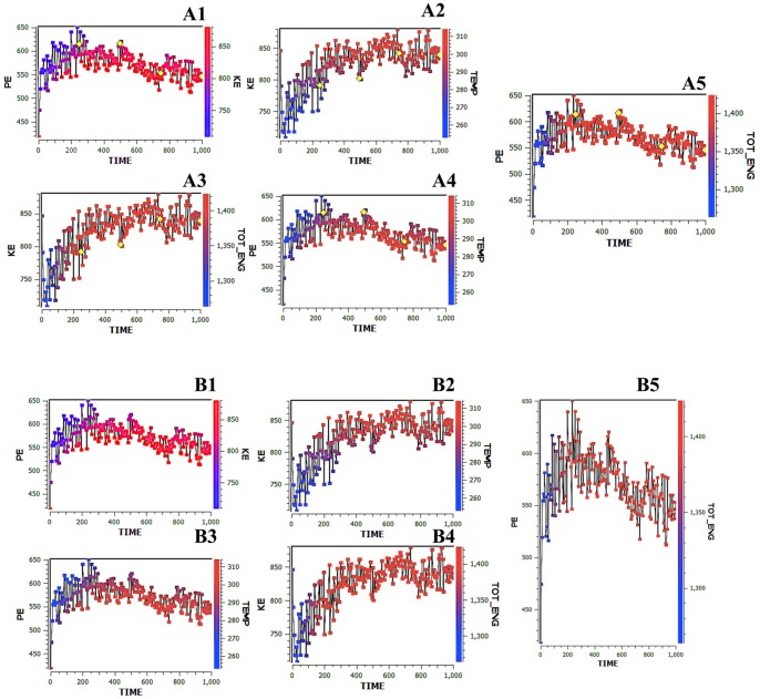
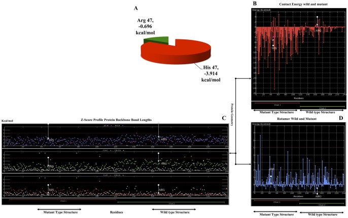
Alzheimer's disease is one of the main causes of dementia among elderly individuals and leads to the neurodegeneration of different areas of the brain, resulting in memory impairments and loss of cognitive functions. Recently, a rare variant that is associated with 3-fold higher risk of Alzheimer's disease onset has been found. The rare variant discovered is a missense mutation in the loop region of exon 2 of Trem2 (rs75932628-T, Arg47His). The aim of this study was to investigate the evidence for potential structural and functional significance of Trem2 gene variant (Arg47His) through molecular dynamics simulations. Our results showed the alteration caused due to the variant in TREM2 protein has significant effect on the ligand binding affinity as well as structural configuration. Based on molecular dynamics (MD) simulation under salvation, the results confirmed that native form of the variant (Arg47His) might be responsible for improved compactness, hence thereby improved protein folding. Protein simulation was carried out at different temperatures. At 300K, the deviation of the theoretical model of TREM2 protein increased from 2.0 Å at 10 ns. In contrast, the deviation of the Arg47His mutation was maintained at 1.2 Å until the end of the simulation (t = 10 ns), which indicated that Arg47His had reached its folded state. The mutant residue was a highly conserved region and was similar to "immunoglobulin V-set" and "immunoglobulin-like folds". Taken together, the result from this study provides a biophysical insight on how the studied variant could contribute to the genetic susceptibility to Alzheimer's disease.


Conflict of interest statement

Competing Interests: The authors have declared that no competing interests exist.

Figures

Figure 1
Figure 1. TREM2 Gene annotations with colored pastilles.
Alternative mRNAs are shown aligned from 5′ to 3′ on a virtual genome where introns have shrunk to a minimal length. The exon size is proportional to the length; the intron height reflects the number of cDNA clones that support each intron. The genes are summarized according to color as follows: gene (pale blue) known to Entrez, disease (red), conservation (brown), interactions (green), and regulation (dark blue). Each arrow represents a gene and covers the extent of the GenBank/dbEST cDNA sequences that belong specifically to the gene and points in the direction of transcription (top strand is up, bottom down).
Figure 2
Figure 2. TREM2 protein domain structure modeling and annotations.
A) TREM2 protein domain conservation, according to the crystal structure of a single chain antibody scA21 against Her2/ErbB2 with a conserved identity of 0.168 and RMSD of 2.63 Å. B) Ribbon diagram representation of a composite model superimposed structure between the conserved domain of TREM2 between the wild type and mutant forms of the loop region at β sheet4: Arg ⇒His47. C) TREM2 protein domain antibody loop in the wild type (yellow) and mutant (violet). D) Predicted altered regions of the TREM2 associated triple risk protein domain. E) Predicted active binding site of wild type Arg (blue) and mutant-type His (violet) at the loop region of TREM2 associated with a triple risk of Alzheimer’s. F) The molecular surfaces of the TREM2 domain model. The molecular surfaces at 2.54 Å RMSD of the active binding sites in wild type (blue) and mutant (violet) forms; the most positive potential is shown in blue, and the most negative is shown in deep violet.
Figure 3
Figure 3. Scanning of residues with functional annotations, prediction of solvent accessibility, and protein stability changes.
A) Compositions of the amino acids in TREM2 protein residues, fixing only the mutant His47 in comparison to the polar and neutral residues for the solvent with a random representative set of non-homologous TREM2 proteins. On the Y-axis is the percentage of amino acids, and on the X-axis is the amino acids regions compared with the functional annotations in different colors as follows: prime energy, SASA (non-polar), stability (gas), total rotatable bonds, SASA (polar), hydropathy, pKa, the SASA (total) interactions, and the solvent-accessible contact area as a percentage of the residue accessibility at the point mutation. B) TREM2 protein domain antibody loop, according to the solvent accessibility compared to the wild type and mutant residue stability. The gray color loop shows the solvent accessibility of the mutant residue (yellow), and the wild type (violet color) residue are shown in violet.
Figure 4
Figure 4. Evaluation of the secondary structures of the TREM2 domain and MD simulation in solvated protein.
Values of median pho (black bars) and phi (yellow bars) ASA for each residue in the extended state (A) with residues sorted according to decreasing total ASA values. Median hydrophilic solvent accessibility as a function of median hydrophobic solvent accessibility of residues was in most of the loop regions. Residues of wild type Arg47 (R) (green) and mutant His (H) (red) are upstream outside of this classification. The distribution of total a solvent accessibility for wild type and mutant (Arg ⇒His47) residues. The values of the median and mean total solvent accessibility are indicated for each of these residues. B) The MD simulations used in the calculations included a water box surrounding the entire protein (middle) and the tertiary structure of the wild type TREM2 protein domain showing triple risk associated mutations, i.e., Arg ⇒His47 (R→H) on β sheet4 is shown in green. The visual inspection also allowed us to identify the side chain of a histidine residue involved in the hydrogen bonding with surrounding molecules and, in that case, the δ nitrogen of the histidine (HSD1-3) was a protonated residue.
Figure 5
Figure 5. Simulated annealing of TREM2 Protein by multiple-time-step molecular dynamics.
Energy refinement calculations of restrained minimizations and dynamics were carried out on the best distance geometry structures using the SYBYL-X program executing the Kollman all-atom force field. The structures of TREM2 were also obtained using simulated annealing on the protein with the distance constraints from the obtained data. Simulated annealing was performed with the Kollman All Atom force field and Kollman charges within Sybyl-X. The molecule was heated to 300 K for 1.0000.00 fs followed by cooling to 200 K during 100.00 fs. These calculation cycles were performed on a super-computing cluster with silicon graphics. The distance residues of geometry simulated parallel annealing and dynamics, as shown as (A1–A5). A1: X-axes of the potential energy (PE) and kinetic energy (KE). A2: X-axis of KE and temperature (T). A3: X-axis of KE and total energy (TOT-ENG). A4: X-axis of PE and (T) temperature. 5: X-axis of PE and TOT-ENG. The highlights are of the distance residues (yellow) and are closed with the Arg47His mutation. Furthermore, the starting structure as a function of the simulation time is given. TREM2 protein domain calculations were performed for the backbone atoms of the respective structure with the dynamics simulation method, as shown in the figure (B1–B5).
Figure 6
Figure 6. Contact energy and protein geometry of the TREM2 domain.
A) Pie chart showing the energy significance for wild-type and mutant residues of Arg⇒His47, i.e., wild-type Arg47 (green) −0.696 kcal/mol and His (red) mutant type −3.914 kcal/mol. B) The constructed TREM2 domains for wild-type and mutant positions of the amino acid residues are shown on the x-axis, while the contact energies are shown on the y-axis. The chain 1 (red) mutant type structure of residue His47 energy was shown on the y-axis is indicated by the arrow shown in the downstream region (white), but chain 2 (green) of wild type residue His47 energy is shown as the x-axis and is indicated by the arrow shown in the upstream region (white). The trends in the variation of the contact energy in most of the parts of the TREM2 domain were in good agreement with that of the X-ray structure of TREM2. C) The rotamer wild type and mutant structure residue positions are shown on the x-axis, although the contact energies are shown on the y-axis. The chain 1 (red) mutant type structure of amino acid residue His47 energy was shown as the y-axis indicated by the arrow shown in the upstream region (white), but chain 2 (green) of wild-type residue His47 energy was shown as the x-axis indicated by the arrow shown in the downstream region (white). D) The protein backbone of both mutants was shown in red (chain-1), and the wild type from is shown in green, with a bond length that is based on the Z-score profile of the protein geometry. The distribution of three portions is given: 1) [PC-N (pink), N-CA (blue), 2) CA-C (green), CB-CA (white), and 3) C = O (red), CB-CA (white).

References

    1. Jonsson T, Stefansson H, Steinberg S, Jonsdottir I, Jonsson PV, et al. (2013) Variant of TREM2 associated with the risk of Alzheimer’s disease. N Engl J Med 368: 107–116. - PMC - PubMed
    1. Guerreiro R, Wojtas A, Bras J, Carrasquillo M, Rogaeva E, et al. (2013) TREM2 variants in Alzheimer’s disease. N Engl J Med 368: 117–127. - PMC - PubMed
    1. Herrup K (2010) Reimagining Alzheimer’s disease–an age-based hypothesis. J Neurosci 30: 16755–16762. - PMC - PubMed
    1. Roy A, Kucukural A, Zhang Y (2010) I-TASSER: a unified platform for automated protein structure and function prediction. Nat Protoc 5: 725–738. - PMC - PubMed
    1. Linding R, Jensen LJ, Diella F, Bork P, Gibson TJ, et al. (2003) Protein disorder prediction: implications for structural proteomics. Structure 11: 1453–1459. - PubMed

Publication types

MeSH terms