. 2014 Mar 24;9(3):e92660.
doi: 10.1371/journal.pone.0092660. eCollection 2014.

uPARAP function in cutaneous wound repair

Affiliations

uPARAP function in cutaneous wound repair

Maryam G Rohani et al. PLoS One. .

Abstract

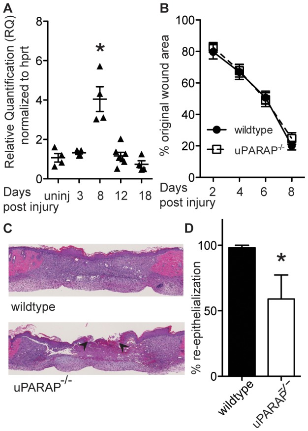
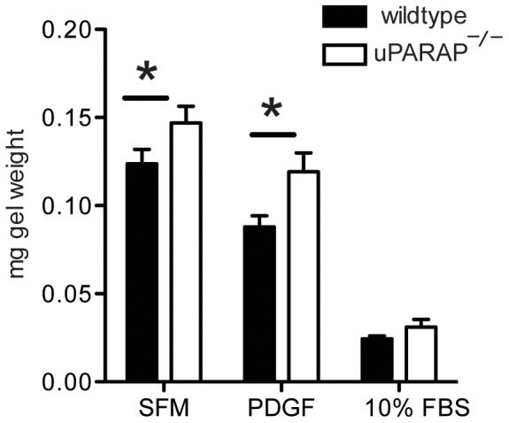
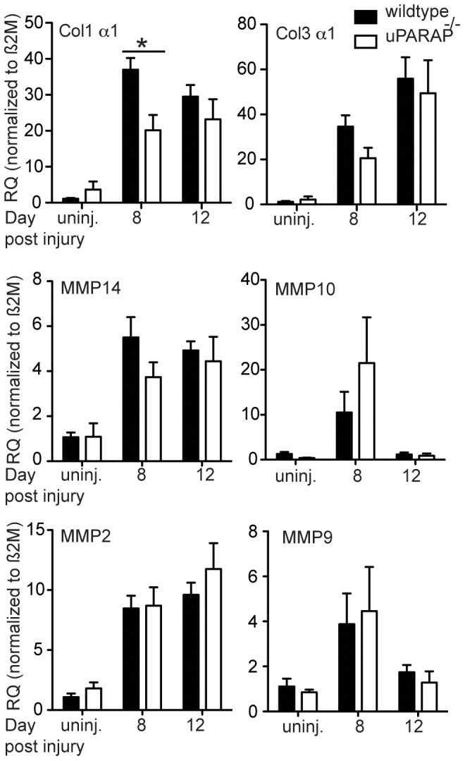
Optimal skin wound healing relies on tight balance between collagen synthesis and degradation in new tissue formation and remodeling phases. The endocytic receptor uPARAP regulates collagen uptake and intracellular degradation. In this study we examined cutaneous wound repair response of uPARAP null (uPARAP-/-) mice. Full thickness wounds were created on dorsal surface of uPARAP-/- or their wildtype littermates. Wound healing evaluation was done by macroscopic observation, histology, gene transcription and biochemical analysis at specific intervals. We found that absence of uPARAP delayed re-epithelialization during wound closure, and altered stiffness of the scar tissue. Despite the absence of the uPARAP-mediated intracellular pathway for collagen degradation, there was no difference in total collagen content of the wounds in uPARAP-/- compared to wildtype mice. This suggests in the absence of uPARAP, a compensatory feedback mechanism functions to keep net collagen in balance.


Conflict of interest statement

Competing Interests: The authors have declared that no competing interests exist.

Figures

Figure 1
Figure 1. Expression of uPARAP during skin wound repair.
(A) Wound samples were collected on indicated days post- injury. mRNA transcription was measured using quantitative real time-PCR. Data were normalized to HPRT expression. Y axis represents fold change relative to uninjured skin. Each point represents an individual mouse. * P <0.05, compared to uninjured skin. Effect of uPARAP on wound closure. (B) On indicated days post-injury, photographs of wounds were taken and percent of wound closure was measured compared to the original wound area using ImageJ analysis (n = 14 wildtype, 13 uPARAP-/-). (C) H&E staining of wound sections on day 8 post-injury shows incomplete re-epithelialization in uPARAP-/- mice. Arrowheads mark the edges of the wound. Section through the midpoint of the wound in wildtype animal shows complete re-epithelialization. (D) Histology assessments of re-epithelialization were quantified (n = 5 wildtype, 4 uPARAP-/- mice), * P<0.05.
Figure 2
Figure 2. Collagen contraction of uPARAP-/- dermal fibroblasts.
Dermal fibroblasts from wildtype or uPARAP-/- mice were grown in collagen gels with serum-free media, PDGF-BB, or 10% FBS for one week, then collagen gels were weighed. Lower weight indicates greater gel contraction. * P<0.05 compared to wildtype. (n = 3/group)
Figure 3
Figure 3. Collagen content in wildtype and uPARAP-/- after wound injury.
(A) Hydroxyproline content was measured in wounds on day 8 and 12 post-injury. (B) Trichrome staining was done on fixed wound samples on day 8 and 12 after wounding. Representative images are shown from at least 4 mice/genotype (C) Ratio of collagen staining area versus total tissue in wound area was quantified in slides stained with trichrome. Data are presented as mean± SEM; n of mice in each group is indicated inside the bars. (D) α-SMA expression in wound tissue homogenates from day 8 post-injury was evaluated by Western blot analysis (top). GAPDH was used as a loading control (bottom), n = 4/genotype. (E) Intensity of bands on immunoblots were quantified and normalized to GAPDH. (F) Representative images of α-SMA staining on fixed wound samples on day 8 post-injury. (G) The area positive for α-SMA was quantified. Each point represents an individual mouse. (H) Area of granulation tissue was traced and quantified in trichrome stained wound samples from day 8. Data are presented as mean± SEM, * P<0.05 compared to wildtype.
Figure 4
Figure 4. Gene expression analysis.
Total RNA was extracted from intact skin or wound samples on day 8 or 12 post-injury. cDNA was made and transcription of target genes was quantified using quantitative real time-PCR method. Data are normalized to β2M housekeeping gene. Data are presented relative to expression in intact skin from wildtype group. * P<0.05, n≥3.
Figure 5
Figure 5. Comparison of stiffness of normal and healed skin in wildtype and uPARAP-/- mice.
(A) Full thickness biopsies collected from intact dorsal surface of wildtype and uPARAP-/- mice were analyzed for mechanical characteristics. n = 6/genotype. (B) Skin wounds on day 12 post-wounding (from the same area as intact skin) were tested for tissue stiffness. * P<0.05, n = 3/genotype.

References

    1. Gurtner GC, Werner S, Barrandon Y, Longaker MT (2008) Wound repair and regeneration. Nature 453: 314–321. - PubMed
    1. Guo S, Dipietro LA (2010) Factors affecting wound healing. J Dent Res 89: 219–229. - PMC - PubMed
    1. Madsen DH, Engelholm LH, Ingvarsen S, Hillig T, Wagenaar-Miller RA, et al. (2007) Extracellular collagenases and the endocytic receptor, urokinase plasminogen activator receptor-associated protein/Endo180, cooperate in fibroblast-mediated collagen degradation. J Biol Chem 282: 27037–27045. - PubMed
    1. Mohamed MM, Sloane BF (2006) Cysteine cathepsins: multifunctional enzymes in cancer. Nat Rev Cancer 6: 764–775. - PubMed
    1. Behrendt N (2004) The urokinase receptor (uPAR) and the uPAR-associated protein (uPARAP/Endo180): membrane proteins engaged in matrix turnover during tissue remodeling. Biol Chem 385: 103–136. - PubMed

Publication types

LinkOut - more resources