. 2014 May;113(5):1827-35.
doi: 10.1007/s00436-014-3829-5. Epub 2014 Mar 25.

Intraperitoneal development of the filarial nematode Brugia malayi in the Mongolian jird (Meriones unguiculatus)

Affiliations

Intraperitoneal development of the filarial nematode Brugia malayi in the Mongolian jird (Meriones unguiculatus)

Yasen Mutafchiev et al. Parasitol Res. 2014 May.

Abstract

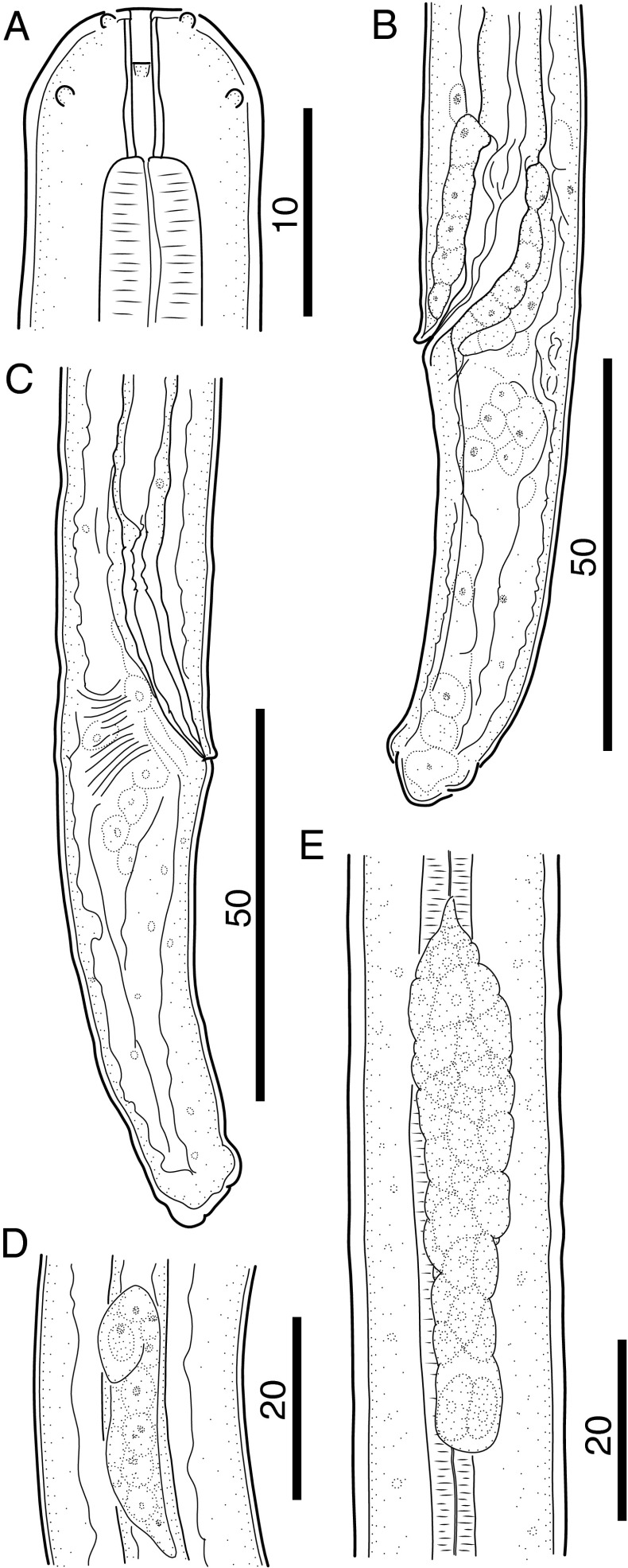
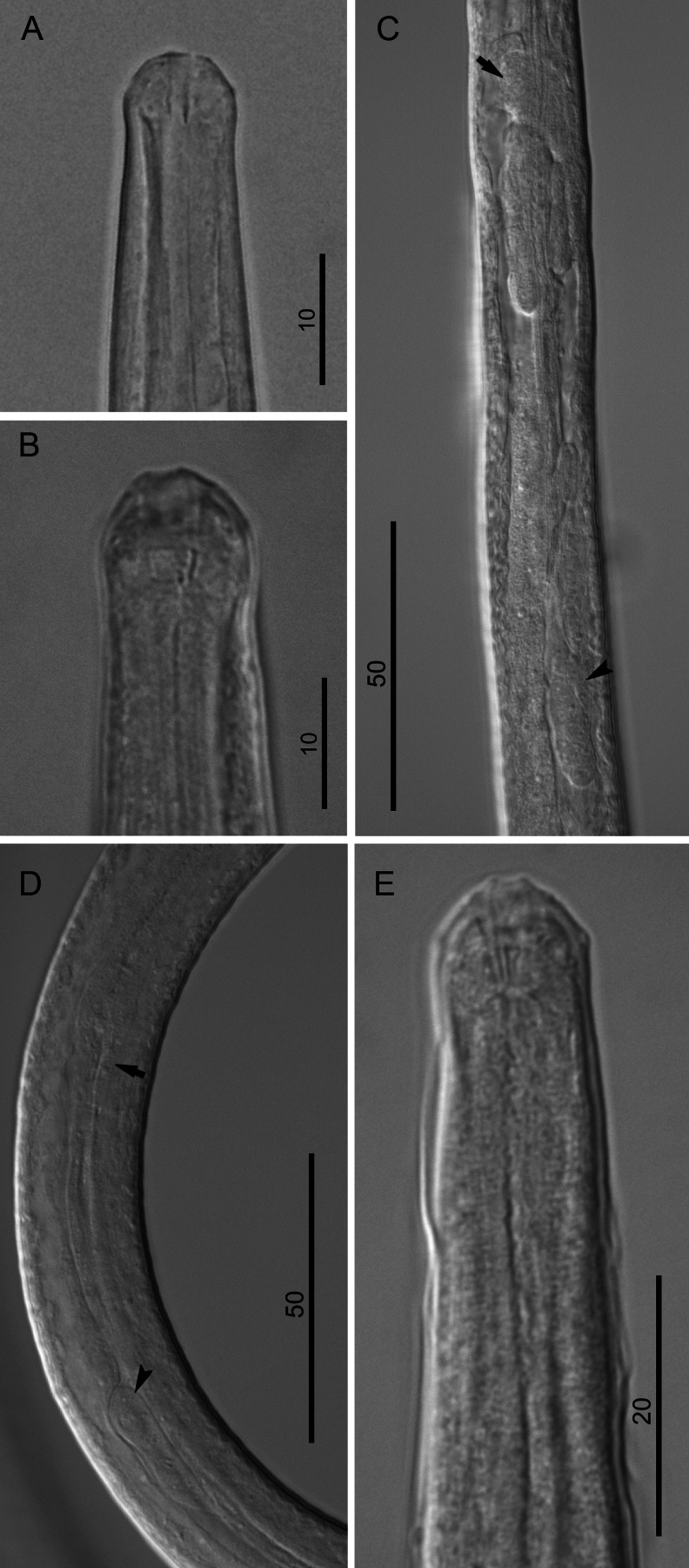
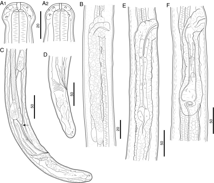
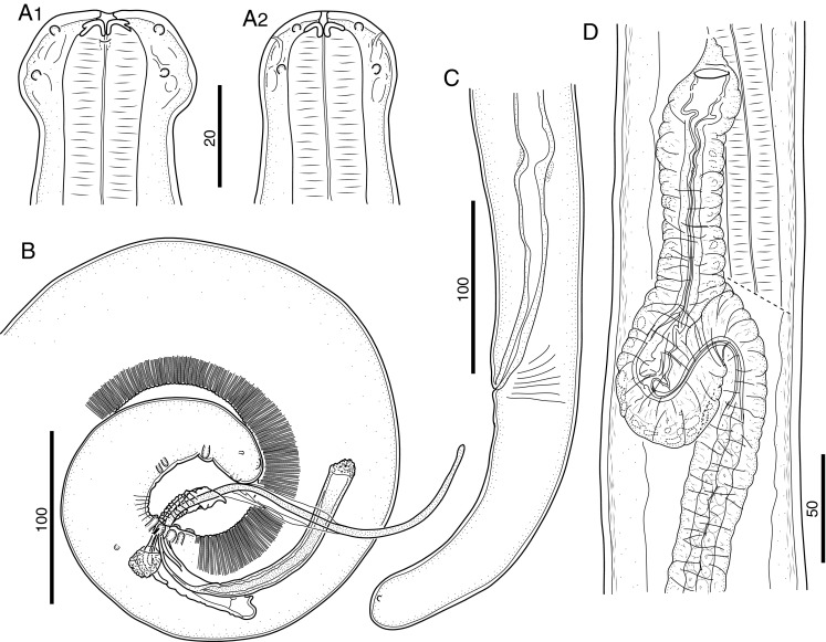
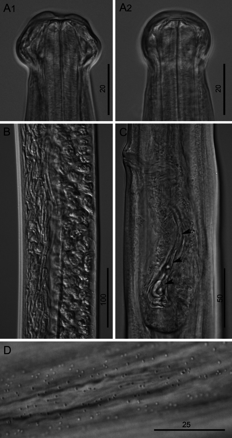
In the present study, we describe intraperitoneal development of the FR3 strain of Brugia malayi in Mongolian jirds (Meriones unguiculatus). The third molt for male worms occurred between 4 and 7 days postinfection (dpi) and between 4 and 8 dpi for females. The fourth and final molt occurred between days 21 and 29 for males and 25 and 34 for females, considerably earlier than the times reported for subcutaneous infection models using cats and jirds. The timing of the third molt coincided largely with reports for subcutaneous Brugia pahangi infections of cats and jirds, but the final molt occurred considerably later and lasted longer than those reported for subcutaneous B. pahangi models. Spermatogenesis occurred by at least 50 dpi in adult males, and insemination of females likely occurred between 50 and 60 dpi. Microfilariae were observed in the uteri and ovejectors of adult females at 65 dpi.


Figures

Fig. 1
Fig. 1
Morphology of third-stage larvae of B. malayi. a Cephalic extremity of 3 dpi larva, lateral view. b Posterior body end of 5 dpi male larva, lateral view. c Posterior body end of 5 dpi female larva, lateral view. d Anlage of reproductive system of 5 dpi male larva, lateral view. e Anlage of reproductive system of 7 dpi female larva, ventral view. Scale bars in micrometers
Fig. 2
Fig. 2
Morphology of third-stage larvae of B. malayi. a Cephalic extremity of 3 dpi larva, lateral view. b Cephalic extremity of 5 dpi larva, lateral view; note hyaline tissue around buccal cavity. c Anlage of reproductive system of 5 dpi female larva, right lateral view; note nerve ring (arrow) and genital anlage (arrowhead). d Anlage of reproductive system of 5 dpi male larva, lateral view; note esophago-intestinal junction (arrow) and genital anlage (arrowhead). e Anterior body end 7 dpi male larva, lateral view; note detached third-stage cuticle. Scale bars in micrometers
Fig. 3
Fig. 3
Morphology of L4 of B. malayi. a Cephalic extremity of 21 dpi larva, lateral and dorsoventral view, respectively. b Reproductive system of 9 dpi female larva, lateral view. c Posterior body end of 21 dpi male larva, lateral view; note developing vas deferens attached to rectum (arrow). d Posterior body end of 21 dpi female larva, lateral view. e Stage of development of vagina, 21 dpi larva, lateral view. f Stage of development of vagina, 31 dpi larva, lateral view. Scale bars in micrometers
Fig. 4
Fig. 4
Morphology of L4 of B. malayi. a Cephalic extremity of 9 dpi, male larva, lateral view. b Region of vulva of 9 dpi larva, ventral view; note future canal of vagina (arrow). c Developing female reproductive system of 9 dpi larva, ventral view; note region of vulva (arrow) and precursor cell of ovary (arrowhead). d Cephalic extremity of 21 dpi, male larva, lateral view; note hyaline tissue around buccal cavity. e Cephalic extremity of 21 dpi, female larva, lateral view. f Posterior body end of 27 dpi male larva, lateral view; note development of caudal papillae (arrowheads) and right spicule (arrows). Scale bars in micrometers
Fig. 5
Fig. 5
Morphology of adults of B. malayi. a Anterior body end of 65 dpi female, lateral and dorsoventral view, respectively. b Posterior body end of 176 dpi male, lateral view. c Posterior body end of 65 dpi female, lateral view. d Vagina of 65 dpi female, ventral view. Scale bars in micrometers
Fig. 6
Fig. 6
Morphology of 65 dpi female of B. malayi. a Cephalic extremity, lateral and dorsoventral view, respectively. b Region of uteri containing microfilariae and eggs. c Vagina with microfilaria (arrowheads), lateral view. d Dotted ornamentation on cuticle surface at posterior body end. Scale bars in micrometers
Fig. 7
Fig. 7
Brugia spp. development varies by host species and route of infection. Bm B. malayi, Bp B. pahangi, IP intraperitoneal, SQ subcutaneous. IP development of B. pahangi has not been reported

References

    1. Ash LR. Chronic Brugia pahangi and Brugia malayi infections in Meriones unguiculatus. J Parasitol. 1973;59:442–447. doi: 10.2307/3278769. - DOI - PubMed
    1. Ash LR, Riley JM. Development of subperiodic Brugia malayi in the jird, Meriones unguiculatus, with notes on infections in other rodents. J Parasitol. 1970;56:969–973. doi: 10.2307/3277515. - DOI - PubMed
    1. Ash LR, Riley JM. Development of Brugia pahangi in the jird, Meriones unguiculatus, with notes on infections in other rodents. J Parasitol. 1970;56:962–968. doi: 10.2307/3277514. - DOI - PubMed
    1. Buckley JJ. On Brugia gen. nov. for Wuchereria spp. of the ‘malayi’ group, i.e., W. malayi (Brug, 1927), W. pahangi Buckley and Edeson, 1956, and W. patei Buckley, Nelson and Heisch, 1958. Ann Trop Med Parasitol. 1960;54:75–77. - PubMed
    1. Buckley JJ, Edeson JF. On the adult morphology of Wuchereria sp. (malayi?) from a monkey (Macaca irus) and from cats in Malaya, and on Wuchereria pahangi n.sp. from a dog and a cat. J Helminthol. 1956;30:1–20. doi: 10.1017/S0022149X00032922. - DOI - PubMed

Publication types

LinkOut - more resources