. 2014 Mar 24;9(3):e92581.
doi: 10.1371/journal.pone.0092581. eCollection 2014.

CCHCR1 interacts specifically with the E2 protein of human papillomavirus type 16 on a surface overlapping BRD4 binding

Affiliations

CCHCR1 interacts specifically with the E2 protein of human papillomavirus type 16 on a surface overlapping BRD4 binding

Mandy Muller et al. PLoS One. .

Abstract

The Human Papillomavirus E2 proteins are key regulators of the viral life cycle, whose functions are commonly mediated through protein-protein interactions with the host cell proteome. We identified an interaction between E2 and a cellular protein called CCHCR1, which proved highly specific for the HPV16 genotype, the most prevalent in HPV-associated cancers. Further characterization of the interaction revealed that CCHCR1 binds the N-terminal alpha helices of HPV16 E2 N-terminal domain. On this domain, the CCHCR1 binding interface overlaps that of BRD4, a key mediator of E2 transcriptional activity. Consequently a physical competition occurs between the two proteins for the binding to HPV16 E2, and CCHCR1 interferes with BRD4-mediated enhancement of E2-dependent transcription. In addition, we showed that the interaction with CCHCR1 induced a massive redistribution of HPV16 E2, from a predominantly nuclear to a cytoplasmic localization in dot-like structures, where E2 perfectly co-localizes with CCHCR1. Such a cytoplasmic docking likely interferes with the nuclear functions of HPV16 E2. Upon co-expression of BRD4 and CCHCR1, E2 accumulates both in the nucleus and in the cytoplasm, indicating that for HPV16, both sub-cellular localization and transcriptional functions of E2 may depend on the proportion of both factors within the cell. We provided evidence of a strong induction of the keratinocyte differentiation marker K10 by HPV16 E2, and showed that this activation is compromised by the interaction with CCHCR1. The specific interaction described here could thus impact on the pathogenesis of HPV16. We propose that it could underlie some specific traits of HPV16 infection, such as an enhanced propensity to give rise to lesions evolving toward cancer.


Conflict of interest statement

Competing Interests: The authors have declared that no competing interests exist.

Figures

Figure 1
Figure 1. Interaction between HPV16 E2 and CCHCR1.
(A) Schematic representation of the GPCA technique. Two proteins A and B are co-expressed in 293 T cells as fusions with two inactive and complementary fragments of the Gaussia princeps luciferase. An interaction between A and B proteins reconstitutes the Gaussia enzymatic activity by bringing in close proximity both fragments. The interaction level is estimated from a NLR (Normalized Luminescence Ratio) corresponding the Gaussia luciferase activity measured when both fusion proteins are expressed divided by the sum of background activities generated by each fusion protein expressed with the empty complementary vector (see for further details). (B) CCHCR1 binding to a panel of E2 proteins. The interactions between 12 E2 proteins and CCHCR1 were measured in GPCA. **, p<0.01 versus the interaction between HPV16 E2 and CCHCR1. (C) Interactions between HPV16 E2 and a panel of known HPV16 E2 interacting partners. The interactions between HPV16 E2 and 13 literature-curated known interactors of this E2 protein were assessed in GPCA. **, p<0.01 versus the interaction between HPV16 E2 and CCHCR1.
Figure 2
Figure 2. Mapping of the CCHCR1 binding interface on HPV16 E2.
(A) top: schematic representation of HPV16 E2 N-terminal domain picturing the position of point mutations used. The E39 amino acid is shown with a dot line since it faces the opposite side of the helices. H1, H2, H3 are respectively alpha-helix 1, 2 and 3. Bottom: diagrams of the HPV16 E2 deletion mutants. (B) Interactions between HPV16 E2 deletion mutants and CCHCR1, BRD4 and TAX1BP1 tested by GPCA. Results are represented relative to the interaction with wild type HPV16 E2. **, p<0.01; ***, p<0.001 versus the interaction with the wild type HPV16 E2. (C) Interaction between HPV16 E2 point mutants and CCHCR1, BRD4 and TBP tested by GPCA. Results are represented as relative to the interaction with the wild type HPV16 E2 protein. ***, p<0.001 versus the interaction with the wild type HPV16 E2. (D) 293 T cells were co-transfected with expression plasmids for Flag-CCHCR1 and GFP-HPV16 E2 WT or I73A as indicated. Cell were lysed and subjected to immunoprecipitation (IP) using anti FLAG antibody followed by western Blotting (WB) with anti FLAG or anti GFP antibodies as indicated. (E) Interaction between HPV16 E2 or HPV11 E2 and BRD4 was assessed in GPCA, in the presence of CCHCR1 as a challenging protein where indicated. Results are reported to NLR values obtained when an empty plasmid was used instead of the challenging protein. ***, p<0.001 versus the interaction without CCHCR1 as a challenger. (F) HaCaT cells were transfected with an E2-reponsive luciferase reporter plasmid (pTK6E2BS) in the presence of HPV16 E2, BRD4 plus CCHCR1 where indicated. Fold activation are given relative to promoter activity without E2; *, p<0.05 versus experiments without CCHCR1.
Figure 3
Figure 3. CCHCR1 inhibits the activation of early differentiation by HPV16 E2.
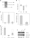
(A) Schematic representation of the differentiation of a keratinized epithelium, with studied markers outlined. (B) HaCaT cells were transfected by a CCHCR1 expression plasmid, and the mRNA levels of three differentiation markers were subsequently monitored by qRT-PCR. Results are represented as relative to mock- transfected cells (empty plasmid). *, p<0.05; **, p<0.01 versus mock-transfected cell experiments. (C) HaCaT cells were infected by Ad-GFP16E2 and the mRNA levels of three differentiation markers were subsequently monitored by qRT-PCR. Results are represented as relative to cells infected with Ad-GFP. *, p<0.05; **, p<0.01 versus cells infected with Ad-GFP. (D) HaCaT cells were infected by Ad-GFP16E2 or Ad-GFP18E2 and mRNA levels of K10 were subsequently monitored by qRT-PCR. Results are represented as relative to Ad-GFP expressing cells. ***, p<0.001 versus cells infected with Ad-GFP16E2. (E) HaCaT cells were transfected by 3XFLAG-HPV16 E2 and CCHCR1 expression plasmids as indicated and the subsequent effect of the mRNA levels of K10 was monitored by qRT-PCR. Results are represented relative to mock-transfected cells. The dotted line represents the K10 mRNA levels expected by simply combining the levels obtained in the presence of each E2 and CCHCR1 protein separately. ***, p<0.001 versus cells infected with HPV16 E2 only. (F) HaCaT cells were transfected as in (E) and western blots were performed on total cell lysate with anti FLAG or anti CCHCR1 antibodies, then with anti-tubulin antibody as loading control.
Figure 4
Figure 4. CCHCR1 docks HPV16 E2 into cytoplasmic dot structures.
HaCaT cells were co-transfected with the expression plasmids for GFP or the indicated GFP-E2 proteins, and mCherry-CCHCR1 or mCherry alone. After fixation, the cells were subjected to fluorescence microscopy after counterstaining of the nucleus with DAPI. From top to bottom: Ectopic expression of CCHCR1 shows punctate staining (red) in the cytoplasm while HPV16 E2 displays a diffuse pattern both in the nucleus and in the cytoplasm. When co-expressed, the two protein signals overlap in cytoplasmic dots typical of CCHCR1 expression. The non-interacting E2 protein mutated HPV16 E2 I73A showed no delocalization from the nucleus.
Figure 5
Figure 5. CCHCR1 does not affect the distribution of non-interacting E2 proteins.
HaCaT cells were co-transfected with the expression plasmids for GFP or the indicated GFP-E2 proteins, and mCherry-CCHCR1 or mCherry alone and processed as in Figure 4. Four E2 proteins (HPV1, 5, 11 and 18) not able to interact with CCHCR1 were studied, and no change in their subcellular localization could be observed in the presence of CCHCR1.
Figure 6
Figure 6. Intermediate pattern of HPV16 E2 distribution in the presence of CCHCR1 and BRD4.
HaCaT cells were transfected with GFP-16E2 and mCherry-CCHCR1, untagged BRD4 or both together. After fixation, the cells were subjected to fluorescence microscopy after counterstaining of the nucleus with DAPI. HPV16 E2 subcellular localization is not affected by the expression of BRD4. In presence of both CCHCR1 and BRD4, HPV16 E2 distributed both in the nucleus and in CCHCR1-containing cytoplasmic dots.

References

    1. zur Hausen H (2009) Papillomaviruses in the causation of human cancers - a brief historical account. Virology 384: 260–265. - PubMed
    1. Doorbar J (2006) Molecular biology of human papillomavirus infection and cervical cancer. Clin Sci (Lond) 110: 525–541. - PubMed
    1. D'Abramo CM, Archambault J (2011) Small molecule inhibitors of human papillomavirus protein - protein interactions. Open Virol J 5: 80–95. - PMC - PubMed
    1. Muller M, Demeret C (2012) The HPV E2-Host Protein-Protein Interactions: A Complex Hijacking of the Cellular Network. Open Virol J 6: 173–189. - PMC - PubMed
    1. Asumalahti K, Laitinen T, Itkonen-Vatjus R, Lokki ML, Suomela S, et al. (2000) A candidate gene for psoriasis near HLA-C, HCR (Pg8), is highly polymorphic with a disease-associated susceptibility allele. Hum Mol Genet 9: 1533–1542. - PubMed

Publication types

MeSH terms

Substances