. 2014 Mar 24;9(3):e92586.
doi: 10.1371/journal.pone.0092586. eCollection 2014.

Endoplasmic reticulum stress-mediated activation of p38 MAPK, Caspase-2 and Caspase-8 leads to abrin-induced apoptosis

Affiliations

Endoplasmic reticulum stress-mediated activation of p38 MAPK, Caspase-2 and Caspase-8 leads to abrin-induced apoptosis

Ritu Mishra et al. PLoS One. .

Abstract

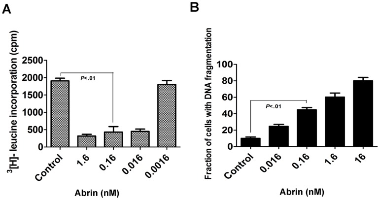
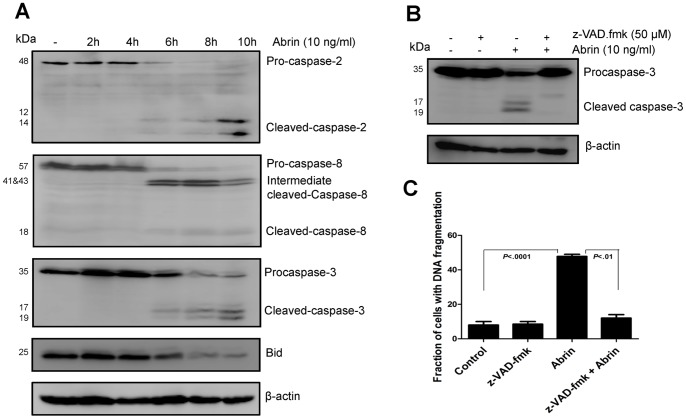
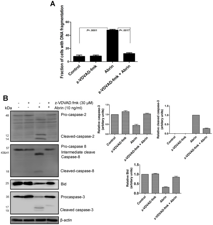
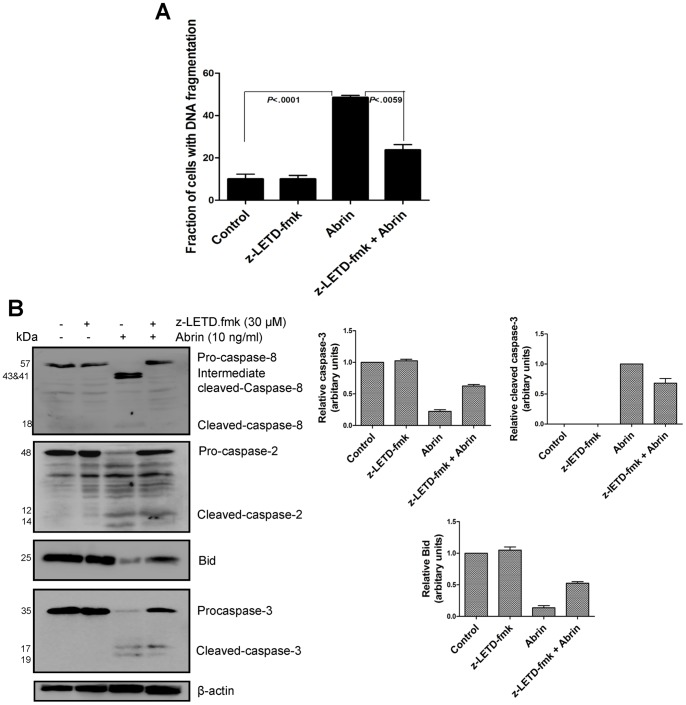
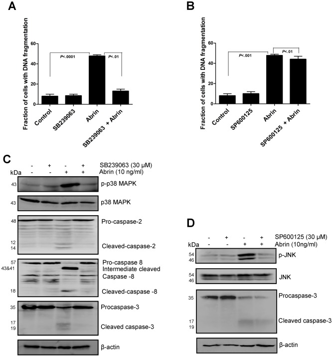
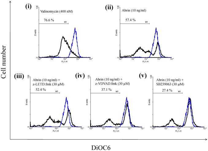
Abrin from Abrus precatorius plant is a potent protein synthesis inhibitor and induces apoptosis in cells. However, the relationship between inhibition of protein synthesis and apoptosis is not well understood. Inhibition of protein synthesis by abrin can lead to accumulation of unfolded protein in the endoplasmic reticulum causing ER stress. The observation of phosphorylation of eukaryotic initiation factor 2α and upregulation of CHOP (CAAT/enhancer binding protein (C/EBP) homologous protein), important players involved in ER stress signaling by abrin, suggested activation of ER stress in the cells. ER stress is also known to induce apoptosis via stress kinases such as p38 MAPK and JNK. Activation of both the pathways was observed upon abrin treatment and found to be upstream of the activation of caspases. Moreover, abrin-induced apoptosis was found to be dependent on p38 MAPK but not JNK. We also observed that abrin induced the activation of caspase-2 and caspase-8 and triggered Bid cleavage leading to mitochondrial membrane potential loss and thus connecting the signaling events from ER stress to mitochondrial death machinery.


Conflict of interest statement

Competing Interests: The authors have declared that no competing interests exist.

Figures

Figure 1
Figure 1. Abrin mediated protein synthesis inhibition and apoptosis in Jurkat cells.
(A) Jurkat cells were treated with different concentrations of abrin for 8 h and protein synthesis was measured by incorporation of [3H]-leucine. (B) Jurkat cells were treated with varying concentrations (16 nM – 0.016 nM) of abrin for 12 h. After the treatments cells were harvested, fixed with 70% ethanol, stained with propidium iodide and quantified for apoptotic population. Each bar is presented as mean ± SE of triplicate samples.
Figure 2
Figure 2. Abrin induces ER stress in Jurkat cells.
(A) After the treatment of Jurkat cells with abrin (10 ng/ml) for different time intervals (0–10 h), whole cell lysates were prepared and analysed by Western blot for the total and phosphorylated levels of eIF2α, JNK and p38 MAPK using specific antibodies (B) Jurkat cells were pretreated with 50 μM z-VAD.fmk for 2 h followed by abrin (10 ng/ml) for 10 h. After the treatments, whole cell lysates were prepared and analysed by Western blot for the total and phosphorylated level of eIF2α, JNK and p38 MAPK using specific antibodies.
Figure 3
Figure 3. Abrin-induced apoptosis activates caspase -2, -8 and -3.
(A) Jurkat cells were treated with abrin (10 ng/ml) for different time intervals (0–10 h). After the treatment, whole cell lysates were prepared and analysed by Western blot for cleaved caspase-2, -8, -3 and Bid using specific antibodies. Equal protein loading was checked by stripping and re-probing the membranes for β-actin. (B) Jurkat cells were pretreated with 50 μM z-VAD.fmk for 2 h followed by abrin (10 ng/ml) for 10 h. After the treatments whole cell lysates were prepared and analysed by Western blot for cleaved caspase-3 using specific antibody. Equal protein loading was checked by stripping and re-probing the membranes for β-actin. (C) After similar treatments with 50 μM z-VAD.fmk and abrin, cells were harvested, fixed with 70% ethanol, stained with propidium iodide and quantified for apoptotic population. Each bar is presented as mean ± SE of triplicate samples.
Figure 4
Figure 4. Role of caspase-2 in abrin-induced apoptosis.
Jurkat cells were pretreated with caspase-2 inhibitor, z-VDVAD.fmk for 2 h and then with abrin for 10 h. Abrin-induced apoptosis in the presence of inhibitor were quantified by flow cytometry (A) after the treatments cells were harvested, fixed with 70% ethanol, stained with propidium iodide and quantified for apoptotic population. After the similar treatments whole cell lysates were prepared and analysed by Western blot for (B) cleaved caspase-2, 8, 3, and full length Bid. Equal protein loading was checked by stripping and re-probing the membranes for β-actin. Densitometry analysis was performed for two to three Western blots. Each bar is presented as mean ± SE of triplicate samples.
Figure 5
Figure 5. Role of caspase-8 in abrin-induced apoptosis.
Jurkat cells were pretreated with caspase-8 inhibitor, z-LETD.fmk for 2 h and then with abrin for 10 h. Abrin-induced apoptosis in the presence of inhibitor was quantified by flow cytometry (A) after the treatments cells were harvested, fixed with 70% ethanol, stained with propidium iodide and quantified for apoptotic population. After the similar treatments whole cell lysates were prepared and analysed by Western blot for (B) cleaved caspase-8, 2, 3, and full length Bid. Equal protein loading was checked by stripping and re-probing the membranes for β-actin. Densitometry analysis was performed for two to three Western blots. Each bar is presented as mean ± SE of triplicate samples.
Figure 6
Figure 6. Abrin causes activation of p38 MAPK pathway which leads to apoptosis.
Jurkat cells were pretreated for 2(10 ng/ml) for 10 h. (A&B) After the treatments, Cells were harvested fixed with 70% ethanol, stained with propidium iodide and quantified for apoptotic population. Each bar is presented as mean ± SE of triplicate samples. After the similar treatments,whole cell lysates were prepared and analysed by Western blot for (C) p-p38 MAPK, total p38 MAPK caspase-2, 8 and 3. (D) For p-JNK, total JNK and caspase-3. Equal protein loading was checked by stripping and re-probing the membranes for β-actin.
Figure 7
Figure 7. Role of p38MAPK, caspase-2 and caspase-8 on abrin-induced mitochondrial membrane potential (MMP) loss in Jurkat cells.
(A) Jurkat cells treated with abrin alone or along with inhibitors were harvested and stained with DiOC6 dye and the percentage of cells positive for green fluorescence were analyzed by flow cytometry as described in Materials and methods. The blue line indicates untreated Jurkat cells and the black line represents Jurkat cells treated as indicated in each panel. Also, percentage in each panel denotes the loss of mitochondrial membrane potential.
Figure 8
Figure 8. Abrin activates DNA damage signaling pathway.
Phosphorylation of H2AX (γH2AX) was analysed in Jurkat cells (A) treated with abrin (10 ng/ml) for different time intervals (0–10 h) (B) pretreated with broad spectrum caspase inhibitor, z-VAD.fmk for 2 h and then with abrin or (C) with gamma radiation (20 Gy). Equal protein loading was checked by stripping and re-probing the membranes for β-actin.

References

    1. Hartley MR, Lord JM (2004) Cytotoxic ribosome-inactivating lectins from plants. Biochimica et Biophysica Acta (BBA) - Proteins and Proteomics 1701: 1–14. - PubMed
    1. Nielsen K, Boston RS (2001) Ribosome-inactivating proteins: A plant perspective. Annual Review of Plant Physiology and Plant Molecular Biology 52: 785–816. - PubMed
    1. Peumans WJ, Hao Q, Van Damme EJM (2001) Ribosome-inactivating proteins from plants: more than RNA N-glycosidases? The FASEB Journal 15: 1493–1506. - PubMed
    1. Bolognesi A, Tazzari PL, Olivieri F, Polito L, Falini B, et al. (1996) Induction of apoptosis by ribosome-inactivating proteins and related immunotoxins. Int J Cancer 68: 349–355. - PubMed
    1. Narayanan S, Surendranath K, Bora N, Surolia A, Karande AA (2005) Ribosome inactivating proteins and apoptosis. FEBS Letters 579: 1324–1331. - PubMed

Publication types

MeSH terms