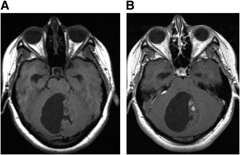
Review
. 2014 Apr;19(4):403-13.
doi: 10.1634/theoncologist.2013-0345. Epub 2014 Mar 24.

Low-grade gliomas

Affiliations
Review

Low-grade gliomas

Deborah A Forst et al. Oncologist. 2014 Apr.

Abstract

Low-grade gliomas (LGGs) are a diverse group of primary brain tumors that often arise in young, otherwise healthy patients and generally have an indolent course with longer-term survival in comparison with high-grade gliomas. Treatment options include observation, surgery, radiation, chemotherapy, or a combined approach, and management is individualized based on tumor location, histology, molecular profile, and patient characteristics. Moreover, in this type of brain tumor with a relatively good prognosis and prolonged survival, the potential benefits of treatment must be carefully weighed against potential treatment-related risks. We review in this article current management strategies for LGG, including surgery, radiotherapy, and chemotherapy. In addition, the importance of profiling the genetic and molecular properties of LGGs in the development of targeted anticancer therapies is also reviewed. Finally, given the prevalence of these tumors in otherwise healthy young patients, the impact of treatment on neurocognitive function and quality of life is also evaluated.

Keywords: Astrocytoma; Glioma; Oligodendroglioma; Radiation; Surgery.


Conflict of interest statement

Disclosures of potential conflicts of interest may be found at the end of this article.

Figures

Figure 1.
Figure 1.
Imaging features of low-grade glioma. The grade 2 oligoastrocytoma pictured in these magnetic resonance images appears as a relatively homogeneous region of high signal intensity on T2/FLAIR-weighted images (A) and low signal intensity on T1 precontrast images (B). There is faint contrast enhancement on the T1 postcontrast images (C).
Figure 2.
Figure 2.
Histopathologic features of low-grade glioma. (A): Oligodendrogliomas are characterized by uniform-appearing, infiltrating cells with perinuclear clearing in a honeycomb pattern. Hematoxylin and eosin stain. Magnification, 400×. (B): Astrocytoma, consisting of fibrillary neoplastic astrocytes on a loose tumor matrix background. Hematoxylin and eosin stain. Magnification, 400×. (C): Oligoastrocytoma, containing a mixture of both tumor cell types. Hematoxylin and eosin stain. Magnification, 400×. Images courtesy of Declan McGuone, neuropathology fellow, Department of Neuropathology, Massachusetts General Hospital.
Figure 3.
Figure 3.
Noninvasive detection of genetic mutations. One-dimensional magnetic resonance spectroscopy with use of specialized spectral-editing sequence for the detection of 2HG as demonstrated in a secondary glioblastoma patient with IDH1R132H mutation (A), in comparison with the spectra from a healthy volunteer with wt IDH1 (B). Figure reprinted (adapted) with permission from Andronesi OC, Kim GS, Gerstner E et al. Detection of 2HG in IDH-mutated glioma patients by in vivo spectral editing and two-dimensional correlation magnetic resonance spectroscopy. Sci Transl Med 2012;4:116ra4. Abbreviations: a.u., arbitrary units; GABA, γ-aminobutyric acid; Gln, glutamine; Glu, glutamate; 2HG, 2-hydroxyglutarate; I, signal intensity; MM, denotes contamination of GABA signal with macromolecule signal; wt, wild-type.
Figure 4.
Figure 4.
Brainstem glioma. The diffuse pontine glioma pictured in this T2/FLAIR-weighted magnetic resonance image appears as an expansile, hyperintense lesion centered within the pons.
Figure 5.
Figure 5.
Juvenile pilocytic astrocytoma. The juvenile pilocytic astrocytoma pictured in these magnetic resonance images appears as a mainly cystic neoplasm centered in the cerebellar vermis. There is a dominant cyst and smaller mural solid component, as seen on T1 precontrast images (A), with small areas of nodular enhancement within the mural component, as seen on T1 postcontrast images (B). The tumor fills and obstructs the fourth ventricle, causing noncommunicating hydrocephalus.

References

    1. et al. In: WHO Classification of Tumours of the Central Nervous System. Louis DN, Ohgaki H, Wiestler OD, editors; Lyon, France: IARC Press; 2007. - PMC - PubMed
    1. Central Brain Tumor Registry of the United States . Statistical Report: Primary Brain and Central Nervous System Tumors Diagnosed in the United States in 2004-2008 (March 23, 2012 Revision) Hinsdale, IL: CBTRUS; 2012.
    1. Schwartzbaum JA, Fisher JL, Aldape KD, et al. Epidemiology and molecular pathology of glioma. Nat Clin Pract Neurol. 2006;2:494–503; quiz 1, 516. - PubMed
    1. Pouratian N, Asthagiri A, Jagannathan J, et al. Surgery insight: The role of surgery in the management of low-grade gliomas. Nat Clin Pract Neurol. 2007;3:628–639. - PubMed
    1. Pirzkall A, Nelson SJ, McKnight TR, et al. Metabolic imaging of low-grade gliomas with three-dimensional magnetic resonance spectroscopy. Int J Radiat Oncol Biol Phys. 2002;53:1254–1264. - PubMed