. 2014 Jun;82(6):2329-36.
doi: 10.1128/IAI.01616-13. Epub 2014 Mar 24.

Modulation of vacuolar pH is required for replication of Edwardsiella ictaluri in channel catfish macrophages

Affiliations

Modulation of vacuolar pH is required for replication of Edwardsiella ictaluri in channel catfish macrophages

Wes A Baumgartner et al. Infect Immun. 2014 Jun.

Abstract

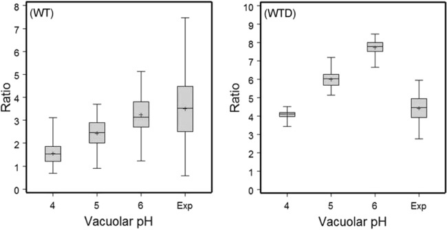
Previous in vitro work demonstrated that Edwardsiella ictaluri produces an acid-activated urease that can modulate environmental pH through the production of ammonia from urea. Additional work revealed that expression of the E. ictaluri type III secretion system (T3SS) is upregulated by acidic pH. Both the urease and the T3SS were previously shown to be essential to intracellular replication. In this work, fluorescence microscopy with LysoTracker Red DND-99 (LTR) indicated that E. ictaluri-containing vacuoles (ECV) became acidified following ingestion by head kidney-derived macrophages (HKDM). In vivo ratiometric imaging demonstrated a lowered ECV pH, which fell to as low as pH 4 but subsequently increased to pH 6 or greater. Inhibition of vacuolar H(+)-ATPases by use of the specific inhibitor bafilomycin A1 abrogated both ECV acidification and intracellular replication in HKDM. Failure of an E. ictaluri urease knockout mutant to increase the ECV pH in the in vivo ratiometric assay suggests that ammonia produced by the urease reaction mediates the pH increase. Additionally, when the specific arginase inhibitor l-norvaline was used to treat E. ictaluri-infected HKDM, the ECV failed to neutralize and E. ictaluri was unable to replicate. This indicates that the HKDM-encoded arginase enzyme produces the urea used by the E. ictaluri urease enzyme. Failure of the ECV to acidify would prevent both upregulation of the T3SS and activation of the urease enzyme, either of which would prevent E. ictaluri from replicating in HKDM. Failure of the ECV to neutralize would result in a vacuolar pH too low to support E. ictaluri replication.


Figures

FIG 1
FIG 1
Intracellular survival and replication of wild-type E. ictaluri (WT) and the Oregon Green 514-stained WT (OG-WT) in channel catfish HKDM. Both WT and OG-WT levels increased approximately 20-fold by 10 hpi. Results for the WT and OG-WT were not significantly different from one another at any time point (P ≤ 0.05), indicating that OG staining of E. ictaluri does not affect replication in HKDM. Results are presented as means and standard errors of the means for three wells per treatment and are representative of three independent experiments.
FIG 2
FIG 2
Fluorescence microscopy with HKDM stained with LysoTracker Red (LTR) and E. ictaluri stained with Oregon Green 514 (OG). (A) DIC image with a box visualizing the area depicted in frames B, C, and D. (B) E. ictaluri stained with OG. (C) E. ictaluri vacuoles stained red with LTR, indicating acidification. (D) Colocalization of OG-stained E. ictaluri and acidified vacuoles. Panel A also shows the location of E. ictaluri within the HKDM.
FIG 3
FIG 3
Percentages of bacteria contained within acidified vacuoles in HKDM stained with LTR from 30 to 90, 90 to 150, and 150 to 210 mpi. Bars represent mean numbers of E. ictaluri bacteria colocalized in acidic vacuoles for three independent experiments plus standard errors. Asterisks indicate significant differences (P ≤ 0.05).
FIG 4
FIG 4
In vivo determination of the pH of the ECV in E. ictaluri-infected channel catfish HKDM at 150 mpi, using WT and heat-killed WT (WTD) bacteria. Plots show data ranges, interquartile ranges (middle 50% of data points; gray boxes), and means (+). Vacuolar pHs 4, 5, and 6 represent the vacuolar pHs established using ionophore calibration solutions to generate fluorescence ratio values. Exp, measurement of fluorescence ratios of the ECV when macrophages were bathed in neutral saline. Ratios represent the relative fluorescence of OG at 510 and 450 nm. Results from three independent experiments were combined.
FIG 5
FIG 5
Acidification of HKDM vacuoles containing heat-killed E. ictaluri and exposed to LTR from 150 to 210 min postinfection. The vacuolar H+-ATPase inhibitor bafilomycin A1 was either removed at 90 min postinfection or maintained throughout the 210 min of the experiment. The results show that bafilomycin A1 prevents vacuolar acidification and that the effect is reversible if bafilomycin is removed. Asterisks indicate a significant difference from cells in which the LTR was removed after 90 min (P ≤ 0.01).
FIG 6
FIG 6
Fold increase of E. ictaluri in HKDM treated or not treated with the vacuolar H+-ATPase inhibitor bafilomycin A1. Treated HKDM were exposed to 100 nM bafilomycin A1 for the full duration of infection (Continual), or bafilomycin A1 was added at 110 mpi or 210 mpi for the remainder of the assay. Data represent mean fold increases at 10 h postinfection for 8 replicate wells plus standard errors. Asterisks indicate a significant difference from untreated cells (P < 0.01).
FIG 7
FIG 7
Replication of E. ictaluri in HKDM treated with the vacuolar H+-ATPase inhibitor bafilomycin A1. Head kidney-derived macrophages were exposed to 100 nM bafilomycin A1 until the gentamicin killing dose was removed or were exposed for the full duration of infection. The results indicate that acidification is required for intracellular replication of E. ictaluri. Bars represent mean fold increases at 10 h postinfection for three independent experiments plus standard errors. Asterisks indicate a significant difference from untreated cells (P ≤ 0.01).
FIG 8
FIG 8
In vivo determination of vacuolar pH, comparing a urease mutant (ΔureG) to wild-type E. ictaluri at 150 to 180 mpi. Plots show data ranges, interquartile ranges (middle 50% of data points; gray boxes), and means (+). Vacuolar pHs 4, 5, and 6 represent vacuolar pHs obtained using ionophore calibration solutions to generate fluorescence ratio values. Exp, measurement of fluorescence ratios of the ECV when macrophages were bathed in neutral saline. Ratios represent the relative fluorescence of OG at 510 and 450 nm. Results were combined from two independent experiments and indicate that the E. ictaluri urease enzyme is required for ECV neutralization.
FIG 9
FIG 9
In vivo determination of the pH of the ECV in channel catfish HKDM with and without 10 mM of the specific arginase inhibitor l-norvaline for 60 mpi. Plots showing data range (whiskers), interquartile range (the mid-50% of data points; box), and the mean (designated +). Vacuolar pH at 4, 5, and 6 represents vacuolar pH using ionophore calibration solutions to generate fluorescent ratio values. “Exp” represents the measurement of fluorescent ratios of the ECV when macrophages were bathed in neutral saline. “Ratio” represents the relative fluorescence of OG at 510 and 450 nm. Results are combined from 2 independent experiments and indicate that the HKDM arginase enzyme is required for ECV neutralization.
FIG 10
FIG 10
Intracellular survival and replication of E. ictaluri in HKDM and l-norvaline-treated HKDM. The results show that the WT increased over 15-fold after 10 h, while norvaline treatment to block arginase activity and prevent ECV neutralization also prevented WT replication. l-Norvaline treatment did not affect WT survival. Results are presented as means plus standard errors for three wells per treatment per time point and are representative of three independent experiments. Asterisks indicate a significant difference from untreated cells (P ≤ 0.001).
FIG 11
FIG 11
Urea concentrations in HKDM at 30 mpi and 3 h 30 mpi in uninfected HKDM and HKDM infected with the WT or urease knockout strain of E. ictaluri. Asterisks indicate a significant difference from uninfected cells: **, P ≤ 0.01; and ***, P ≤ 0.001.

References

    1. Hawke JP. 1979. A bacterium associated with disease of pond cultured catfish, Ictalurus punctatus. J. Fish. Res. Board Can. 36:1508–1512. 10.1139/f79-219 - DOI
    1. USDA. 2010. Catfish 2010 part I: reference of catfish health and production practices in the United States, 2009. USDA-APHIS-VS, CEAH, Ft. Collins, CO
    1. Thune RL, Stanley LA, Cooper RK. 1993. Bacterial diseases of catfishes, p 511–520 In Stoskopf MK. (ed), Fish medicine. W B Saunders Company, Philadelphia, PA
    1. Booth NJ, El Kamel A, Thune RL. 2006. Intracellular replication of Edwardsiella ictaluri in channel catfish macrophages. J. Aquat. Anim. Health 18:101–108. 10.1577/H05-025.1 - DOI
    1. Thune RL, Fernandez DH, Benoit JL, Kelly-Smith M, Rogge ML, Booth NJ, Landry CA, Bologna RA. 2007. Signature-tagged mutagenesis of Edwardsiella ictaluri identifies virulence-related genes, including a salmonella pathogenicity island 2 class of type III secretion systems. Appl. Environ. Microbiol. 73:7934–7946. 10.1128/AEM.01115-07 - DOI - PMC - PubMed

MeSH terms

LinkOut - more resources