

Cadherin 5 is regulated by corticosteroids and associated with central serous chorioretinopathy
- PMID: 24665005
- PMCID: PMC4215937
- DOI: 10.1002/humu.22551
Cadherin 5 is regulated by corticosteroids and associated with central serous chorioretinopathy
Abstract
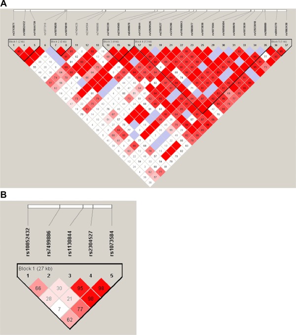
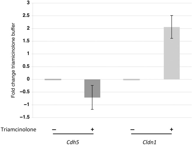
Central serous chorioretinopathy (CSC) is characterized by leakage of fluid from the choroid into the subretinal space and, consequently, loss of central vision. The disease is triggered by endogenous and exogenous corticosteroid imbalance and psychosocial stress and is much more prevalent in men. We studied the association of genetic variation in 44 genes from stress response and corticosteroid metabolism pathways with the CSC phenotype in two independent cohorts of 400 CSC cases and 1,400 matched controls. The expression of cadherin 5 (CDH5), the major cell-cell adhesion molecule in vascular endothelium, was downregulated by corticosteroids which may increase permeability of choroidal vasculature, leading to fluid leakage under the retina. We found a significant association of four common CDH5 SNPs with CSC in male patients in both cohorts. Two common intronic variants, rs7499886:A>G and rs1073584:C>T, exhibit strongly significant associations with CSC; P = 0.00012; odds ratio (OR) = 1.5; 95%CI [1.2;1.8], and P = 0.0014; OR = 0.70; 95%CI [0.57;0.87], respectively. A common haplotype was present in 25.4% male CSC cases and in 35.8% controls (P = 0.0002; OR = 0.61, 95% CI [0.47-0.79]). We propose that genetically predetermined variation in CDH5, when combined with triggering events such as corticosteroid treatment or severe hormonal imbalance, underlie a substantial proportion of CSC in the male population.
Keywords: CDH5; Cadherin 5; central serous chorioretinopathy; genetic association; retinal disease.
© 2014 The Authors. *Human Mutation published by Wiley Periodicals, Inc.
Figures
References
-
- Abramoff MD, Magelhaes PJ, Ram SJ. Image processing with ImageJ. Biophoton Int. 2004;11:36–42.
-
- Ahnoux-Zabsonre A, Quaranta M, Mauget-Faysse M. Prevalence of Helicobacter pylori in central serous chorioretinopathy and diffuse retinal epitheliopathy: a complementary study. J Fr Ophtalmol. 2004;27:1129–1133. - PubMed
-
- Carvalho-Recchia CA, Yannuzzi LA, Negrao S, Spaide RF, Freund KB, Rodriguez-Coleman H, Lenharo M, Iida T. Corticosteroids and central serous chorioretinopathy. Ophthalmology. 2002;109:1834–1837. - PubMed
-
- Dejana E, Orsenigo F, Lampugnani MG. The role of adherens junctions and VE-cadherin in the control of vascular permeability. J Cell Sci. 2008;121(Pt 13):2115–2122. - PubMed
Publication types
MeSH terms
Substances
Grants and funding
LinkOut - more resources
Full Text Sources
Other Literature Sources
Medical
Miscellaneous
