. 2013 Apr 1;1(2):e24333.
doi: 10.4161/tisb.24333.

Interleukin-4 and interleukin-13 cause barrier dysfunction in human airway epithelial cells

Affiliations

Interleukin-4 and interleukin-13 cause barrier dysfunction in human airway epithelial cells

Bahman Saatian et al. Tissue Barriers. .

Abstract

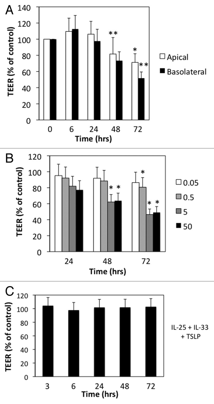
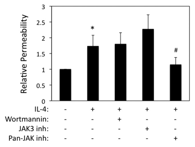
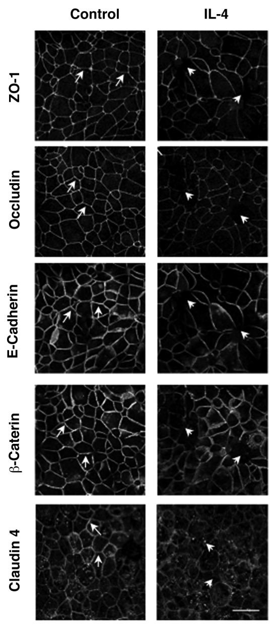
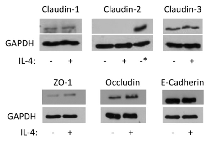
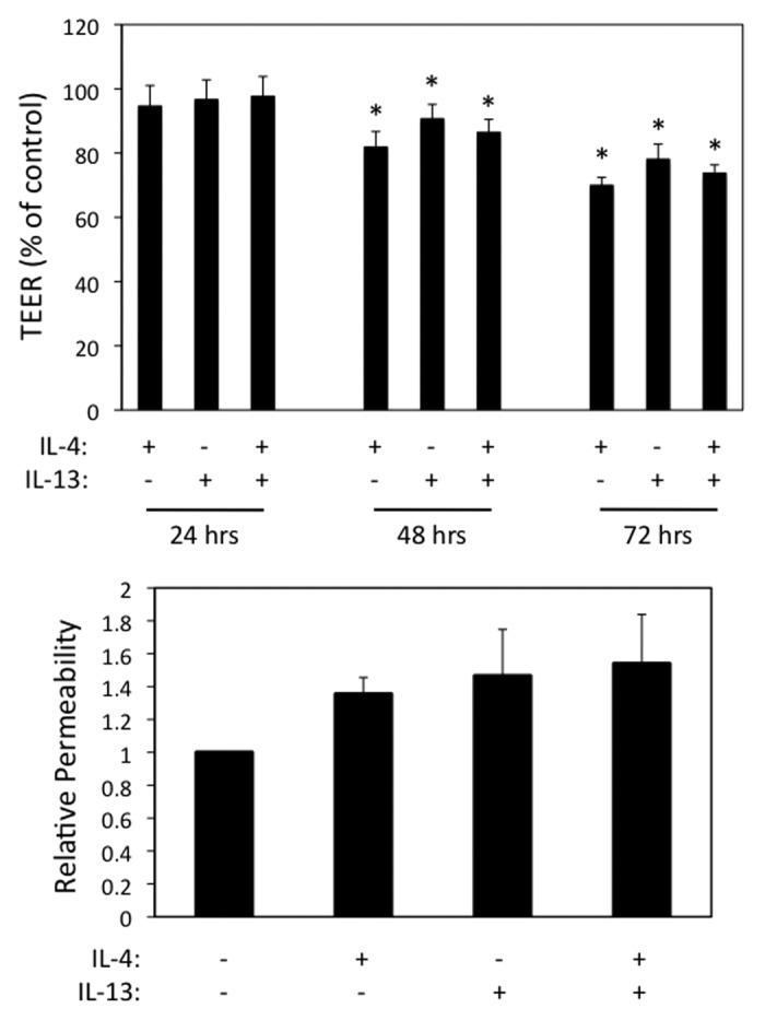
Emerging evidence indicates that airway epithelial barrier function is compromised in asthma, a disease characterized by Th2-skewed immune response against inhaled allergens, but the mechanisms involved are not well understood. The purpose of this study was to investigate the effects of Th2-type cytokines on airway epithelial barrier function. 16HBE14o- human bronchial epithelial cells monolayers were grown on collagen coated Transwell inserts. The basolateral or apical surfaces of airway epithelia were exposed to human interleukin-4 (IL-4), IL-13, IL-25, IL-33, thymic stromal lymphopoietin (TSLP) alone or in combination at various concentrations and time points. We analyzed epithelial apical junctional complex (AJC) function by measuring transepithelial electrical resistance (TEER) and permeability to FITC-conjugated dextran over time. We analyzed AJC structure using immunofluorescence with antibodies directed against key junctional components including occludin, ZO-1, β-catenin and E-cadherin. Transepithelial resistance was significantly decreased after both basolateral and apical exposure to IL-4. Permeability to 3 kDa dextran was also increased in IL-4-exposed cells. Similar results were obtained with IL-13, but none of the innate type 2 cytokines examined (TSLP, IL-25 or IL-33) significantly affected barrier function. IL-4 and IL-13-induced barrier dysfunction was accompanied by reduced expression of membrane AJC components but not by induction of claudin- 2. Enhanced permeability caused by IL-4 was not affected by wortmannin, an inhibitor of PI3 kinase signaling, but was attenuated by a broad spectrum inhibitor of janus associated kinases. Our study indicates that IL-4 and IL-13 have disruptive effect on airway epithelial barrier function. Th2-cytokine induced epithelial barrier dysfunction may contribute to airway inflammation in allergic asthma.

Keywords: IL-13; IL-25; IL-33; IL-4; Janus associated kinase (JAK); TSLP; Th2 cytokines; adherens junction; airway epithelial cell; allergy; apical junctional complex; asthma; barrier dysfunction; tight junction.


Figures

None
Figure 1. IL-4 selectively increases permeability of model airway epithelial cell monolayers. (A) 16 HBE cells were incubated with IL-4 (50 ng/ml) applied to the apical (open bars) or basolateral (closed bars) surface for different time points. (B) Dose response for the indicated concentrations of IL-4 (ng/ml) over time. (C) 16HBE cells were incubated with a cocktail of TSLP+IL-25+IL-33 (50 ng/ml each) for the indicated time points followed by analysis of TEER. All data are expressed relative to TEER measured in control cells (typical values > 550 Ω × cm2), which was set as 100%, and are the mean ± SEM of n = 5−12 experiments; *p < 0.05, **p < 0.001.
None
Figure 2. IL-4 enhances paracellular permeability in a Jak-dependent manner. 16HBE cells were grown to confluence and then incubated with IL-4 (50 ng/ml for 72 h), followed by analysis of paracellular flux to 3-kDa-FITC-dextran added to the apical chamber. Cells were also incubated with wortmannin or different JAK inhibitors as indicated. Data are the mean ± SEM of n = 3−4 experiments. *p < 0.05 compared with control cells. #p < 0.05 compared with cells treated with IL-4 alone.
None
Figure 3. IL-4 disrupts apical junctional complex structures. 16HBE cells were incubated with IL-4 (50 ng/ml for 72 h) followed by analysis of AJC structure using immunofluorescence and antibodies directed against ZO-1, Occludin, E-Cadherin, β-Catenin or Claudin- 4. Results are from one experiment representative of n = 2. Scale bar = 30 microns.
None
Figure 4. IL-4 does not induce claudin- 2 expression or major changes in AJC protein expression. 16HBE cells were incubated without (-) or with (+) IL-4 (50 ng/ml) for 72 h, and then whole cell lysates were analyzed by western blot for expression of the indicated junctional proteins. GAPDH was used as lane loading control. The asterisk in the Claudin-2 immunoblot represents lysates from Caco-2 cells used as a positive control. Results are from one experiment in representative of n = 2−3.
None
Figure 5. IL-13 induces airway epithelial barrier dysfunction similar to IL-4. (A) 16HBE cells were incubated with IL-4 or IL-13 (50 ng/ml each) alone or in combination followed by analysis of TEER over time (A) or paracellular permeability to 3-kDa dextran after 72 h (B). Results in (A) are normalized to control TEER, which was set as 100. Data are the mean ± SEM of n = 3 experiments. * indicates p < 0.05 compared with the control cells.

References

    1. Trautmann A, Kruger K, Akdis M, Muller-Wening D, Akkaya A, Brocker EB, et al. Apoptosis and loss of adhesion of bronchial epithelial cells in asthma. Int Arch Allergy Immunol. 2005;138:142–50. doi: 10.1159/000088436. - DOI - PubMed
    1. Xiao C, Puddicombe SM, Field S, Haywood J, Broughton-Head V, Puxeddu I, et al. Defective epithelial barrier function in asthma. J Allergy Clin Immunol. 2011;128:549–56, e1-12. doi: 10.1016/j.jaci.2011.05.038. - DOI - PubMed
    1. Rezaee F, Meednu N, Emo JA, Saatian B, Chapman TJ, Naydenov NG, et al. Polyinosinic:polycytidylic acid induces protein kinase D-dependent disassembly of apical junctions and barrier dysfunction in airway epithelial cells. J Allergy Clin Immunol. 2011;128:1216–24, e11. doi: 10.1016/j.jaci.2011.08.035. - DOI - PMC - PubMed
    1. Niessen CM. Tight junctions/adherens junctions: basic structure and function. J Invest Dermatol. 2007;127:2525–32. doi: 10.1038/sj.jid.5700865. - DOI - PubMed
    1. Schulzke JD, Fromm M. Tight junctions: molecular structure meets function. Ann N Y Acad Sci. 2009;1165:1–6. doi: 10.1111/j.1749-6632.2009.04925.x. - DOI - PubMed