Review
. 2013 Jul 1;1(3):e25502.
doi: 10.4161/tisb.25502. Epub 2013 Jun 24.

Claudins reign: The claudin/EMP/PMP22/γ channel protein family in C. elegans

Affiliations
Review

Claudins reign: The claudin/EMP/PMP22/γ channel protein family in C. elegans

Jeffrey S Simske. Tissue Barriers. .

Abstract

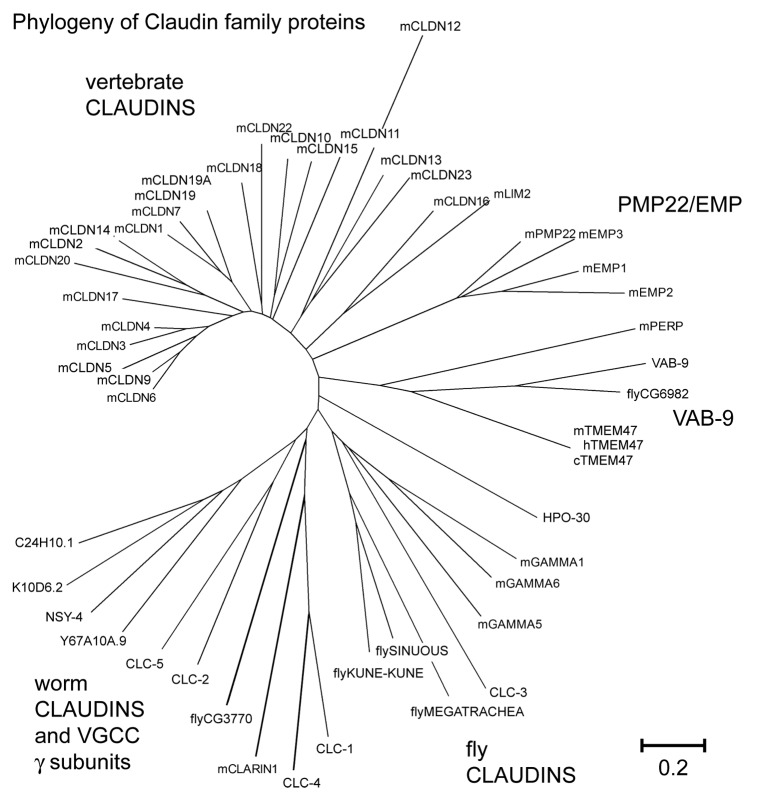
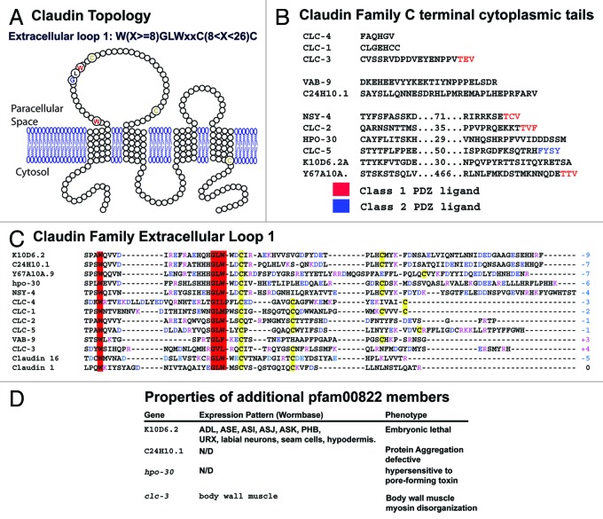
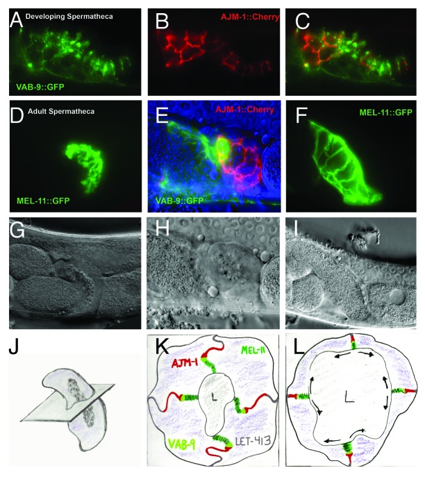
The claudin family of integral membrane proteins was identified as the major protein component of the tight junctions in all vertebrates. Since their identification, claudins, and their associated pfam00822 superfamily of proteins have been implicated in a wide variety of cellular processes. Claudin homologs have been identified in invertebrates as well, including Drosophila and C. elegans. Recent studies demonstrate that the C. elegans claudins, clc-1-clc- 5, and similar proteins in the greater PMP22/EMP/claudin/voltage-gated calcium channel γ subunit family, including nsy-4, and vab-9, while highly divergent at a sequence level from each other and from the vertebrate claudins, in many cases play roles similar to those traditionally assigned to their vertebrate homologs. These include regulating cell adhesion and passage of small molecules through the paracellular space, channel activity, protein aggregation, sensitivity to pore-forming toxins, intercellular signaling, cell fate specification and dynamic changes in cell morphology. Study of claudin superfamily proteins in C. elegans should continue to provide clues as to how claudin family protein function has been adapted to perform diverse functions at specialized cell-cell contacts in metazoans.

Keywords: C. elegans; CLC-1; NSY-4; VAB-9; actomyosin; claudin; epithelia; junctions; morphogenesis; neuronal symmetry.


Figures

None
Figure 1. Schematic of various cell junction arrangements in vertebrates, Drosophila, and C. elegans epithelia. Corresponding cell junction regions either in relative location, function, and/or molecular make-up are indicated with similar colors. Green indicates the tight junction in vertebrates the SAR-like region in C. elegans, the sub-apical region (SAR) in Drosophila. Blue indicates the cadherin-based adherens junction. Red indicates the septate junction in Drosophila and the AJM-1/DLG-1 region in C. elegans. Vertebrates have no precisely analogous structure to the septate junction in epithelia, but share a molecularly similar barrier at the paranodal junction. Vertebrate desmosomes are shown in yellow (A). Lateral views (cutaways) show the nature of the junctional structures in the membranes. Adherens junctions appear as solid bands in the membrane, pleated septate junctions are characterized by regular wave-like strands, and tight junctions appear as irregular but connected anastomosing strands. For simplicity, only the composition of cell junction components in the paracellular space is shown. (B) The most common cellular junctions and representative protein components are listed. Transmembrane proteins are indicated in color (bold) and components unique to vertebrates (other than desmosomes) are underlined.
None
Figure 2. pfam00822 proteins from vertebrates and invertebrates are highly divergent. Bootstrap analysis of pfam00822 proteins using the MEGA (Molecular Evolutionary Genetic Analysis) software program available at http://www.megasoftware.net. The phylogenetic tree resulting from ClustalW2 analysis is shown. Several different subgroup clades are observed. Note that NSY-4 clusters with other putative gamma subunits and C. elegans claudins, while the vertebrate VGCC gamma subunits are loosely associated with fly claudins. Scale bar indicates amino acid substitutions per residue. Accession numbers for proteins included in the alignments are: NSY-4 (NP_500189.4), K10D6.2(NP_505843), R04F11.1 (NP_506087), C24H10.1(NP_508863), Y67A10A.9(NP_502746.1), mGamma5 (voltage-dependent calcium channel gamma subunit 5) (NP_542375), mGamma1 (NP_031608), mGamma6 (NP_573446.1), mGamma3 (NP_062303,)CLC-1(NP_509847), CLC-2(NP_509257), CLC-3 isoform a(NP_001024993), CLC-4 (NP_509800), CLC-5(NP_509258), VAB-9 (NP_495836), CG6982 (dVAB-9) (NP_001097876), mTMEM47(NP_620090), cTMEM47 (NP_001003045.1), hTm47 (NP_113630.1), xtmem47 (NP_001085134.1), mclaudin1 (NP_057883), mclaudin2 (NP_057884), mclaudin3 (NP_034032), mclaudin4 (NP_034033), mclaudin5 (NP_038833), mclaudin6 (NP_0247), mclaudin7 (NP_058583), mclaudin9 (NP_064689), mclaudin10 (a) (NP_076367), mclaudin11 (NP_032796), mclaudin12 (NP_075028), mclaudin13 (NP_065250), mclaudin14 (NP_001159398), mclaudin15 (NP_068365), mclaudin16 (NP_444471), claudin17 (NP_852467), mclaudin18(NP_062789), mclaudin19(1)(NP_001033679), Claudin19(2)(NP_694745), mclaudin20 (NP_001095030), mclaudin22 (NP_083659), mclaudin23 (NP_082274), fly_Sinuous (a) (NP_647971), fly_Megatrachea (NP_726742), fly_CG3770 (NP_611985.), fly_Kune-kune (NP_610179), mEMP2 (NP_031955), mPMP22 (NP_032911), mEMP3(NP_001139818), mEMP1(NP_034258), mPERP(NP_071315), LIM2 (NP_808361)
None
Figure 3. Sequence conservation and motifs in C. elegans pfam00822 proteins. (A) The predicted membrane spanning topology of VAB-9 is shown; pfam00822 proteins have the same membrane-spanning topology. Highly conserved residues among family members are indicated in enlarged, colored circles. (B) The C-terminal cytoplasmic tails of the C. elegans pfam00822 proteins are shown, from the end of the final transmembrane domain to the final residue. Sequences of longer proteins are abbreviated; numbers indicate intervening residues not shown. Terminal sequences conforming to class 1 and 2 PDZ binding domain ligand rules are indicated in red and blue, respectively. (C) The first extracellular loop domain of C. elegans proteins and murine claudin are shown. Postiviely charged residues are indicated in red, negative charged residues in blue. The sum of extracellular loop charge is indicated at the end of the sequence. Cysteines are indicated in yellow and the conserved tryptophan and GLW motif is indicated in red. (D) Additional pfam00822 proteins, for which some preliminary information is known, are shown. K10D6.2 expression, C24H10 function in protein aggregation, hpo-30 function in toxin resistance, and clc-3 expression and phenotype are referenced in , , , , and respectively. Additional unpublished results, particularly regarding clc-3, can be found at http://wormbase.org.
None
Figure 4. Cell junctions in the spermatheca. VAB-9::GFP, AJM-1::Cherry and MEL-11::GFP expression decorate distinct junctional regions of the spermathecal membrane. (A-C) During development, VAB-9::GFP (A) localizes to more a more apical (luminal) position relative to AJM-1::Cherry (B). The merged image is presented in (C). (D-E) In adult spermatheca, MEL-11::GFP (D) appears to localize to the most apical, folded regions of the lateral membranes (corresponding to the pleated septate junctions), while AJM-1::Cherry (E) decorates the more basal membranes (corresponding to the electron dense adherens junction and smooth or continuous cell junction). LET-413 (gray) is localized along the most basal portion of the lateral surface. VAB-9::GFP expression is reduced in the adult spermatheca, but is increased in the spermatheca-uterine valve (E). During ovulation (F), the MEL-11::GFP-decorated pleated septate junctions “unzip” to allow the passage of the oocyte. (G-H) show the matching DIC images for (D-F), and (J-L) are representative illustrations of (G-I). The plane passing through the center of the spermatheca in (J) is projected in (K) and (L). Arrows in (L) indicate the expanding apical (luminal) surface of spermathecal cells at the expense of the lateral (pleated junctional) domain. Similar expansion may take place reducing the smooth/continuous junction in favor of basolateral surface are (not shown).

References

    1. Furuse M, Fujita K, Hiiragi T, Fujimoto K, Tsukita S. Claudin-1 and -2: novel integral membrane proteins localizing at tight junctions with no sequence similarity to occludin. J Cell Biol. 1998;141:1539–50. doi: 10.1083/jcb.141.7.1539. - DOI - PMC - PubMed
    1. Furuse M, Sasaki H, Fujimoto K, Tsukita S. A single gene product, claudin-1 or -2, reconstitutes tight junction strands and recruits occludin in fibroblasts. J Cell Biol. 1998;143:391–401. doi: 10.1083/jcb.143.2.391. - DOI - PMC - PubMed
    1. Morita K, Sasaki H, Furuse M, Tsukita S. Endothelial claudin: claudin-5/TMVCF constitutes tight junction strands in endothelial cells. J Cell Biol. 1999;147:185–94. doi: 10.1083/jcb.147.1.185. - DOI - PMC - PubMed
    1. Nitta T, Hata M, Gotoh S, Seo Y, Sasaki H, Hashimoto N, et al. Size-selective loosening of the blood-brain barrier in claudin-5-deficient mice. J Cell Biol. 2003;161:653–60. doi: 10.1083/jcb.200302070. - DOI - PMC - PubMed
    1. Gow A, Davies C, Southwood CM, Frolenkov G, Chrustowski M, Ng L, et al. Deafness in Claudin 11-null mice reveals the critical contribution of basal cell tight junctions to stria vascularis function. J Neurosci. 2004;24:7051–62. doi: 10.1523/JNEUROSCI.1640-04.2004. - DOI - PMC - PubMed

LinkOut - more resources