. 2014 Sep;20(17-18):2455-62.
doi: 10.1089/ten.TEA.2013.0464. Epub 2014 Jun 18.

An autologous bone marrow mesenchymal stem cell-derived extracellular matrix scaffold applied with bone marrow stimulation for cartilage repair

Affiliations

An autologous bone marrow mesenchymal stem cell-derived extracellular matrix scaffold applied with bone marrow stimulation for cartilage repair

Cheng Tang et al. Tissue Eng Part A. 2014 Sep.

Abstract

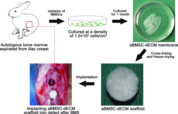
Purpose: It is well known that implanting a bioactive scaffold into a cartilage defect site can enhance cartilage repair after bone marrow stimulation (BMS). However, most of the current scaffolds are derived from xenogenous tissue and/or artificial polymers. The implantation of these scaffolds adds risks of pathogen transmission, undesirable inflammation, and other immunological reactions, as well as ethical issues in clinical practice. The current study was undertaken to evaluate the effectiveness of implanting autologous bone marrow mesenchymal stem cell-derived extracellular matrix (aBMSC-dECM) scaffolds after BMS for cartilage repair.

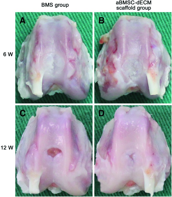
Methods: Full osteochondral defects were performed on the trochlear groove of both knees in 24 rabbits. One group underwent BMS only in the right knee (the BMS group), and the other group was treated by implantation of the aBMSC-dECM scaffold after BMS in the left knee (the aBMSC-dECM scaffold group).

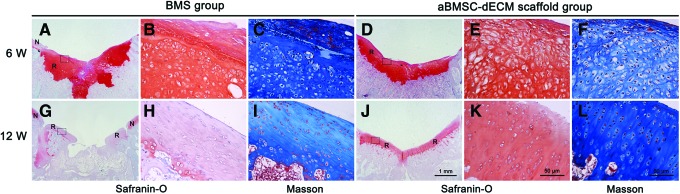
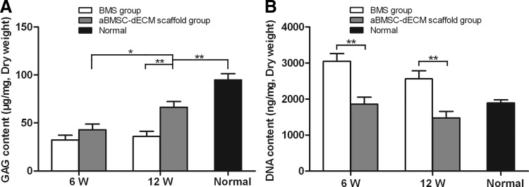
Results: Better repair of cartilage defects was observed in the aBMSC-dECM scaffold group than in the BMS group according to gross observation, histological assessments, immunohistochemistry, and chemical assay. The glycosaminoglycan and DNA content, the distribution of proteoglycan, and the distribution and arrangement of type II and I collagen fibers in the repaired tissue in the aBMSC-dECM scaffold group at 12 weeks after surgery were similar to that surrounding normal hyaline cartilage.

Conclusions: Implanting aBMSC-dECM scaffolds can enhance the therapeutic effect of BMS on articular cartilage repair, and this combination treatment is a potential method for successful articular cartilage repair.


Figures

<b>FIG. 1.</b>
FIG. 1.
Procedure for preparation and implantation of autologous bone marrow mesenchymal stem cell–derived extracellular matrix (aBMSC-dECM) scaffolds. Bone marrow was aspirated from the iliac crest. Then, the isolated mononuclear cells were cultured at high density for 1 month. The aBMSC-dECM membrane was collected from the bottom of the culture plate. The aBMSC-dECM scaffold was fabricated by crosslinking and freeze-drying, and then implanted into a cartilage defect after bone marrow stimulation (BMS). Color images available online at www.liebertpub.com/tea
<b>FIG. 2.</b>
FIG. 2.
Macroscopic evaluation of the repaired cartilage tissue in both the BMS group and the aBMSC-dECM scaffold group at 6 and 12 weeks after surgery. At 6 weeks, the repaired tissue of the BMS group (A) and the aBMSC-dECM scaffold group (B) partially covered the cartilage defect. At 12 weeks, compared with the BMS group (C), the area of repaired tissue in the aBMSC-dECM scaffold group (D) was wider. Color images available online at www.liebertpub.com/tea
<b>FIG. 3.</b>
FIG. 3.
Safranin-O (A, B, D, E, G, H, J, K) and Masson's Trichrome (C, F, I, L) staining of the repaired cartilage tissue in both the BMS group and the aBMSC-dECM scaffold group at 6 and 12 weeks after surgery. All defects were filled mostly with nonhyaline cartilage tissue in the BMS group (A–C) and the aBMSC-dECM scaffold group (D–F) at 6 weeks. However, after 12 weeks, the strength of metachromatic staining in the aBMSC-dECM scaffold group (J–L) resembled that of normal cartilage, and chondrocytes formed mature lacunas and were perpendicularly aligned with subchondral bone. A, D, G, and J:×10, B, C, E, F, H, I, K, and L:×400. N, native cartilage; R, repaired cartilage. Color images available online at www.liebertpub.com/tea
<b>FIG. 4.</b>
FIG. 4.
The distribution and arrangement of collagen fibers were observed by immunohistochemical analysis of type II and I collagen, and Sirius red staining. At 6 weeks after surgery, no organized orientation pattern could be found in either the BMS group (A–C, G–I) or the aBMSC-dECM scaffold group (D–F, J–L). However, at 12 weeks, the distribution and arrangement of collagen fibers in the aBMSC-dECM scaffold group (P–R, V–X) were distributed more evenly and vertically aligned similar to normal cartilage. A–F and M–R:×100; G–L and S–X:×400. N, native cartilage; R, repaired cartilage. Color images available online at www.liebertpub.com/tea
<b>FIG. 5.</b>
FIG. 5.
The glycosaminoglycan (GAG) (A) and DNA (B) contents of repaired cartilage tissue in both the BMS group and the aBMSC-dECM scaffold group. The GAG contents in the aBMSC-dECM scaffold group rapidly increased with time. At 12 weeks after surgery, the GAG content of repaired tissue in the aBMSC-dECM scaffold group reached 70% that of normal hyaline cartilage and was significantly higher than that in the BMS group. The DNA content in the aBMSC-dECM scaffold group was similar to that of normal hyaline cartilage at 6 and 12 weeks after surgery. Results are presented as mean±SD. *p<0.05, **p<0.01.

References

    1. Chang C.H., Kuo T.F., Lin F.H., Wang J.H., Hsu Y.M., Huang H.T., et al. . Tissue engineering-based cartilage repair with mesenchymal stem cells in a porcine model. J Orthop Res 29,1874, 2011 - PubMed
    1. Lee C.H., Cook J.L., Mendelson A., Moioli E.K., Yao H., and Mao J.J.Regeneration of the articular surface of the rabbit synovial joint by cell homing: a proof of concept study. Lancet 376,440, 2010 - PMC - PubMed
    1. Gomoll A.H., Farr J., Gillogly S.D., Kercher J., and Minas T.Surgical management of articular cartilage defects of the knee. J Bone Joint Surg Am 92,2470, 2010 - PubMed
    1. Mithoefer K., McAdams T.R., Scopp J.M., and Mandelbaum B.R.Emerging options for treatment of articular cartilage injury in the athlete. Clin Sports Med 28,25, 2009 - PubMed
    1. Kalson N.S., Gikas P.D., and Briggs T.W.Current strategies for knee cartilage repair. Int J Clin Pract 64,1444, 2010 - PubMed

Publication types

MeSH terms