Characterization of an anti-Bla g 1 scFv: epitope mapping and cross-reactivity
- PMID: 24667070
- PMCID: PMC4097036
- DOI: 10.1016/j.molimm.2014.02.003
Characterization of an anti-Bla g 1 scFv: epitope mapping and cross-reactivity
Abstract
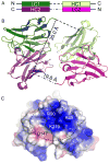
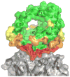
Bla g 1 is a major allergen from Blatella germanica and one of the primary allergens used to assess cockroach allergen exposure. The epitope of an anti-Bla g 1 scFv was mapped in order to better understand cross reactivity with other group 1 cockroach allergens and patient IgE epitopes. X-ray crystallography was used to determine the structure of the scFv. The scFv epitope on Bla g 1 was located by alanine scanning site-directed mutagenesis and ELISA. Twenty-six rBla g 1-GST alanine mutants were evaluated for variations in binding to the scFv compared to the wild type allergen. Six mutants showed a significant difference in scFv binding affinity. These mutations clustered to form a discontinuous epitope mainly comprising two helices of Bla g 1. The allergen-scFv complex was modeled based on the results, and the epitope region was found to have low sequence similarity with Per a 1, especially among the residues identified as functionally important for the scFv binding to Bla g 1. Indeed, the scFv failed to bind Per a 1 in American cockroach extract. The scFv was unable to inhibit the binding of IgE antibodies from a highly cockroach allergic patient to Bla g 1. Based on the surface area of Bla g 1 occluded by the scFv, putative regions of patient IgE-Bla g 1 interactions can be inferred. This scFv could be best utilized as a capture antibody in an IgE detection ELISA, or to differentiate Bla g 1 from Per a 1 in environmental exposure assays.
Keywords: Allergen; Bla g 1; Cockroach; Epitope; Structure; scFv.
Published by Elsevier Ltd.
Figures


Similar articles
-
IgE binding reactivity of peptide fragments of Bla g 4, a major German cockroach allergen.Korean J Parasitol. 2009 Mar;47(1):31-6. doi: 10.3347/kjp.2009.47.1.31. Epub 2009 Mar 12. Korean J Parasitol. 2009. PMID: 19290089 Free PMC article.
-
Mechanisms of allergen-antibody interaction of cockroach allergen Bla g 2 with monoclonal antibodies that inhibit IgE antibody binding.PLoS One. 2011;6(7):e22223. doi: 10.1371/journal.pone.0022223. Epub 2011 Jul 15. PLoS One. 2011. PMID: 21789239 Free PMC article.
-
IgE-binding epitope analysis of Bla g 5, the German cockroach allergen.Protein Pept Lett. 2010 May;17(5):573-7. doi: 10.2174/092986610791112765. Protein Pept Lett. 2010. PMID: 20044919
-
Recombinant allergens for diagnosis of cockroach allergy.Curr Allergy Asthma Rep. 2014 Apr;14(4):428. doi: 10.1007/s11882-014-0428-6. Curr Allergy Asthma Rep. 2014. PMID: 24563284 Free PMC article. Review.
-
Investigating cockroach allergens: aiming to improve diagnosis and treatment of cockroach allergic patients.Methods. 2014 Mar 1;66(1):75-85. doi: 10.1016/j.ymeth.2013.07.036. Epub 2013 Aug 2. Methods. 2014. PMID: 23916425 Free PMC article. Review.
Cited by
-
Interfaces between allergen structure and diagnosis: know your epitopes.Curr Allergy Asthma Rep. 2015 Apr;15(8):506. doi: 10.1007/s11882-014-0506-9. Curr Allergy Asthma Rep. 2015. PMID: 25750181 Free PMC article.
-
Cross-reaction between Formosan termite (Coptotermes formosanus) proteins and cockroach allergens.PLoS One. 2017 Aug 2;12(8):e0182260. doi: 10.1371/journal.pone.0182260. eCollection 2017. PLoS One. 2017. PMID: 28767688 Free PMC article.
-
Current (Food) Allergenic Risk Assessment: Is It Fit for Novel Foods? Status Quo and Identification of Gaps.Mol Nutr Food Res. 2018 Jan;62(1):1700278. doi: 10.1002/mnfr.201700278. Epub 2017 Dec 11. Mol Nutr Food Res. 2018. PMID: 28925060 Free PMC article. Review.
-
New Insights into Cockroach Allergens.Curr Allergy Asthma Rep. 2017 Apr;17(4):25. doi: 10.1007/s11882-017-0694-1. Curr Allergy Asthma Rep. 2017. PMID: 28421512 Free PMC article. Review.
-
Recent Developments in Engineering Non-Paralytic Botulinum Molecules for Therapeutic Applications.Toxins (Basel). 2024 Apr 3;16(4):175. doi: 10.3390/toxins16040175. Toxins (Basel). 2024. PMID: 38668600 Free PMC article. Review.
References
-
- Aalberse RC, Crameri R. IgE-binding epitopes: a reappraisal. Allergy. 2011;66:1261–1274. - PubMed
-
- Arruda LK, Vailes LD, Mann BJ, Shannon J, Fox JW, Vedvick TS, Hayden ML, Chapman MD. Molecular-cloning of a major cockroach (Blattella-Germanica) allergen, Bla-G-2—sequence homology to the aspartic proteases. J Biol Chem. 1995;270:19563–19568. - PubMed
-
- Benjamin DC, Berzofsky JA, East IJ, Gurd FR, Hannum C, Leach SJ, Margoliash E, Michael JG, Miller A, Prager EM, et al. The antigenic structure of proteins: a reappraisal. Annu Rev Immunol. 1984;2:67–101. - PubMed
Publication types
MeSH terms
Substances
Grants and funding
LinkOut - more resources
Full Text Sources
Other Literature Sources
Research Materials

