

Characterization of chikungunya virus induced host response in a mouse model of viral myositis
- PMID: 24667237
- PMCID: PMC3965460
- DOI: 10.1371/journal.pone.0092813
Characterization of chikungunya virus induced host response in a mouse model of viral myositis
Abstract
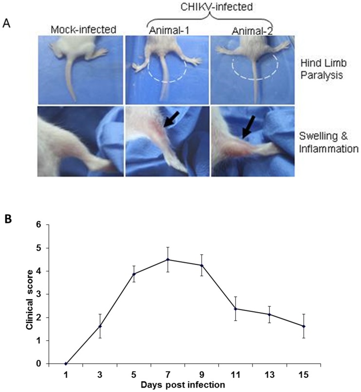
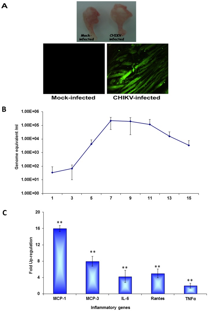
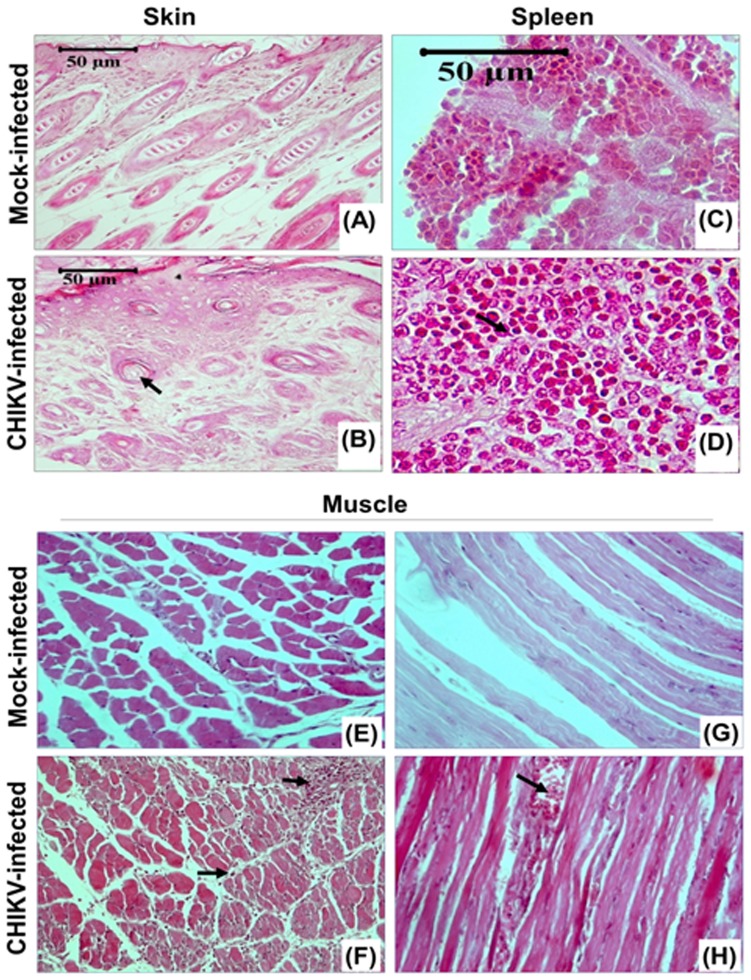
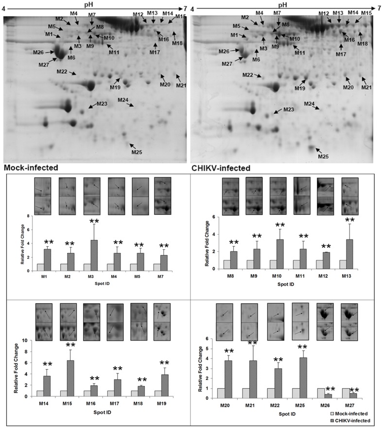
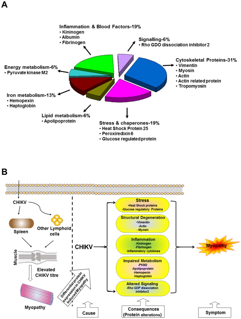
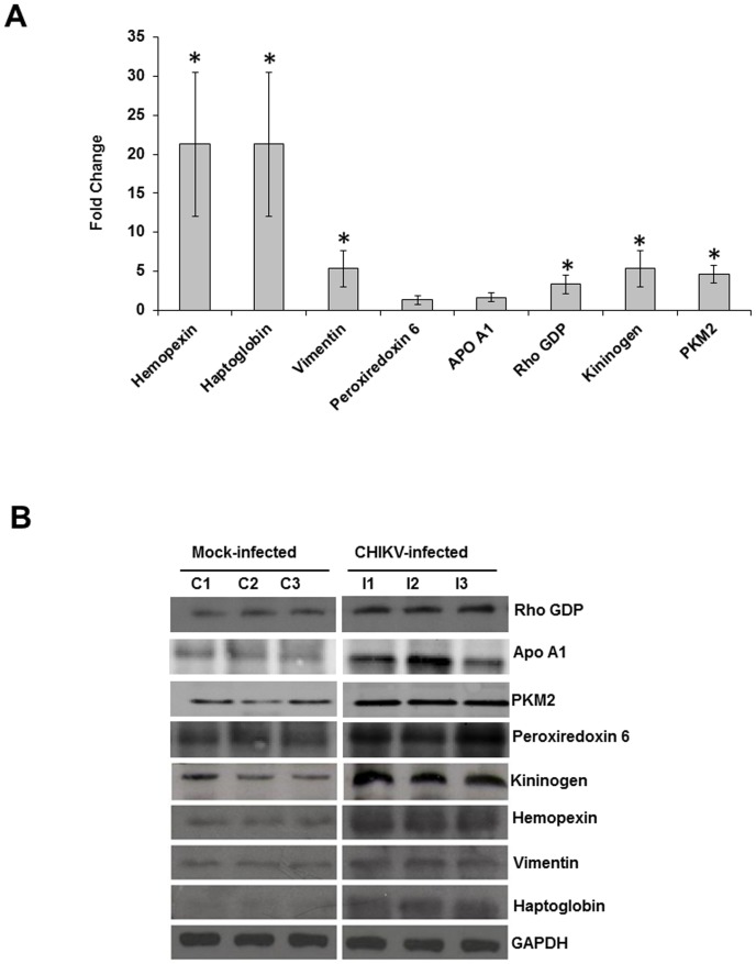
While a number of studies have documented the persistent presence of chikungunya virus (CHIKV) in muscle tissue with primary fibroblast as the preferable cell target, little is known regarding the alterations that take place in muscle tissue in response to CHIKV infection. Hence, in the present study a permissive mouse model of CHIKV infection was established and characterized in order to understand the pathophysiology of the disease. The two dimensional electrophoresis of muscle proteome performed for differential analysis indicated a drastic reprogramming of the proteins from various classes like stress, inflammation, cytoskeletal, energy and lipid metabolism. The roles of the affected proteins were explained in relation to virus induced myopathy which was further supported by the histopathological and behavioural experiments proving the lack of hind limb coordination and other loco-motor abnormalities in the infected mice. Also, the level of various pro-inflammatory mediators like IL-6, MCP-1, Rantes and TNF-α was significantly elevated in muscles of infected mice. Altogether this comprehensive study of characterizing CHIKV induced mouse myopathy provides many potential targets for further evaluation and biomarker study.
Conflict of interest statement
Figures
Similar articles
-
Proteomic profiling of chikungunya virus-infected human muscle cells: reveal the role of cytoskeleton network in CHIKV replication.J Proteomics. 2014 Aug 28;108:445-64. doi: 10.1016/j.jprot.2014.06.003. Epub 2014 Jun 14. J Proteomics. 2014. PMID: 24933005
-
Distinct Roles of Interferon Alpha and Beta in Controlling Chikungunya Virus Replication and Modulating Neutrophil-Mediated Inflammation.J Virol. 2019 Dec 12;94(1):e00841-19. doi: 10.1128/JVI.00841-19. Print 2019 Dec 12. J Virol. 2019. PMID: 31619554 Free PMC article.
-
A mouse model of chikungunya virus-induced musculoskeletal inflammatory disease: evidence of arthritis, tenosynovitis, myositis, and persistence.Am J Pathol. 2011 Jan;178(1):32-40. doi: 10.1016/j.ajpath.2010.11.018. Epub 2010 Dec 23. Am J Pathol. 2011. PMID: 21224040 Free PMC article.
-
Chikungunya virus pathogenesis and immunity.Vector Borne Zoonotic Dis. 2015 Apr;15(4):241-9. doi: 10.1089/vbz.2014.1710. Vector Borne Zoonotic Dis. 2015. PMID: 25897810 Review.
-
Chikungunya Virus-Induced Arthritis: Role of Host and Viral Factors in the Pathogenesis.Viral Immunol. 2017 Dec;30(10):691-702. doi: 10.1089/vim.2017.0052. Epub 2017 Sep 14. Viral Immunol. 2017. PMID: 28910194 Review.
Cited by
-
Mosquito-bite infection of humanized mice with chikungunya virus produces systemic disease with long-term effects.PLoS Negl Trop Dis. 2021 Jun 9;15(6):e0009427. doi: 10.1371/journal.pntd.0009427. eCollection 2021 Jun. PLoS Negl Trop Dis. 2021. PMID: 34106915 Free PMC article.
-
The DEAD-box RNA helicase Dhx15 controls glycolysis and arbovirus replication in Aedes aegypti mosquito cells.PLoS Pathog. 2022 Nov 28;18(11):e1010694. doi: 10.1371/journal.ppat.1010694. eCollection 2022 Nov. PLoS Pathog. 2022. PMID: 36441781 Free PMC article.
-
Loss of TLR3 aggravates CHIKV replication and pathology due to an altered virus-specific neutralizing antibody response.EMBO Mol Med. 2015 Jan;7(1):24-41. doi: 10.15252/emmm.201404459. EMBO Mol Med. 2015. PMID: 25452586 Free PMC article.
-
High throughput proteomic analysis and a comparative review identify the nuclear chaperone, Nucleophosmin among the common set of proteins modulated in Chikungunya virus infection.J Proteomics. 2015 Apr 29;120:126-41. doi: 10.1016/j.jprot.2015.03.007. Epub 2015 Mar 14. J Proteomics. 2015. PMID: 25782748 Free PMC article. Review.
-
Telmisartan Restricts Chikungunya Virus Infection In Vitro and In Vivo through the AT1/PPAR-γ/MAPKs Pathways.Antimicrob Agents Chemother. 2022 Jan 18;66(1):e0148921. doi: 10.1128/AAC.01489-21. Epub 2021 Nov 8. Antimicrob Agents Chemother. 2022. PMID: 34748384 Free PMC article.
References
-
- Enserink M (2008) Entomology. A mosquito goes global. Science 320: 864–866. - PubMed
-
- Rezza G, Nicoletti L, Angelini R, Romi R, Finarelli AC, et al. (2007) Infection with chikungunya virus in Italy: an outbreak in a temperate region. Lancet 370: 1840–1846. - PubMed
-
- Santhosh SR, Dash PK, Parida M, Khan M, Tiwari M, et al. (2008) Comparative full genome analysis revealed E1:A226V shift in 2007 Indian Chikungunya virus isolates. Virus Research 135: 36–41. - PubMed
-
- Jain M, Rai S, Chakravarti A (2008) Chikungunya: a review. Trop Doct 38: 70–72. - PubMed
-
- Arankalle VA, Shrivastava S, Cherian S, Gunjikar RS, Walimbe AM (2007) Genetic divergence of Chikungunya viruses in India (1963–2006) with special reference to the 2005–2006 explosive epidemic. J Gen Virol 88: 1967–1976. - PubMed
Publication types
MeSH terms
Substances
LinkOut - more resources
Full Text Sources
Other Literature Sources
Medical
Molecular Biology Databases
Miscellaneous
