. 2014 Mar 25;9(3):e91422.
doi: 10.1371/journal.pone.0091422. eCollection 2014.

Effects of high affinity leptin antagonist on prolactin receptor deficient male mouse

Affiliations

Effects of high affinity leptin antagonist on prolactin receptor deficient male mouse

Nadège Carré et al. PLoS One. .

Abstract

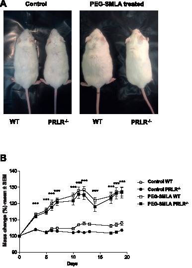
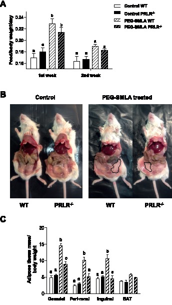
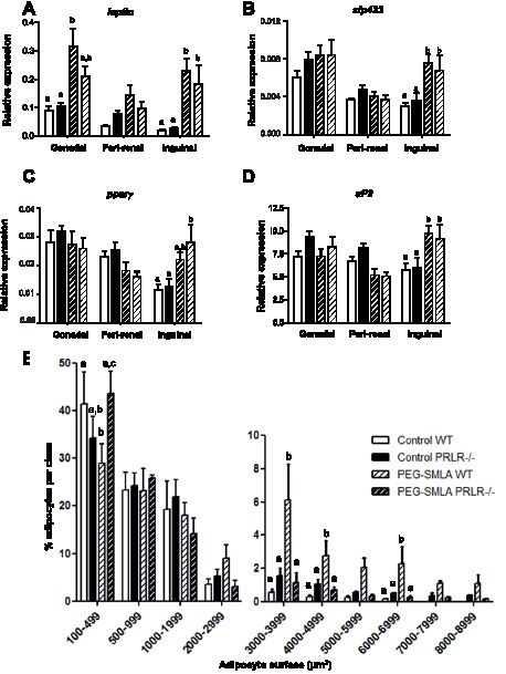
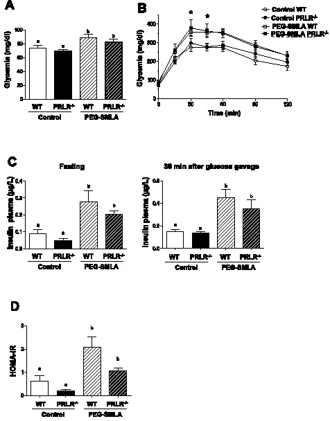
Hyperprolactinemia occurs during gestation and lactation with marked hyperphagia associated with leptin resistance. Prolactin (PRL) induces the expression of orexigenic neuropeptide Y (NPY) in hypothalamic dorsomedial nucleus (DMH) leading to hyperphagia. Along this line prolactin receptor deficient (PRLR-/-) mice are resistant to obesity under high fat diet due to increased energy expenditure. As these mice have an altered food intake, our objective was to test whether leptin is responsible for these characteristics. PRLR-/- male mice and control littermates were injected subcutaneously every other day with 12 mg/kg pegylated superactive mouse leptin antagonist (PEG-SMLA) for 3 weeks. We tested the effect of PEG-SMLA on body weight, food intake and metabolic parameters. The antagonist led to a rapid increase in body weight (20%) but increased adipose mass in PEG-SMLA treated mice was less pronounced in PRLR-/- than in WT mice. Food intake of PEG-SMLA-injected animals increased during the first week period of the experiment but then declined to a similar level of the control animals during the second week. Interestingly, PRLR-/- mice were found to have the same bone volume than those of control mice although PEG-SMLA increased bone mass by 7% in both strains. In addition, PEG-SMLA led to insulin resistance and glucose intolerance as well as an altered lipid profile in treated mice. Altogether, these results suggest that PRLR-/- mice respond to leptin antagonist similarly to the control mice, indicating no interaction between the actions of the two hormones.


Conflict of interest statement

Competing Interests: The authors have declared that no competing interests exist.

Figures

Figure 1
Figure 1. Effect of PEG-SMLA treatment on weight gain of WT and PRLR−/− mice.
(A) Representative dorsal view of male mice after 20 days of saline (control) or PEG-SMLA injections. (B) Comparison of PEG-SMLA effects on weight gain of WT (open squares) or PRLR−/− (black squares) male mice. PEG-SMLA was injected daily at 6 mg/kg. ***, p<0.005 between non-treated (Cont) and PEG-SMLA treated groups.
Figure 2
Figure 2. Effects of PEG-SMLA treatment on food intake and adipose tissues mass.
(A) Food consumption (g) is calculated reported to body weight of each mouse on first or second week of treatment. PEG-SMLA injections are represented within hatched bars. b, p<0.001 related to Control WT mice. (B) At the end of treatment, the intact abdominal fat depot (delimited lines) is shown on both genotypes. (C) Ratio of adipose depot mass per body weight. b, p<0.001 and c, p<0.01 related to Control WT mice. (n = 7–10/group).
Figure 3
Figure 3. Expression of key adipose genes.
(A–D) Leptin, Zfp423, PPAR and aP2 gene expression was quantified by qPCR in each gonadal, peri-renal, inguinal adipose depot of WT and PRLR−/− non-treated mice (Control) and treated animals (PEG-SMLA, hatched bars) respectively (n = 5–8/group) b, p<0.01. (E) Distribution of adipocyte surfaces in inguinal adipose tissue in untreated WT and PRLR−/− (Control) and treated animals (PEG-SMLA, hatched bars). Statistical analysis was performed. b, p<0.01, c, p<0.001.
Figure 4
Figure 4. PEG-SMLA treatment induced glucose intolerance and insulin resistance on WT and PRLR−/− animals.
(A) Fasting glycemia in saline treated and PEG-SMLA treated mice. b, *p<0.05. (B) Oral glucose tolerance test (2g/kg) was performed in WT and PRLR−/− mice PEG-SMLA treated or not. (C) Fasting (left panel) and 30 minutes after glucose gavage (right panel) plasma insulin levels. (D) HOMA-IR index reflecting insulin resistance is calculated as follows: fasting plasma glucose (mg/dl) × fasting insulin (mU/L)/405.

References

    1. Goffin V, Binart N, Touraine P, Kelly PA (2002) Prolactin: the new biology of an old hormone. Annu Rev Physiol 64: 47–67 10.1146/annurev.physiol.64.081501.131049 - DOI - PubMed
    1. Ben-Jonathan N, LaPensee CR, LaPensee EW (2008) What can we learn from rodents about prolactin in humans? Endocr Rev 29: 1–41 10.1210/er.2007-0017 - DOI - PMC - PubMed
    1. Sauvé D, Woodside B (2000) Neuroanatomical specificity of prolactin-induced hyperphagia in virgin female rats. Brain Res 868: 306–314. - PubMed
    1. Chen P, Smith MS (2004) Regulation of hypothalamic neuropeptide Y messenger ribonucleic acid expression during lactation: role of prolactin. Endocrinology 145: 823–829 10.1210/en.2003-1255 - DOI - PubMed
    1. Ling C, Hellgren G, Gebre-Medhin M, Dillner K, Wennbo H, et al. (2000) Prolactin (PRL) receptor gene expression in mouse adipose tissue: increases during lactation and in PRL-transgenic mice. Endocrinology 141: 3564–3572. - PubMed

Publication types