Review
. 2014 Jun 15;68(1):134-9.
doi: 10.1016/j.ymeth.2014.03.014. Epub 2014 Mar 22.

Assays to monitor autophagy in Drosophila

Affiliations
Review

Assays to monitor autophagy in Drosophila

Caroline Mauvezin et al. Methods. .

Abstract

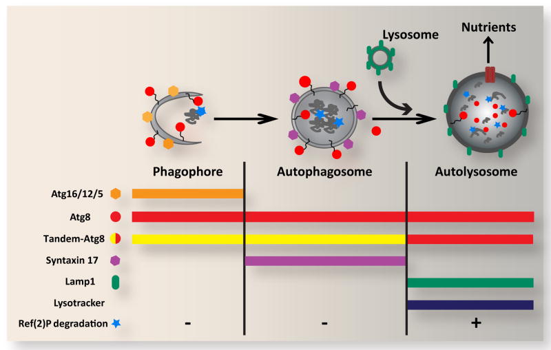
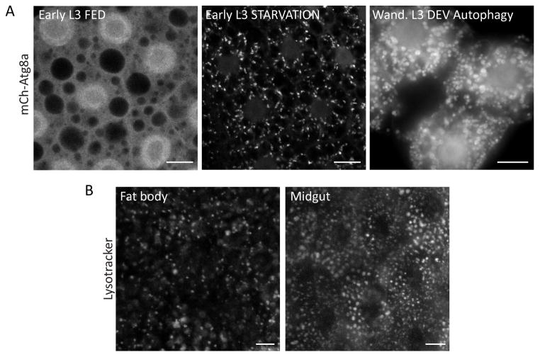
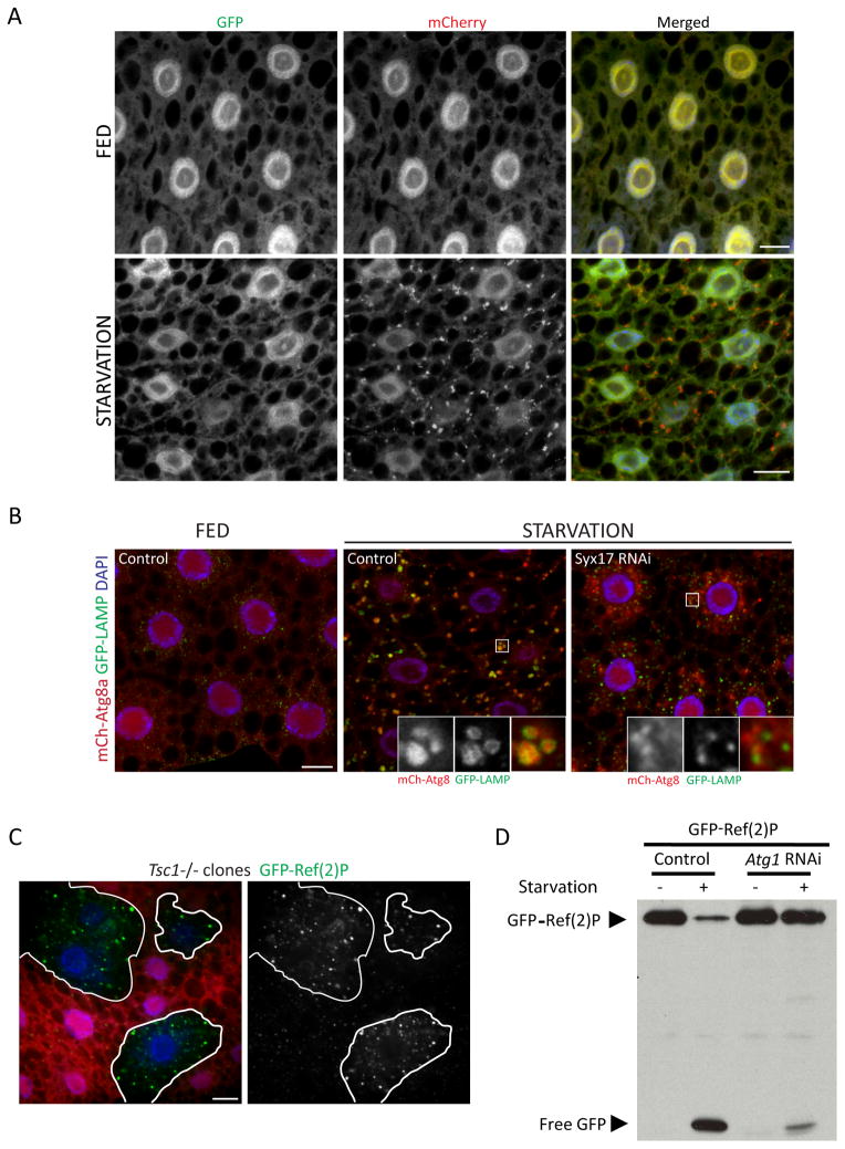
The term autophagy refers to the engulfment and degradation of cytoplasmic components within the lysosome. This process can benefit cells and organisms by removing damaged, superfluous, or harmful cellular components, and by generating a supply of recycled macromolecules that can support biosynthesis or energy production. Recent interest in autophagy has been driven by its potential role in several disease-related phenomena including neurodegeneration, cancer, immunity and aging. Drosophila provides a valuable animal model context for these studies, and work in this system has also begun to identify novel developmental and physiological roles of autophagy. Here, we provide an overview of methods for monitoring autophagy in Drosophila, with a special emphasis on the larval fat body. These methods can be used to investigate whether observed vesicles are of autophagic origin, to determine a relative rate of autophagic degradation, and to identify specific step(s) in the autophagic process in which a given gene functions.

Keywords: Atg8; Autophagosome; Autophagy; Drosophila; Fat body; Ref(2)P.


Figures

Figure 1
Figure 1. Overview of autophagic structures, markers and assays
In response to cellular stress and other inductive signals, cytoplasm, protein aggregates and organelles are engulfed within double-membraned autophagosomes, which form through growth of the cup-shaped phagophore/isolation membrane. Fusion of the outer autophagosomal membrane with endosomes (not shown) and lysosomes results in exposure of the sequestered material to the hydrolytic lysosomal compartment. Degraded materials are ultimately exported to the cytoplasm as nutrients for energy and biosynthesis. Each of these steps can be identified and distinguished based on their specific constellation of associated proteins, as well as by pH and degradative properties. Atg5, Atg12 and Atg16 form a complex on the outer surface of the phagophore, and are removed upon its closure to form the autophagosome. Syntaxin 17 is recruited to the mature autophagosome, and is released upon fusion with the lysosome. In contrast, Atg8 is associated with the entire autophagic pathway from phagophore to autolysosome. Autophagosome-lysosome fusion can be monitored through loss of GFP fluorescence from the tandem GFP-mCherry-Atg8 marker. General lysosomal markers such as Lamp1 and LysoTracker can also be used to identify later autophagic vesicles. The degradative capacity of autophagy can be monitored through the cytoplasmic levels or specific cleavage of the autophagy substrate Ref(2)P.
Figure 2
Figure 2. Static markers of autophagic vesicles
A. Localization of Atg8a in the larval fat body. During the early L3 instar, autophagy proceeds at a low basal rate in well-fed animals, and mCherry-Atg8a is distributed evenly throughout the cell, with accumulation in the nucleus and exclusion from lipid droplets evident. In response to starvation, Atg8a is recruited to autophagic vesicles, forming discrete punctae of varying size and shape. In wandering stage L3 larvae, Atg8a is constitutively associated with autophagic vesicles, which are larger and rounder than the structures observed at earlier developmental stages. Scale bars, 10μm. B. The acidophilic dye LysoTracker can be used to visualize the growth and activation of the lysosomal compartment in response to autophagy induction. Fat body and midgut cells display a distinct distribution pattern and appearance of autolysosomes, which in these images were induced in response to the oxidative stressors hydrogen peroxide and paraquat, respectively. Scale bars, 10μm.
Figure 3
Figure 3. Assays of autophagic flux in the larval fat body
A. Tandem-tagged GFP-mCherry-Atg8a can reveal fusion of autophagosomes with lysosomes. Under fed conditions, the green and red fluorescence of this marker display a nearly identical diffuse pattern. Upon autophagy induction by starvation, localization of this marker to punctate autophagic vesicles is evident in the red channel but not in the green channel, due to quenching of GFP fluorescence within the acidic autolysosome. Scale bars, 10μm. B. Co-localization of Atg8a with the lysosomal marker GFP-LAMP can also be used to assess autophagosome-lysosome fusion. In these images, induction of autophagy by starvation results in accumulation of mCherry-Atg8a within vesicles marked with GFP-LAMP. Co-localization of these markers is inhibited by depletion of Syntaxin 17, a SNARE required for autophagosome-lysosome fusion. Scale bar, 10μm. C. Accumulation of the autophagy substrate Ref(2)P can reveal a disruption of autophagy. The level of GFP-Ref(2)P within clones of Tsc1 mutant cells (marked by lack of dsRed marker; outlined in white) is increased relative to surrounding control cells, reflecting autophagy suppression in response to activated TOR signaling. Scale bar, 10μm. D. Autophagy-dependent proteolytic cleavage of GFP-Ref(2)P, assayed by anti-GFP immunoblotting of fat body extracts. Starvation-induced autophagy results in a loss of full-length GFP-Ref(2)P and a concomitant accumulation of free GFP. Both responses are inhibited in cells depleted of Atg1.

References

    1. Meléndez A, Neufeld TP. The cell biology of autophagy in metazoans: a developing story. Development. 2008;135:2347–2360. - PMC - PubMed
    1. Zirin J, Perrimon N. Drosophila as a model system to study autophagy. Semin Immunopathol. 2010;32:363–372. - PMC - PubMed
    1. Mizushima N, Yamamoto A, Matsui M, Yoshimori T, Ohsumi Y. In vivo analysis of autophagy in response to nutrient starvation using transgenic mice expressing a fluorescent autophagosome marker. Mol Biol Cell. 2004;15:1101–1111. - PMC - PubMed
    1. Kroemer G, Mariño G, Levine B. Autophagy and the integrated stress response. Mol Cell. 2010;40:280–293. - PMC - PubMed
    1. Klionsky DJ, Abdalla FC, Abeliovich H, Abraham RT, Acevedo-Arozena A, Adeli K, Zuckerbraun B. Guidelines for the use and interpretation of assays for monitoring autophagy. Autophagy. 2012;8:445–544. - PMC - PubMed

LinkOut - more resources