. 2014 Mar 25;9(3):e93293.
doi: 10.1371/journal.pone.0093293. eCollection 2014.

Peripheral CD4CD8 double positive T cells with a distinct helper cytokine profile are increased in rheumatoid arthritis

Affiliations

Peripheral CD4CD8 double positive T cells with a distinct helper cytokine profile are increased in rheumatoid arthritis

Dagmar Quandt et al. PLoS One. .

Abstract

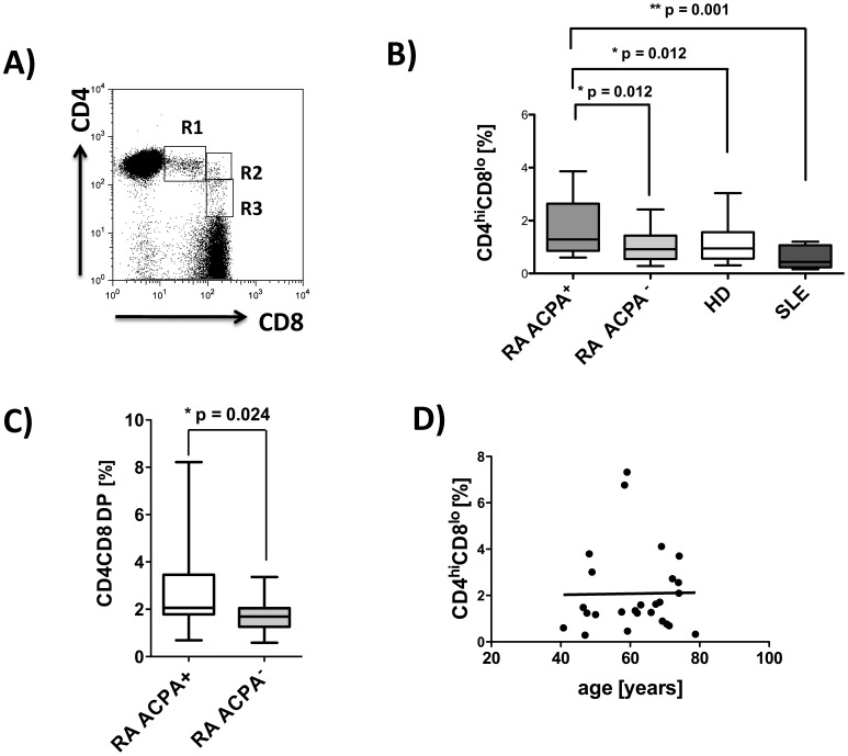
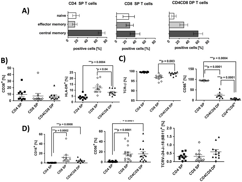
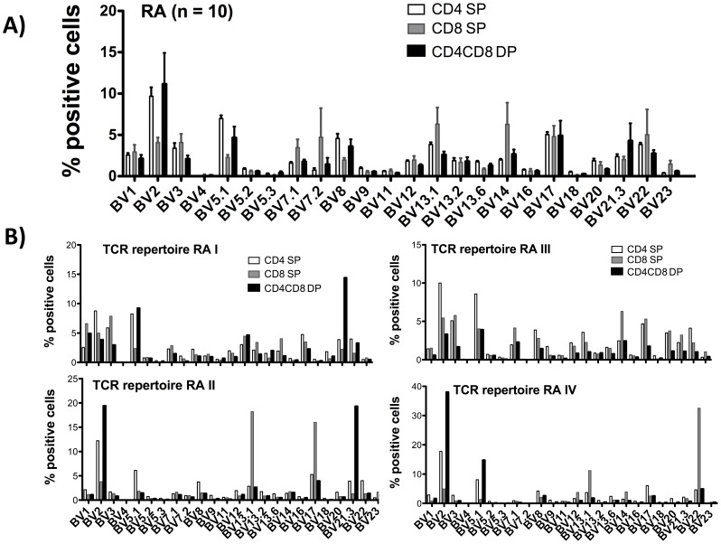
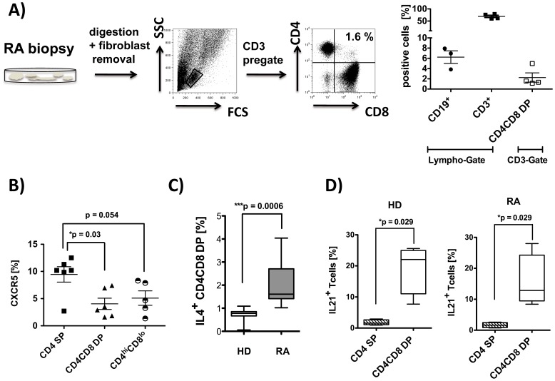
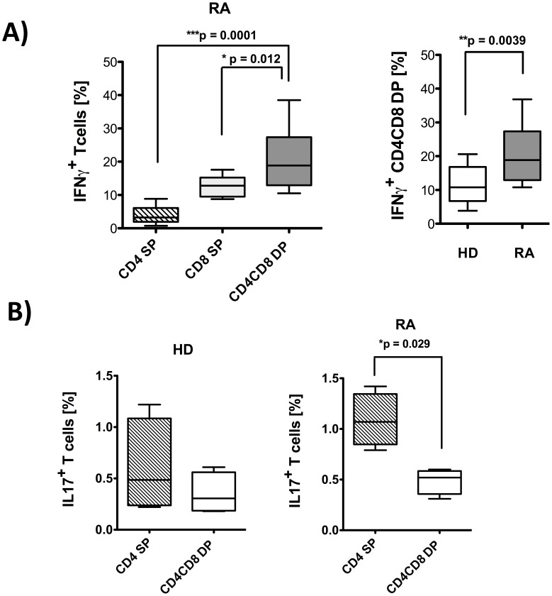
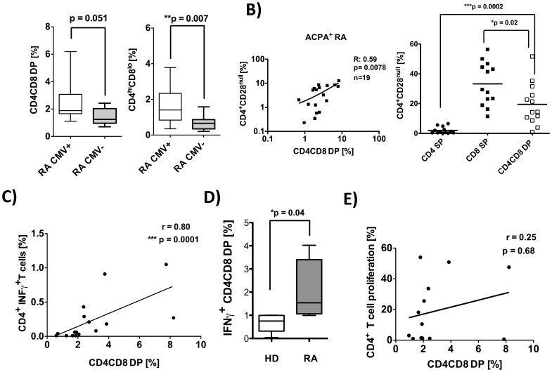
Peripheral CD4CD8 double positive (DP) T cells have been reported to play a role in several autoimmune diseases, virus infections and cancer. In rheumatoid arthritis (RA), both CD4 and CD8 single positive (SP) T cells are known to be involved in the pathogenesis, but the role of peripheral CD4CD8 DP T cells has not been investigated in detail. Anti cyclic citrullinated antibodies (ACPA) positive and ACPA negative RA patients, patients with systemic lupus erythematodes (SLE) and age matched healthy donors (HD) were enrolled in the analysis. The frequencies and phenotype of DP T cells in PBMC were investigated. In addition, DP T cells were quantified in biopsies from rheumatoid synovium. After in vitro restimulation, the cytokine production of DP T cells was investigated in cultures of PBMC. CMV specific cytokine secretion as well as proliferation was analyzed following antigen specific restimulation after an appropriate culture duration. DP T cells were found more frequently in RA patients than in healthy controls or patients with SLE. These DP T cells express αβ TCRs, are of a memory phenotype and share features of both CD4 as well as CD8 SP T cells. Importantly, DP T cells were found to also be present in the rheumatoid synovium. Further characterization of DP T cells from RA patients revealed increased production of IL-21 and IL-4, implying a possible role as T helper cells. In addition, DP T cells in RA seem to contribute to the inflammatory process, because they produce significantly more IFNγ than counterparts from HD and are increased in CMV+ RA patients. Given their capacity to produce a variety of cytokines (IL4, IL21 and IFNγ), their association with ACPA positive RA and their presence in the synovium, we suggest an important role of double positive T cells in the pathogenesis of rheumatoid arthritis.


Conflict of interest statement

Competing Interests: The authors declare that they have no competing interest.

Figures

Figure 1
Figure 1. Increased frequencies of peripheral CD4CD8 DP T cells in ACPA+ rheumatoid arthritis patients.
PBMC were isolated from RA patients (ACPA+, n = 37, ACPA, n = 20), healthy donors (n = 36), and patients with SLE (n = 8), and FACS analyses from live cells (PI staining used for exclusion of dead cells) were performed. Analyses for CD4CD8 double positive T cells are always pregated on CD3 positive T cells. A) Representative FACS plot from one RA patient, showing CD4CD8 double positive T cells, R1 = CD4hiCD8lo, R2 = CD4hiCD8hi and R3 = CD4l°CD8hi B) Comparison of frequencies of CD4hiCD8loT cells in PBMCs of ACPA+/− RA, HD and SLE patients. C) Total CD4CD8 DP T cells in ACPA+ (n = 19) and ACPA (n = 20) RA patients. D) Correlation analysis for frequencies of CD4hiCD8lo T cells with age in ACPA+ RA patients. Box plots depict median, interquartile range and 10–90 percentile. Significance as given, *p<0.05, **p<0.01.
Figure 2
Figure 2. Phenotypic characterization of CD4CD8 DP T cells in RA patients.
PBMC from RA patients were isolated and FACS analyses from live cells were performed (PI staining used for exclusion of dead cells). CD3 pregated cells were further gated on CD4 single, CD8 single and on CD4CD8 double positive T cells. A) Distribution of CD45ROCCR7+ (naive) CD45RO+CCR7 (effector memory) and CD45RO+CCR7+ (central memory) cells in the cell populations indicated. n = 5 5 B) Percentage of T cells positive for CD38 (n = 10) and HLA-DR (n = 11). C) Percentage of cells bearing a TCR alpha-beta (n = 10) and cells positive for CD8β (n = 9) in the T cell populations indicated. (D) Percentage of cells positive for CD16 (n = 10), CD56 (n = 15) and for TCRVα24-Jα18 (n = 11, marker for iNKT) in the T cell populations indicated. In all graphs, lines represent means and SEM. Significance as given, *p<0.05, **p<0.01, ***p<0.001.
Figure 3
Figure 3. TCR BV distribution in CD4CD8 double positive T cells of RA patients.
PBMC from RA patients were isolated and FACS analyses from live cells were performed (PI staining used for exclusion of dead cells). CD3 pre-gated cells were further gated on CD4 single, CD8 single and on CD4CD8 double positive T cells. Bar charts depict the percentage of cells positive for each BV element in the indicated T cell subpopulations investigated. A) Mean distribution of 24 TCR BV elements in peripheral blood T cells from 10 RA patients. B) Examples of the individual distribution of 24 TCR BV elements in 4 RA patients.
Figure 4
Figure 4. CD4CD8 double positive T cells are present in the synovium of RA patients and produce T helper 2 like cytokines.
A) Single cell suspensions were prepared from synovial biopsies from RA patients (n = 4) and analyzed by flow cytometry using anti-CD19, anti-CD3, anti-CD4 and anti-CD8 monoclonal antibodies (PI staining used for exclusion of dead cells). Representative FACS plots are depicted. Each data point represents one experiment. B) PBMCs from RA patients (n = 6) were isolated and FACS analysis from live cells were performed (PI staining used for exclusion of dead cells). Cells were pre-gated on CD3+ T cells, and CXCR5 expression was analyzed in the subpopulation indicated. C) PBMC were stimulated in vitro for 4 hrs with Cytostim. Subsequently, an IL-4 specific secretion assay was performed, and expression of CD4 and CD8 was determined in vital cells (PI staining used for exclusion of dead cells) pre-gated on CD3. Percentage of IL-4 producing CD4CD8 double positive T cells in HD (n = 7) and RA patients (n = 8) is given. D) PBMCs from HD (n = 4) and RA (n = 4) were stimulated with PMA/Iono for 4 hrs, and intracellular staining for IL-21 was performed. Cells are counterstained with anti-CD3, anti-CD4 and anti-CD8. Depicted is the percentage of cytokine producers in total CD4CD8 DP T cells and in CD4 SP cells. Box plots in C) and D) show median, interquartile range, and 10–90 percentiles. Significance as given, *p<0.05.
Figure 5
Figure 5. CD4CD8 double positive T cells from RA patients produce inflammatory IFNγ.
PBMC from RA and HD were stimulated in(A) or PMA/Iono (B). Subsequently, cytokine specific secretion assays (A) or intracellular stainings (B) were performed and cells were counterstained with anti-CD3, anti-CD4 and anti-CD8. Flow cytometric cytokine analysis were performed on live cells for IFNγ secretion (PI staining used for exclusion of dead cells) or on fixed cells for intra-cellular staining for IL-17, in each case after pre-gating on CD3+ T cells. A) Percentage of IFNγ producing T cells in samples from rheumatoid arthritis patients (n = 12, left panel and grey box in the right panel) and healthy donors (n = 13, clear box in the right panel) among CD4 SP, CD8 SP and CD4CD8 DP T cells. B) Percentage of IL-17 producing CD4 SP and CD4CD8 double positive T cells from RA patients (n = 4) and HD (n = 4). Presented box plots show median, interquartile range, and 10–90 percentiles. Significance as given, *p<0.05, **p<0.01, ***p<0.001.
Figure 6
Figure 6. Frequencies of CD4CD8 double positive T cells are increased in CMV positive RA patients and correlate with CMV specific INFγ producers.
A) PBMC were isolated from CMV+ RA patients (n = 28) and CMV RA patients (n = 12) and analyzed by flow cytometry using anti-CD19, anti-CD3, anti-CD4 and anti-CD8 monoclonal antibodies (PI staining used for exclusion of dead cells). B) Correlation of the frequency of total double positive T cells with CD4+CD28null T cells in ACPA+ RA patients (left graph) and comparison of frequencies of CD28null cells among CD4 SP, CD8 SP and total DP T cells (right graph). C–D) PBMCs were restimulated with CMV-lysate for 4 hrs in vitro. Subsequently, an INFγ specific secretion assay was performed and cells were counterstained with aCD3, aCD4 and aCD8. Flow cytometric cytokine analyses were performed on live cells (PI staining used for exclusion of dead cells) pre-gated on CD3+ T cells. C) Depicted is the correlation of CMV specific INFγ producing CD4 T cells with the frequencies of CD4CD8 DP cells from the same donor (n = 16). D) CMV specific INFγ producing CD4CD8 DP T cells from RA (n = 5) and HD (n = 5) are shown. E) PBMCs were labeled with CFDA-SE and restimulated with CMV-lysat (n = 13) for 7 d in vitro. Depicted is the correlation of CMV specific CD4 T cell proliferation with the frequencies of CD4CD8 DP cells from the same donor. Presented box plots show median, interquartile range, and 10–90 percentiles. Significance as given, *p<0.05, **p<0.01, ***p<0.001.

Similar articles

Cited by

References

    1. Pierer M, Rothe K, Quandt D, Schulz A, Rossol M, et al. (2012) Association of anticytomegalovirus seropositivity with more severe joint destruction and more frequent joint surgery in rheumatoid arthritis. Arthritis Rheum 64(6): 1740–1749. - PubMed
    1. Nascimbeni M, Shin EC, Chiriboga L, Kleiner DE, Rehermann B, et al. (2004) Peripheral CD4(+)CD8(+) T cells are differentiated effector memory cells with antiviral functions. Blood 104(2): 478–486. - PubMed
    1. Parel Y, Chizzolini C (2004) CD4+ CD8+ double positive (DP) T cells in health and disease. Autoimmun Rev 3(3): 215–220. - PubMed
    1. Zloza A, Al-Harthi L (2006) Multiple populations of T lymphocytes are distinguished by the level of CD4 and CD8 coexpression and require individual consideration. J Leukoc Biol 79(1): 4–6. - PubMed
    1. Desfrancois J, Moreau-Aubry A, Vignard V, Godet Y, Khammari A, et al. (2010) Double positive CD4CD8 alphabeta T cells: a new tumor-reactive population in human melanomas. PLoS One 5(1): e8437. - PMC - PubMed

Publication types

MeSH terms