. 2014 May-Jun;152(1-2):127-39.
doi: 10.1016/j.clim.2014.03.007. Epub 2014 Mar 22.

Using immunomic approach to enhance tumor-associated autoantibody detection in diagnosis of hepatocellular carcinoma

Affiliations

Using immunomic approach to enhance tumor-associated autoantibody detection in diagnosis of hepatocellular carcinoma

Liping Dai et al. Clin Immunol. 2014 May-Jun.

Abstract

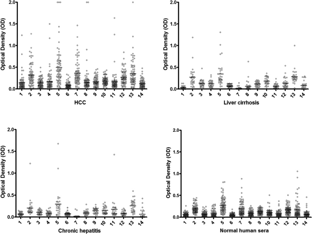
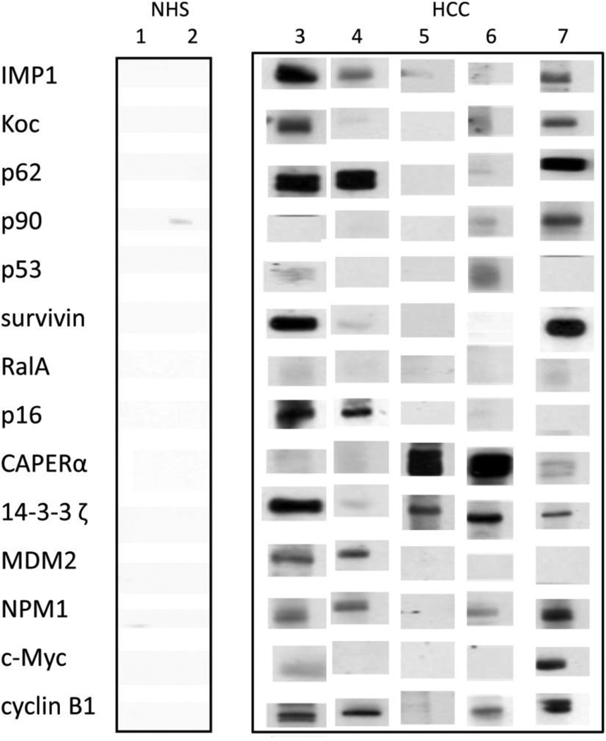
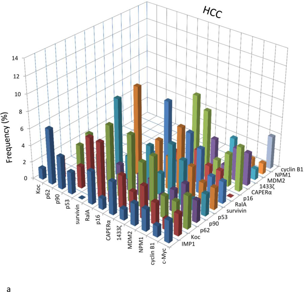
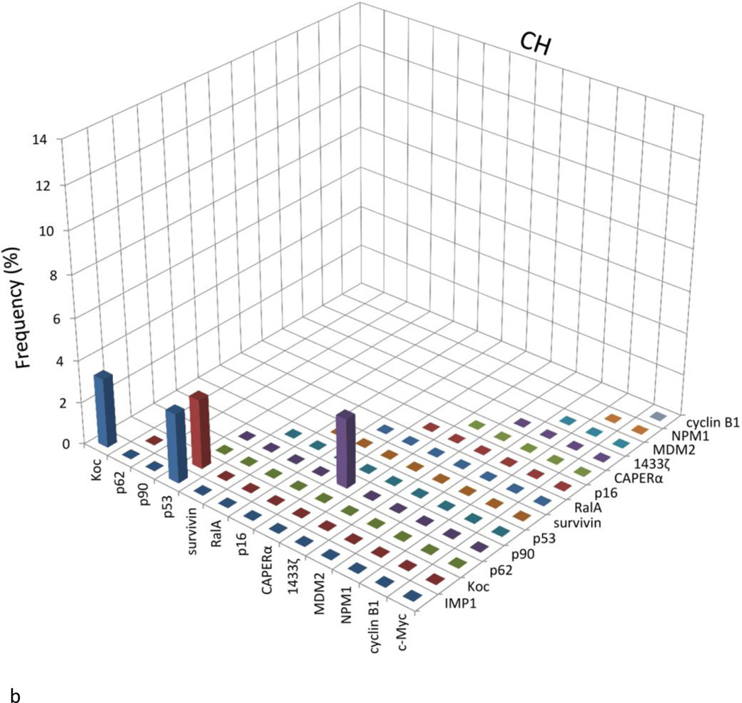
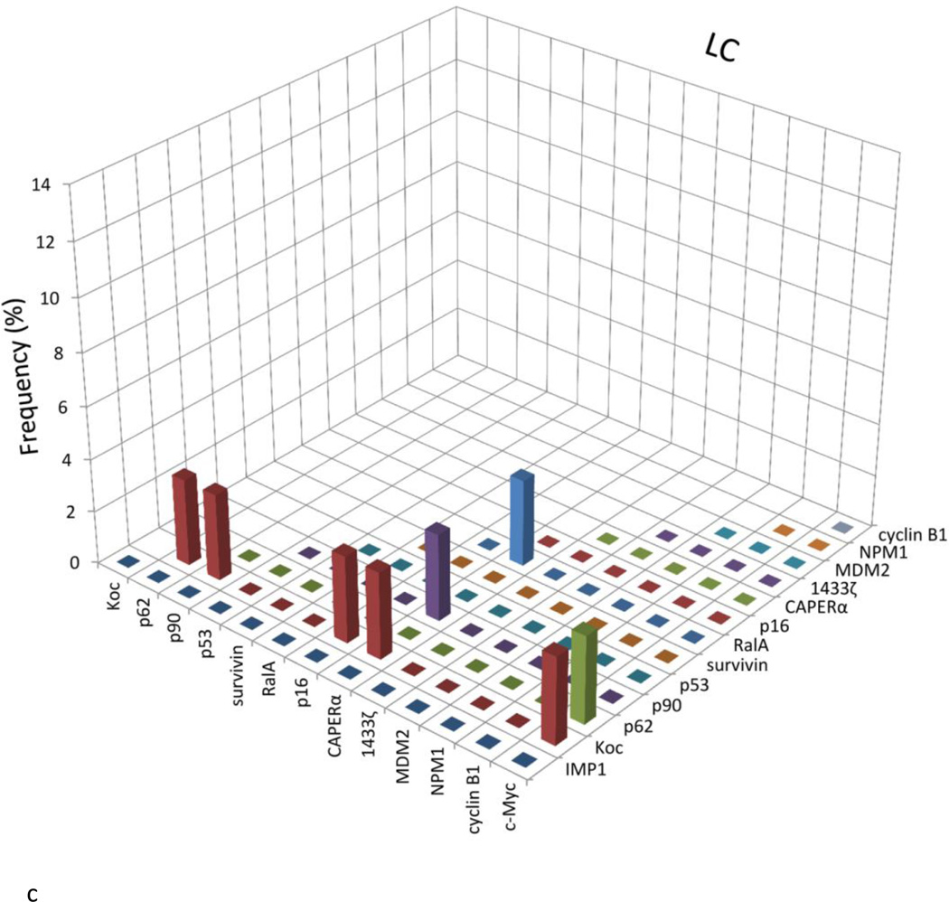
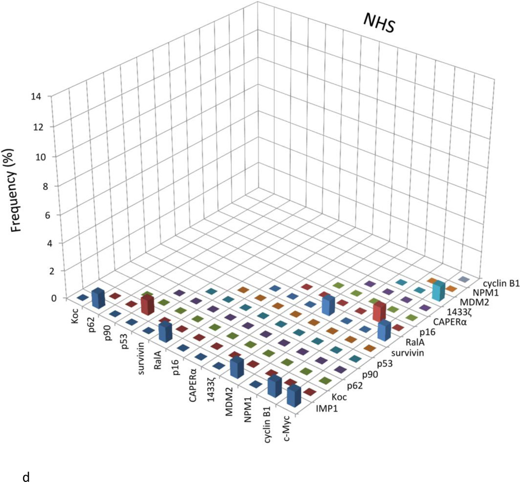
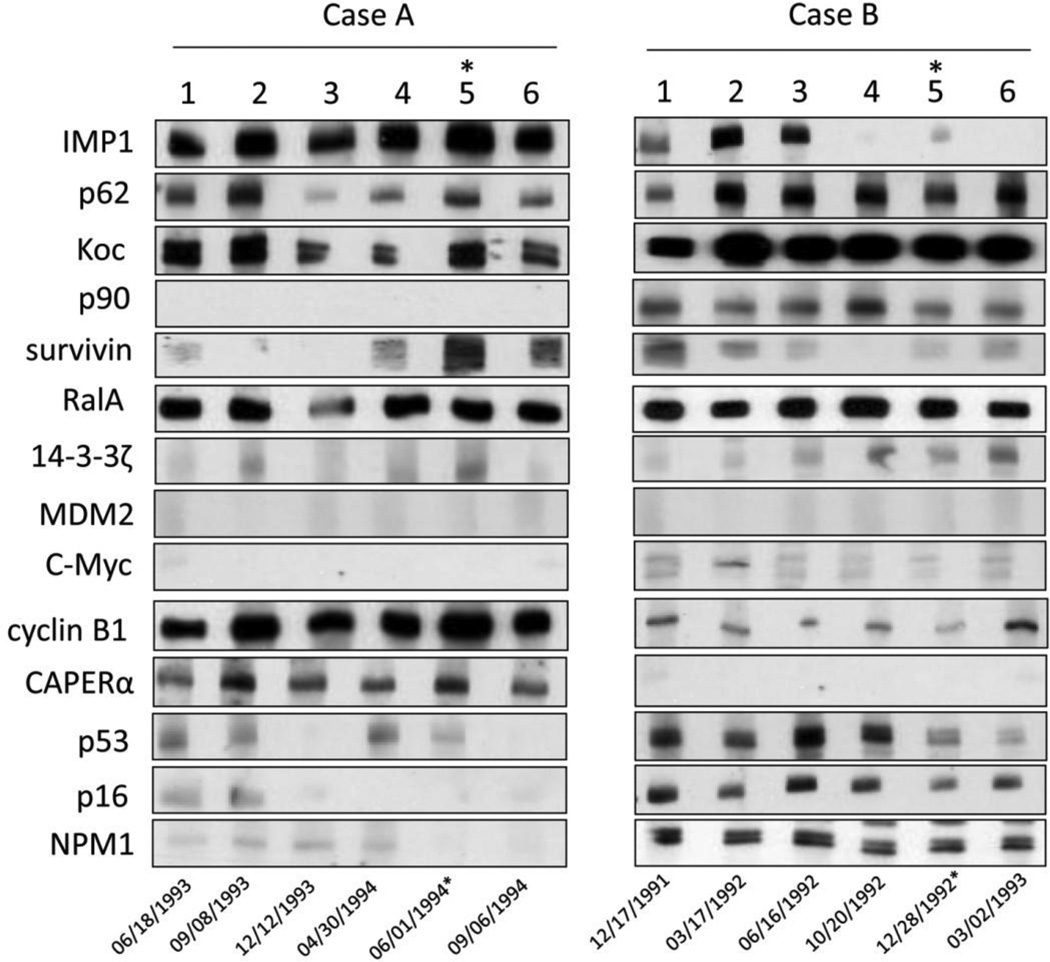
To explore the possibility of using a mini-array of multiple tumor-associated antigens (TAAs) as an approach to the diagnosis of hepatocellular carcinoma (HCC), 14 TAAs were selected to examine autoantibodies in sera from patients with chronic hepatitis, liver cirrhosis and HCC by immunoassays. Antibody frequency to any individual TAA in HCC varied from 6.6% to 21.1%. With the successive addition of TAAs to the panel of TAAs, there was a stepwise increase of positive antibody reactions. The sensitivity and specificity of 14 TAAs for immunodiagnosis of HCC was 69.7% and 83.0%, respectively. This TAA mini-array also identified 43.8% of HCC patients who had normal alpha-fetoprotein (AFP) levels in serum. In summary, this study further supports the hypothesis that a customized TAA array used for detecting anti-TAA autoantibodies can constitute a promising and powerful tool for immunodiagnosis of HCC and may be especially useful in patients with normal AFP levels.

Keywords: Autoantibodies;; Cancer immunodiagnosis;; Hepatocellular carcinoma; Tumor-associated antigens (TAAs);.


Conflict of interest statement

Disclosure of conflict of interest

The authors have no conflict of interest to disclose.

Figures

Figure 1
Figure 1
The range of antibody titers to different tumor associated antigens is expressed as absorbance units obtained from enzyme immunoassays. The mean of optical density (OD) was shown for very kind of condition. The high titer reactivity of HCC sera and the distinct difference between cancer and normal and autoimmune sera are demonstrated in this figure. The X-axis represents the panel of 14 TAAs. 1, survivin; 2, CAPERα; 3, RalA; 4, P62; 5, Koc; 6, MDM2; 7, cyclin B1; 8, p53; 9, 14-3-3 ζ; 10, p90; 11, IMP1; 12, c-Myc; 13, NPM1; 14, p16.
Figure 2
Figure 2
Some representative sera show immunoreactivity to 14 TAAs, which were verified by Western blotting. Lanes 1, 2: NHS; lanes 3–7: HCC sera. Some HCC sera show reaction with multiple antibodies against 14 TAAs, while in contrast, normal human sera show very weak or no reactions and rarely multiple reactions with 14 TAAs.
Figure 3
Figure 3
Analysis to determine the presence or absence of co-expression of antibodies to any combination of two of the 14 TAAs. The height of the bar represents the percentage of sera with co-expression of two antibodies as, e.g., the presence of c-Myc antibody together with IMP1 antibody, c-Myc antibody with Koc antibody, and so on. NHS: Normal human sera; HCC: Hepatocellular carcinoma; CH: Chronic hepatitis; LC: Liver cirrhosis
Figure 3
Figure 3
Analysis to determine the presence or absence of co-expression of antibodies to any combination of two of the 14 TAAs. The height of the bar represents the percentage of sera with co-expression of two antibodies as, e.g., the presence of c-Myc antibody together with IMP1 antibody, c-Myc antibody with Koc antibody, and so on. NHS: Normal human sera; HCC: Hepatocellular carcinoma; CH: Chronic hepatitis; LC: Liver cirrhosis
Figure 4
Figure 4
The expression of autoantibodies for the representative HCC patients with serial bleeding serum samples. Case A and case B are two representative patients with serial serum samples. Number 1–6 stand for six time point of sample collection. For case A, 1: 6/18/1993; 2: 9/8/1993; 3: 12/17/1993; 4: 3/30/1994; 5: 6/1/1994; 6: 9/6/1994. For case B, 1: 12/17/1991; 2: 3/17/1992; 3: 6/16/1992; 4: 10/20/1992; 5: 11/24/1992; 6: 3/2/1993. *: time point of HCC diagnosis

References

    1. Bruix J, Sherman M. Management of hepatocellular carcinoma. Hepatology. 2005;42(5):1208–1236. - PubMed
    1. Houghton AN. Cancer antigens: immune recognition of self and altered self. J. Exp. Med. 1994;180(1):1–4. - PMC - PubMed
    1. Old LJ, Chen YT. New paths in human cancer serology. J. Exp. Med. 1998;187(8):1163–1167. - PMC - PubMed
    1. Tan EM, Zhang J. Autoantibodies to tumor-associated antigens: reporters from the immune system. Immunol. Rev. 2008;222:328–340. - PMC - PubMed
    1. Zhang JY, Tan EM. Autoantibodies to tumor-associated antigens as diagnostic biomarkers in hepatocellular carcinoma and other solid tumors. Expert Rev Mol Diagn. 2010;10(3):321–328. - PMC - PubMed

Publication types

MeSH terms