. 2014 Mar 25;5(2):e00003-14.
doi: 10.1128/mBio.00003-14.

The pathogen Candida albicans hijacks pyroptosis for escape from macrophages

The pathogen Candida albicans hijacks pyroptosis for escape from macrophages

Nathalie Uwamahoro et al. mBio. .

Abstract

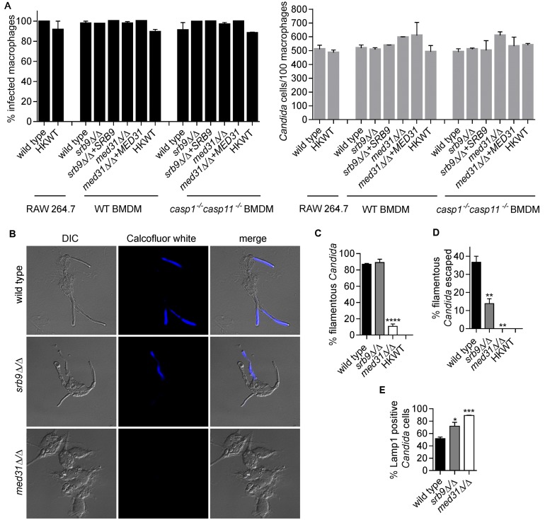
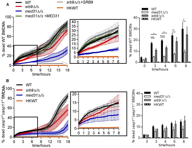
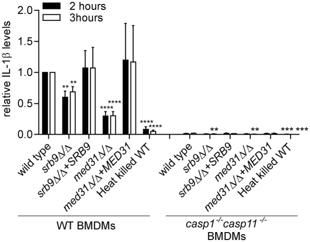
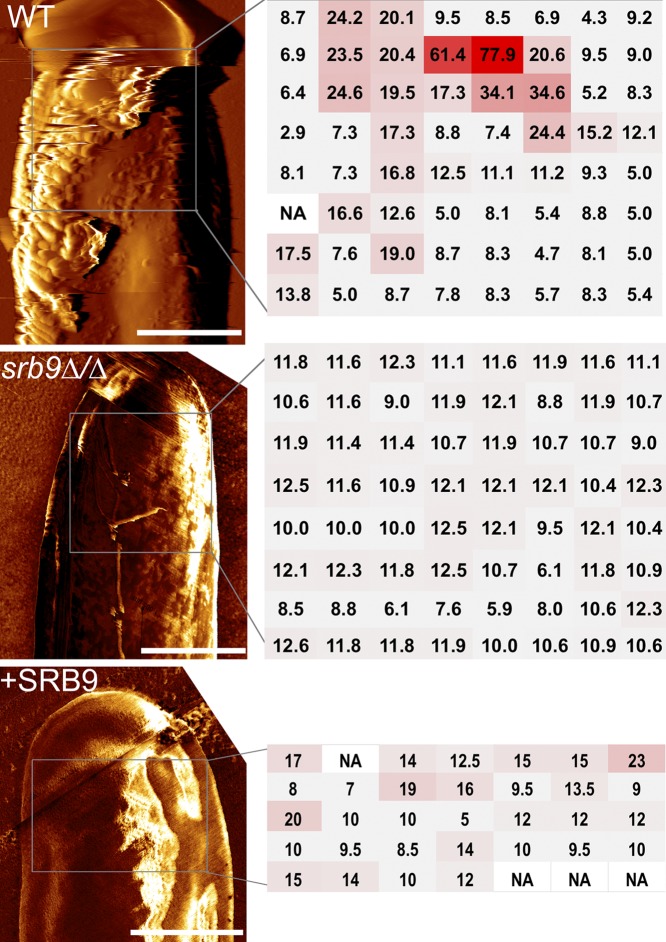
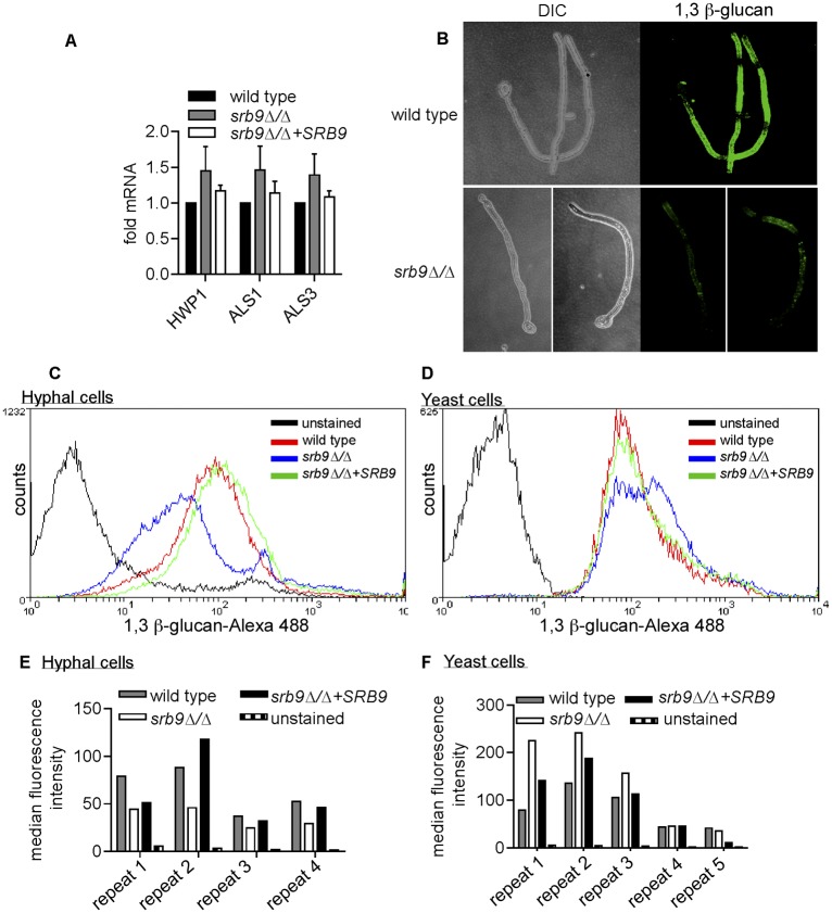
The fungal pathogen Candida albicans causes macrophage death and escapes, but the molecular mechanisms remained unknown. Here we used live-cell imaging to monitor the interaction of C. albicans with macrophages and show that C. albicans kills macrophages in two temporally and mechanistically distinct phases. Early upon phagocytosis, C. albicans triggers pyroptosis, a proinflammatory macrophage death. Pyroptosis is controlled by the developmental yeast-to-hypha transition of Candida. When pyroptosis is inactivated, wild-type C. albicans hyphae cause significantly less macrophage killing for up to 8 h postphagocytosis. After the first 8 h, a second macrophage-killing phase is initiated. This second phase depends on robust hyphal formation but is mechanistically distinct from pyroptosis. The transcriptional regulator Mediator is necessary for morphogenesis of C. albicans in macrophages and the establishment of the wild-type surface architecture of hyphae that together mediate activation of macrophage cell death. Our data suggest that the defects of the Mediator mutants in causing macrophage death are caused, at least in part, by reduced activation of pyroptosis. A Mediator mutant that forms hyphae of apparently wild-type morphology but is defective in triggering early macrophage death shows a breakdown of cell surface architecture and reduced exposed 1,3 β-glucan in hyphae. Our report shows how Candida uses host and pathogen pathways for macrophage killing. The current model of mechanical piercing of macrophages by C. albicans hyphae should be revised to include activation of pyroptosis by hyphae as an important mechanism mediating macrophage cell death upon C. albicans infection. IMPORTANCE Upon phagocytosis by macrophages, Candida albicans can transition to the hyphal form, which causes macrophage death and enables fungal escape. The current model is that the highly polarized growth of hyphae results in macrophage piercing. This model is challenged by recent reports of C. albicans mutants that form hyphae of wild-type morphology but are defective in killing macrophages. We show that C. albicans causes macrophage cell death by at least two mechanisms. Phase 1 killing (first 6 to 8 h) depends on the activation of the pyroptotic programmed host cell death by fungal hyphae. Phase 2 (up to 24 h) is rapid and depends on robust hyphal formation but is independent of pyroptosis. Our data provide a new model for how the interplay between fungal morphogenesis and activation of a host cell death pathway mediates macrophage killing by C. albicans hyphae.


Figures

FIG 1
FIG 1
C. albicans triggers pyroptotic macrophage cell death. (A) Wild-type (WT) C. albicans was incubated with wild-type BMDMs at MOI 1:6 (macrophage:Candida), and macrophage cell death was monitored over time. Shown are averages and standard errors of the means (SEM) of the results from two independent biological experiments. HKWT, heat-killed wild-type C. albicans cells (yeast morphology). (B) Experiments were performed as described for panel A except that the RAW 264.7 macrophage cell line was used. Averages and SEM are shown (n = 2). (C) Yeast and filamentous forms were counted from images from the live-cell microscopy experiments described for panels B and E at 30 min after the 1-h coincubation. A total of 100 phagocytosed Candida cells were counted for each of the independent biological experiments and classified as yeast, germ tubes, or hyphae. Values shown are means ± SEM (n = 2 for the RAW 264.7 cells and n = 3 for BMDMs). (D) Images corresponding to selected time points (h) from the live-cell microscopy of wild-type C. albicans infecting wild-type or casp1−/− casp11−/− BMDMs. (E) Wild-type C. albicans was incubated with wild-type or casp1−/− casp11−/− BMDMs. Averages and SEM of the results of 4 independent experiments are shown. These data and the data in the graph in panel F are the same as those determined in the wild-type Candida control experiments represented in Fig. 3. They are shown here separately for clarity of the results. (F) Graphs show means and SEM for percentages of macrophage cell death at selected time points from the curves shown in panel E. **, P <0.01; *, P <0.05. Representative live-cell microscopy movies from the macrophage-killing experiments represented in this figure are shown in Videos S1 to S3 in the supplemental material. (G) BMDMs were infected with live or heat-killed wild-type (HKWT) Candida at MOI 1:6 (macrophage:Candida) or treated with cycloheximide (CHX; 50 µg/ml) for 3 h, and the generation of cleaved caspase 3 was detected by immune blotting. Loading was visualized by Ponceau staining. Cycloheximide treatment served as a positive control.
FIG 2
FIG 2
Mediator regulates morphogenesis and escape of C. albicans from macrophages. (A) Percentages of infected macrophages and numbers of Candida cells/100 macrophages were determined from images from the live-cell microscopy experiments represented in Fig. 1B and 3. Three independent biological experiments were performed for BMDMs and two for the RAW 264.7 cell line, and a total of 200 macrophages were counted in each of the experiments. Values are means ± SEM. HKWT, heat-killed wild-type cells. (B and C) Wild-type C. albicans and med31∆/ and srb9∆/ strains were used to infect wild-type BMDMs, and fungal cell morphology was assessed by microscopy and quantified at 3 h postphagocytosis by counting at least 200 cells/strain. Averages and SEM of the results of 3 independent experiments are shown. ****, P ≤ 0.0001. DIC, differential interference contrast. (D) Percentages of escaped C. albicans hyphae and calcofluor white (CW)-stained hyphae were determined by counting total cells in macrophages from bright-field images (see images in panel B). CW stains only fungal cells that are outside the macrophages, while the phagocytosed cells are protected. At least 200 cells/strain were counted. Data represent averages and SEM (n = 3). **, P ≤ 0.01. (E) Association of C. albicans with late phagosomes in wild-type BMDMs was monitored by immunofluorescence by staining for the phagosomal marker Lamp1. Lamp1-positive C. albicans cells were scored by microscopy (images are shown in Fig. S1 in the supplemental material) at the 2-h time point (following the 1-h coincubation). Three independent experiments were performed, and at least 50 Candida cells were counted in each. Averages and SEM are shown. *, P <0.05; ***, P <0.001.
FIG 3
FIG 3
Roles of Mediator and Candida morphogenesis in pyroptosis-dependent and -independent macrophage death. (A and B) Wild-type C. albicans, the med31∆/ and srb9∆/ mutants, and the complemented strains were used to infect wild-type (A) or casp1−/− casp11−/− (B) BMDMs, as described for Fig. 1. The controls for these experiments with wild-type C. albicans were the same as those described for Fig. 1E and F. Each of the experiments was performed with the wild type and with both mutants of C. albicans, infecting wild-type or casp1−/− casp11−/− mutant BMDMs, all assayed together to allow direct comparisons of the effects of host and pathogen mutations. The wild-type C. albicans results are presented separately in Fig. 1 for clarity of the Results section. For simplicity, the results from wild-type BMDMs and those from casp1−/− casp11−/− BMDMs are presented in separate graphs. The experiments were performed 3 independent times (for the med31∆/ mutant) or 4 independent times (for the srb9∆/ mutant). The means and SEM for percentages of dead macrophages are shown. Graphs are for means and SEM for individual time points with statistical significance. All numerical P values for the differences between wild-type and mutant strains are shown in Fig. S2 in the supplemental material. P values are indicated as follows: *, <0.05; **, <0.01; ***, <0.001; ****, <0.0001. Videos of mutant Candida are shown in Videos S4 and S5.
FIG 4
FIG 4
Effects of Mediator subunits on IL-1β secretion from macrophages. BMDMs were pretreated with LPS and infected with C. albicans, and IL-1β levels were determined from supernatants after 2 or 3 h after the 1 h coincubation as described in Materials and Methods. The experiment was performed 3 independent times (4 for the srb9Δ/Δ mutant), and fold differences were calculated, with IL-1β levels induced by wild-type C. albicans in wild-type BMDMs set to 1. Averages and SEM of the results of the independent experiments are shown. The individual experiments with IL-1β concentrations in supernatants (pg/ml) are shown in Fig. S4 in the supplemental material. Figure S4 also shows rescaled relative levels for IL-1β in casp1−/− casp11−/− BMDMs for ease of comparison. P values were calculated for the comparisons between wild-type and mutant C. albicans and between wild-type and heat-killed C. albicans in either WT BMDMs (left side of the graph) or casp1−/− casp11−/− BMDMs (right side). **, P ≤ 0.01; ***, P ≤ 0.001; ****, P ≤ 0.0001.
FIG 5
FIG 5
srb9∆/ mutant hyphae display a breakdown of cell surface architecture. AFM (atomic force microscopy) was performed on hyphae grown in vitro under conditions that mimic those of the macrophage experiments (RPMI media, 37°C). Deflection images of hyphal tips from wild-type and srb9∆/ mutant hyphae are presented on the left and force measurements on the right. The regions in which force measurements were done were squares of the following sizes: 1.7 µm-by-1.7 µm for the wild type, 1.2 µm-by-1.2 µm for srb9∆/, and 1.5 µm-by-1 µm for the complemented strain). The adhesion forces, extracted from force-distance curves, were measured in an 8-by-8-matrix for the wild-type and mutant strains, or a 7-by-5 matrix for the complemented strain, as shown in the figure (the unit of adhesion force is nN). The measurements are color coded from gray (low intensity) to red (high intensity). Multiple hyphae were measured for each of the strains and gave equivalent results. The scale bar is 1 µm.
FIG 6
FIG 6
Srb9 regulates 1,3 β-glucan exposure on the cell surface of hyphae. (A) Quantitative PCR of the expression levels of the cell wall adhesins ALS1, ALS3, and HWP1 after phagocytosis by macrophages in wild-type, srb9∆/ mutant, and SRB9 complemented C. albicans strains. The experiment was performed on 3 separate occasions. 18S rRNA was used for normalization. Averages and SEM of the results from the 3 biological repeats are shown. (B) Wild-type and mutant hyphae stained with the 1,3 β-glucan antibody were visualized by confocal microscopy. Image stacks were used to create 3D renditions of wild-type and srb9∆/ mutant hyphae grown in vitro under conditions that mimicked the macrophage experiments (RPMI media, 37°C). (C to F) Hyphal growth of wild-type, srb9∆/ mutant, and complemented strains was induced in Spider media at 37°C for 3 h. Yeast cells were grown in YPD at 30° C. Exposed 1,3 β-glucan was stained using the 1,3 β-glucan antibody. Flow cytometry experiments were performed with several independent biological repeats, assayed on separate occasions. The flow cytometry curves (C and D) are from one representative experiment, and the bar graphs (E and F) show the median fluorescence obtained for the individual biological repeats.

References

    1. Brown GD, Denning DW, Gow NA, Levitz SM, Netea MG, White TC. 2012. Hidden killers: human fungal infections. Sci. Transl. Med. 4:165rv113. 10.1126/scitranslmed.3004404 - DOI - PubMed
    1. Gow NA, van de Veerdonk FL, Brown AJ, Netea MG. 2012. Candida albicans morphogenesis and host defence: discriminating invasion from colonization. Nat. Rev. Microbiol. 10:112–122. 10.1038/nrmicro2711 - DOI - PMC - PubMed
    1. Miramón P, Kasper L, Hube B. 2013. Thriving within the host: Candida spp. interactions with phagocytic cells. Med. Microbiol. Immunol. 202:183–195. 10.1007/s00430-013-0288-z - DOI - PubMed
    1. Lo HJ, Köhler JR, DiDomenico B, Loebenberg D, Cacciapuoti A, Fink GR. 1997. Nonfilamentous C. albicans mutants are avirulent. Cell 90:939–949. 10.1016/S0092-8674(00)80358-X - DOI - PubMed
    1. McKenzie CG, Koser U, Lewis LE, Bain JM, Mora-Montes HM, Barker RN, Gow NA, Erwig LP. 2010. Contribution of Candida albicans cell wall components to recognition by and escape from murine macrophages. Infect. Immun. 78:1650–1658. 10.1128/IAI.00001-10 - DOI - PMC - PubMed

Publication types