. 2014 Mar 25;5(2):e00047-14.
doi: 10.1128/mBio.00047-14.

A mouse model for Betacoronavirus subgroup 2c using a bat coronavirus strain HKU5 variant

A mouse model for Betacoronavirus subgroup 2c using a bat coronavirus strain HKU5 variant

Sudhakar Agnihothram et al. mBio. .

Abstract

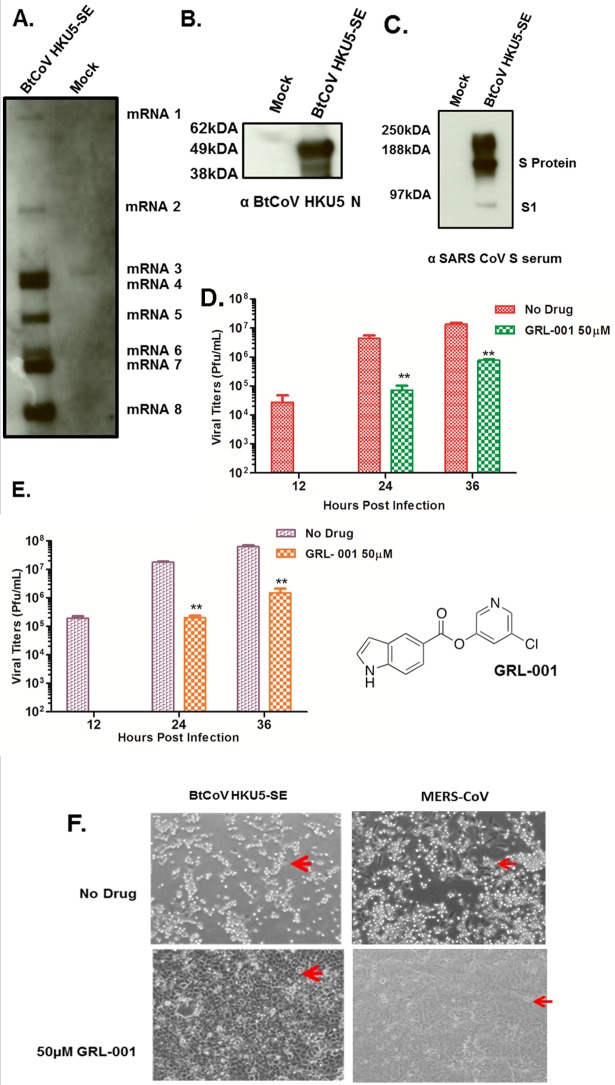
Cross-species transmission of zoonotic coronaviruses (CoVs) can result in pandemic disease outbreaks. Middle East respiratory syndrome CoV (MERS-CoV), identified in 2012, has caused 182 cases to date, with ~43% mortality, and no small animal model has been reported. MERS-CoV and Pipistrellus bat coronavirus (BtCoV) strain HKU5 of Betacoronavirus (β-CoV) subgroup 2c share >65% identity at the amino acid level in several regions, including nonstructural protein 5 (nsp5) and the nucleocapsid (N) protein, which are significant drug and vaccine targets. BtCoV HKU5 has been described in silico but has not been shown to replicate in culture, thus hampering drug and vaccine studies against subgroup 2c β-CoVs. We report the synthetic reconstruction and testing of BtCoV HKU5 containing the severe acute respiratory syndrome (SARS)-CoV spike (S) glycoprotein ectodomain (BtCoV HKU5-SE). This virus replicates efficiently in cell culture and in young and aged mice, where the virus targets airway and alveolar epithelial cells. Unlike some subgroup 2b SARS-CoV vaccines that elicit a strong eosinophilia following challenge, we demonstrate that BtCoV HKU5 and MERS-CoV N-expressing Venezuelan equine encephalitis virus replicon particle (VRP) vaccines do not cause extensive eosinophilia following BtCoV HKU5-SE challenge. Passage of BtCoV HKU5-SE in young mice resulted in enhanced virulence, causing 20% weight loss, diffuse alveolar damage, and hyaline membrane formation in aged mice. Passaged virus was characterized by mutations in the nsp13, nsp14, open reading frame 5 (ORF5) and M genes. Finally, we identified an inhibitor active against the nsp5 proteases of subgroup 2c β-CoVs. Synthetic-genome platforms capable of reconstituting emerging zoonotic viral pathogens or their phylogenetic relatives provide new strategies for identifying broad-based therapeutics, evaluating vaccine outcomes, and studying viral pathogenesis. IMPORTANCE The 2012 outbreak of MERS-CoV raises the specter of another global epidemic, similar to the 2003 SARS-CoV epidemic. MERS-CoV is related to BtCoV HKU5 in target regions that are essential for drug and vaccine testing. Because no small animal model exists to evaluate MERS-CoV pathogenesis or to test vaccines, we constructed a recombinant BtCoV HKU5 that expressed a region of the SARS-CoV spike (S) glycoprotein, thereby allowing the recombinant virus to grow in cell culture and in mice. We show that this recombinant virus targets airway epithelial cells and causes disease in aged mice. We use this platform to (i) identify a broad-spectrum antiviral that can potentially inhibit viruses closely related to MERS-CoV, (ii) demonstrate the absence of increased eosinophilic immune pathology for MERS-CoV N protein-based vaccines, and (iii) mouse adapt this virus to identify viral genetic determinants of cross-species transmission and virulence. This study holds significance as a strategy to control newly emerging viruses.


Figures

FIG 1
FIG 1
Genome organization and replication of BtCoV HKU5-SE in Vero cells. (A) Schematic of the genome organization of BtCoV HKU5 (top) and the recombinant virus in which the S protein was replaced with the ectodomain of SARS S glycoprotein (SE, yellow). The asterisk indicates the Y436H substitution. The bottom panel represents cDNA fragments A to F encoding the genome, with the T7 start site, the poly(T) tract, and the BglI restriction sites flanking the fragments. (B and C) BtCoV HKU5-SE replication in Vero cells compared with the replication of MERS-CoV Hu/SA-N1/2012 (MERS-CoV SA 1) (B) and SARS-CoV (C). Vero cells were infected with BtCoV HKU5-SE, MERS-CoV, and SARS-CoV at the indicated MOIs. Supernatants were sampled in triplicate at the times indicated, and titers determined on Vero cells by plaque assay. Error bars indicate standard deviations (SD).
FIG 2
FIG 2
Subgenomic mRNA and protein expression in BtCoV HKU5-SE and the 3C-like protease inhibitor study. (A) Northern blot showing subgenomic mRNA expression in BtCoV HKU5-SE-infected cells. (B and C) Western blots showing expression of structural proteins N (B) and S (C) from BtCoV HKU5-SE-infected cell lysates stained with polyclonal serum as indicated below the images. (D and E) Viral titers from cells pretreated with 50 µM of GRL-001 and infected with BtCoV HKU5-SE (D) and MERS-CoV (E) at an MOI of 0.1 PFU/ml. Drug treatment was continued after infection, and virus-containing supernatants were sampled in triplicate. Error bars indicate SD. Asterisks indicate statistical significance (P < 0.05, Student t test). The structure of GRL-001 is shown to the right of panel E. (F) Bright-field images of cells infected with BtCoV HKU5-SE or MERS-CoV at an MOI of 0.1, showing strong cytopathic effects with no drug treatment (top, red arrows) and intact monolayers with 50 µM GRL-001 treatment (bottom, red arrows) at 36 h p.i..
FIG 3
FIG 3
BtCoV HKU5-SE replication in BALB/c mice. (A) Weight loss in young (10-week-old) and aged (1-year-old) BALB/c mice and 1-year-old ACE2−/− mice (BALB/c background) was measured after intranasal infection with 1 × 10⁵ PFU of BtCoV HKU5-SE. (B) Viral replication in mouse lungs at 2 and 4 days p.i. LOD, limit of detection. (C) Lung pathology from hematoxylin-and-eosin (H&E)-stained sections was blind scored for clinical disease features on a scale of 0 to 3 (1, mild; 3, severe). Error bars indicate SD. (D) Representative H&E-stained sections of lungs from a young and an aged mouse harvested at 4 days p.i., showing severe inflammatory infiltration in the aged mouse.
FIG 4
FIG 4
Viral antigen distribution in the lung. Immunohistochemistry was performed using anti-HKU5 N sera, and sections representing different regions of lungs from young and aged mice at 2 days p.i. are shown. Stained sections of lungs from mock-infected mice as controls are indicated on the extreme right in both rows.
FIG 5
FIG 5
Vaccine studies in mice. (A) Weight loss of aged mice (1 year old) vaccinated with SARS-CoV S or mock vaccinated prior to challenge with 105 PFU of BtCoV HKU5-SE. (B) Virus titers in lungs at days 2 and 4 p.i. (C) Antiserum to SARS-CoV S protein but not antiserum to HKU5 S or N protein neutralizes BtCoV HKU5-SE in an in vitro PRNT50 assay. (D) Weight loss of young mice either mock vaccinated or vaccinated with VRP (virus replicon particles) expressing HKU5 N or MERS-CoV N and challenged with BtCoV HKU5-SE. (E) Virus titers in lungs of mice at 2 days p.i. (F) Flow cytometric analysis showing numbers of eosinophils at 4 days p.i. in the lungs of mice either mock vaccinated or vaccinated with BtCoV HKU5 N or MERS-CoV N and then challenged with 105 PFU of BtCoV HKU5-SE. Error bars indicate SD.
FIG 6
FIG 6
BtCoV HKU5-SE MA replication in young and aged BALB/c mice. (A) Weight loss curves of young (10-week-old) and aged (1-year-old) mice infected intranasally with 1 × 10⁵ PFU of BtCoV HKU5-SE MA through 4 days p.i. (B) Viral titers in lungs of mice at days 2 and 4 p.i. Error bars indicate SD. (C) Representative H&E-stained sections of lungs from a young and an aged mouse harvested at 4 days p.i., showing severe inflammatory infiltration and hyaline membrane formation (black arrow) in the aged mouse.
FIG 7
FIG 7
BtCoV HKU5-SE MA genome sequence. (A) Schematics of BtCoV HKU5-SE, depicting all open reading frames (ORFs) (top) and the mouse-adapted mutations as red stick-and-ball symbols at indicated spots in the genome (bottom). (B) Details of the mouse adaptations at the nucleotide and amino acid levels (top), and analysis of viremia in BtCoV HKU5-SE MA-infected aged (1-year-old) BALB/c mice (bottom).

References

    1. Tsushima K, King LS, Aggarwal NR, De Gorordo A, D’Alessio FR, Kubo K. 2009. Acute lung injury review. Intern. Med. 48:621–630. 10.2169/internalmedicine.48.1741 - DOI - PubMed
    1. Masters PS. 2006. The molecular biology of coronaviruses. Adv. Virus Res. 66:193–292. 10.1016/S0065-3527(06)66005-3 - DOI - PMC - PubMed
    1. Graham RL, Baric RS. 2010. Recombination, reservoirs, and the modular spike: mechanisms of coronavirus cross-species transmission. J. Virol. 84:3134–3146. 10.1128/JVI.01394-09 - DOI - PMC - PubMed
    1. Bolles M, Donaldson E, Baric R. 2011. SARS-CoV and emergent coronaviruses: viral determinants of interspecies transmission. Curr. Opin. Virol. 1:624–634. 10.1016/j.coviro.2011.10.012 - DOI - PMC - PubMed
    1. Pyrc K, Sims AC, Dijkman R, Jebbink M, Long C, Deming D, Donaldson E, Vabret A, Baric R, van der Hoek L, Pickles R. 2010. Culturing the unculturable: human coronavirus HKU1 infects, replicates, and produces progeny virions in human ciliated airway epithelial cell cultures. J. Virol. 84:11255–11263. 10.1128/JVI.00947-10 - DOI - PMC - PubMed

Publication types

MeSH terms

LinkOut - more resources