Comparative Study
. 2014 Mar 25;5(2):e00969-14.
doi: 10.1128/mBio.00969-14.

Comparison of widely used Listeria monocytogenes strains EGD, 10403S, and EGD-e highlights genomic variations underlying differences in pathogenicity

Comparative Study

Comparison of widely used Listeria monocytogenes strains EGD, 10403S, and EGD-e highlights genomic variations underlying differences in pathogenicity

Christophe Bécavin et al. mBio. .

Abstract

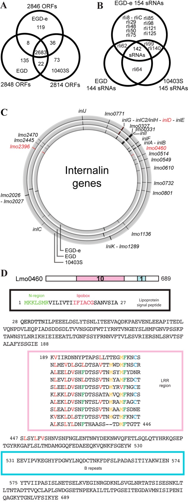
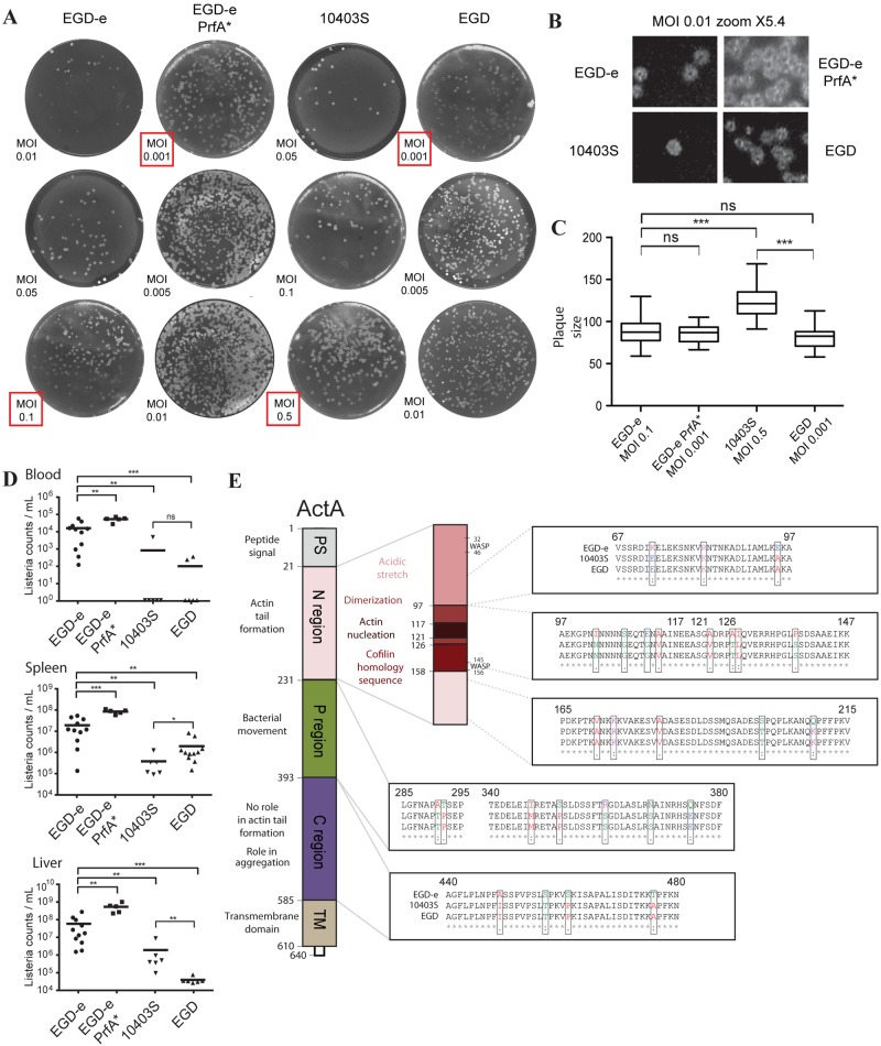
For nearly 3 decades, listeriologists and immunologists have used mainly three strains of the same serovar (1/2a) to analyze the virulence of the bacterial pathogen Listeria monocytogenes. The genomes of two of these strains, EGD-e and 10403S, were released in 2001 and 2008, respectively. Here we report the genome sequence of the third reference strain, EGD, and extensive genomic and phenotypic comparisons of the three strains. Strikingly, EGD-e is genetically highly distinct from EGD (29,016 single nucleotide polymorphisms [SNPs]) and 10403S (30,296 SNPs), and is more related to serovar 1/2c than 1/2a strains. We also found that while EGD and 10403S strains are genetically very close (317 SNPs), EGD has a point mutation in the transcriptional regulator PrfA (PrfA*), leading to constitutive expression of several major virulence genes. We generated an EGD-e PrfA* mutant and showed that EGD behaves like this strain in vitro, with slower growth in broth and higher invasiveness in human cells than those of EGD-e and 10403S. In contrast, bacterial counts in blood, liver, and spleen during infection in mice revealed that EGD and 10403S are less virulent than EGD-e, which is itself less virulent than EGD-e PrfA*. Thus, constitutive expression of PrfA-regulated virulence genes does not appear to provide a significant advantage to the EGD strain during infection in vivo, highlighting the fact that in vitro invasion assays are not sufficient for evaluating the pathogenic potential of L. monocytogenes strains. Together, our results pave the way for deciphering unexplained differences or discrepancies in experiments using different L. monocytogenes strains. IMPORTANCE Over the past 3 decades, Listeria has become a model organism for host-pathogen interactions, leading to critical discoveries in a broad range of fields, including bacterial gene regulation, cell biology, and bacterial pathophysiology. Scientists studying Listeria use primarily three pathogenic strains: EGD, EGD-e, and 10403S. Despite many studies on EGD, it is the only one of the three strains whose genome has not been sequenced. Here we report the sequence of its genome and a series of important genomic and phenotypic differences between the three strains, in particular, a critical mutation in EGD's PrfA, the main regulator of Listeria virulence. Our results show that the three strains display differences which may play an important role in the virulence differences observed between the strains. Our findings will be of critical relevance to listeriologists and immunologists who have used or may use Listeria as a tool to study the pathophysiology of listeriosis and immune responses.


Figures

FIG 1
FIG 1
SNPs, synteny, and sequence type analysis of EGD, EGD-e, and 10403S. (A) SNPs among the EGD, 10403S, and EGD-e reference genomes. Purple indicates synonymous changes, blue indicates nonsynonymous changes, and black indicates intergenic changes. (B) Minimum spanning tree analysis of 360 L. monocytogenes strains based on MLST (multilocus sequence typing) data (adapted from reference 21). The EGD-e, EGD, and 10403S strains are highlighted in red. (C) Linear synteny view of the three strains. Phage BO25 is integrated into EGD in tRNAArg. Phage A118 is integrated inside the comK gene in EGD-e and 10403S. ComK is complete in EGD.
FIG 2
FIG 2
Conservation of ORFs, small RNAs, and internalins in EGD, EGD-e, and 10403S. (A) Venn diagram showing the numbers of ORFs common to the different strains. A bidirectional best-hit search with an E value score lower than 1e−4 was used to determine homologies. (B) Venn diagram of the small RNAs found in the three strains. The percentage of similarity was calculated from BLASTN results. Small RNAs with a percentage lower than 10% were not considered conserved. (C) Genomic locations of 27 internalins in the EGD-e, EGD, and 10403S genomes (using CGView [81]). In red are indicated internalins present only in one or two strains. (D) Lmo0460 amino acid sequence. Lmo0460 is a predicted lipoprotein present only in EGD-e which contains an atypical leucine-rich repeat (LRR) domain.
FIG 3
FIG 3
PrfA* mutation and the overexpression of the PrfA core regulon in EGD. (A) Protein sequence alignment of PrfA in 43 L. monocytogenes strains. The well-known PrfA* mutation G145S (51, 82) is highlighted in red and appears only in the EGD and M7 strains. All other amino acid changes found are drawn showing their positions in the different domains of PrfA (52). HTH, helix turn helix. (B) Schematic representation of the virulence locus synteny in EGD-e, EGD, and 10403S. Amino acid differences from EGD-e’s sequence are displayed. (C) Genome browser view showing tiling array whole-transcriptome coverage of the virulence locus and the inlA-inlB operon in EGD-e, EGD-e PrfA*, and EGD. Each tiled probe indicating expression from the two genomic strands (top for plus strand, bottom for minus strand) is represented as a black dot for EGD-e, an orange dot for EGD-e PrfA*, and a green dot for EGD. (D) Comparison of expression levels of InlA, InlB, and LLO in EGD-e, EGD-e PrfA*, EGD, and L. innocua Clip11262 (used as a nonpathogenic reference bacterium) in whole bacterial lysates or in the cell wall fraction (InlA). (E) Immunofluorescence of InlA and ActA in EGD and EGD-e PrfA* in BHI medium.
FIG 4
FIG 4
Differential bacterial invasion phenotypes in vitro. (A) Colony sizes of the three strains after 24 h of growth on solid BHI agar plates reveal that EGD-e PrfA* has smaller colonies than EGD-e and that EGD has smaller colonies than 10403S. (B) Gentamicin assays at 2 h postinfection of HeLa, JEG3, and Raw264 cells by the four different strains, EGD, EGD-e PrfA*, 10403S, and EGD. ns, not significantly different; *, P value of <0.05; **, P value of <0.005; ***, P value of <0.0005.
FIG 5
FIG 5
Plaque assays, virulence in mice, and ActA amino acid changes. (A) Plaque assays of EGD-e, EGD-e PrfA*, 10403S, and EGD at different MOIs show different sizes depending on the strain. Highlighted in red are the MOIs used for plaque size measurement. (B) Magnifications (×5.4) of the plaques for EGD-e, EGD-e PrfA*, 10403S, and EGD at an MOI of 0.01. (C) Measurement of plaque size (in square pixels) using Icy image analysis software (80). Different MOIs were used for each strain in order to have the same number of plaques in each well. Differences between strains were assessed by unpaired t test. Plaques from 10403S are bigger than the ones from EGD-e and the two PrfA* strains (EGD-e PrfA*, EGD). (D) CFU counts measured 72 h after intravenous infection with EGD-e, EGD-e PrfA*, 10403S, and EGD. Each dot represents the value for one mouse, and asterisks indicate Mann-Whitney statistical test results; results are from two independent experiments. (E) Motifs of the ActA protein (adapted from reference 61) and the different amino acid changes between EGD-e and EGD are shown. ActA has the same amino acid sequence in EGD and 10403S. The two WASP-like sequences of ActA present no differences between EGD-e, EGD, and 10403S.

References

    1. Camejo A, Carvalho F, Reis O, Leitão E, Sousa S, Cabanes D. 2011. The arsenal of virulence factors deployed by Listeria monocytogenes to promote its cell infection cycle. Virulence 2:379–394. 10.4161/viru.2.5.17703 - DOI - PubMed
    1. Hamon M, Bierne H, Cossart P. 2006. Listeria monocytogenes: a multifaceted model. Nat. Rev. Microbiol. 4:423–434. 10.1038/nrmicro1413 - DOI - PubMed
    1. Lecuit M. 2007. Human listeriosis and animal models. Microbes Infect. 9:1216–1225. 10.1016/j.micinf.2007.05.009 - DOI - PubMed
    1. Cossart P, Toledo-Arana A. 2008. Listeria monocytogenes, a unique model in infection biology: an overview. Microbes Infect. 10:1041–1050. 10.1016/j.micinf.2008.07.043 - DOI - PubMed
    1. Cossart P. 2011. Illuminating the landscape of host-pathogen interactions with the bacterium Listeria monocytogenes. Proc. Natl. Acad. Sci. U. S. A. 108:1984–1991. 10.1073/pnas.1112371108 - DOI - PMC - PubMed

Publication types

Associated data