. 2014 Mar 25;9(3):e93327.
doi: 10.1371/journal.pone.0093327. eCollection 2014.

Forced resurgence and targeting of intracellular uropathogenic Escherichia coli reservoirs

Affiliations

Forced resurgence and targeting of intracellular uropathogenic Escherichia coli reservoirs

Matthew G Blango et al. PLoS One. .

Abstract

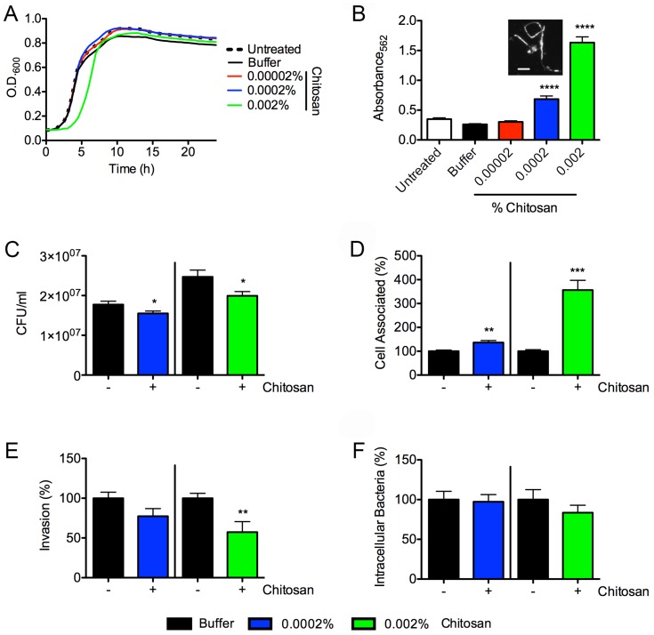
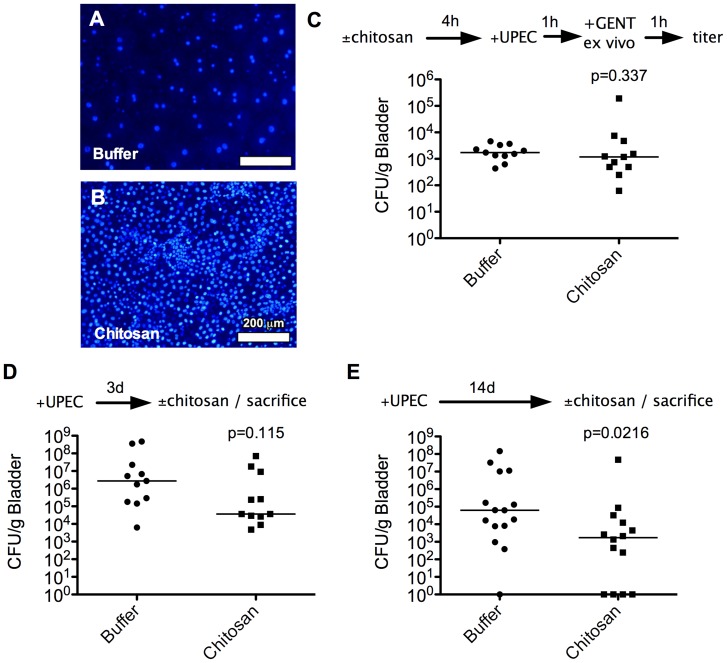
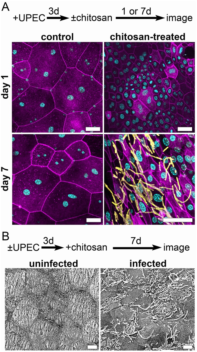
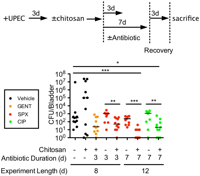
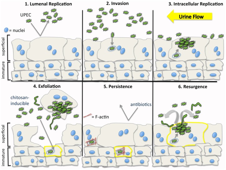
Intracellular quiescent reservoirs of uropathogenic Escherichia coli (UPEC), which can seed the bladder mucosa during the acute phase of a urinary tract infection (UTI), are protected from antibiotic treatments and are extremely difficult to eliminate. These reservoirs are a potential source for recurrent UTIs that affect millions annually. Here, using murine infection models and the bladder cell exfoliant chitosan, we demonstrate that intracellular UPEC populations shift within the stratified layers of the urothelium during the course of a UTI. Following invasion of the terminally differentiated superficial layer of epithelial cells that line the bladder lumen, UPEC can multiply and disseminate, eventually establishing reservoirs within underlying immature host cells. If given access, UPEC can invade the superficial and immature bladder cells equally well. As infected immature host cells differentiate and migrate towards the apical surface of the bladder, UPEC can reinitiate growth and discharge into the bladder lumen. By inducing the exfoliation of the superficial layers of the urothelium, chitosan stimulates rapid regenerative processes and the reactivation and efflux of quiescent intracellular UPEC reservoirs. When combined with antibiotics, chitosan treatment significantly reduces bacterial loads within the bladder and may therefore be of therapeutic value to individuals with chronic, recurrent UTIs.


Conflict of interest statement

Competing Interests: The authors have declared that no competing interests exist.

Figures

Figure 1
Figure 1. Chitosan affects UPEC growth, biofilm formation, and interactions with BECs.
(A) Growth of UTI89 in modified M9 medium containing increasing concentrations of chitosan (0.00002, 0.0002, 0.002%) or phosphate buffer (pH 4.5). Data shown are representative of three independent experiments carried out in triplicate. (B) Graph shows chitosan effects on biofilm production by UTI89 relative to untreated controls, as measured by microtiter plate assays. Data represent mean results ± SEM from three separate experiments performed in quadruplicate. Inset shows an example of filamentous bacteria in 0.002% chitosan; scale bar, 10 μm. Chitosan (0.0002 or 0.002%) effects on (C) UTI89 growth in RPMI medium during a 2-h incubation with 5637 BECs, (D) bacterial attachment to BECs, (E) bacterial invasion of BECs, and (F) bacterial survival within BECs over a 14-h period were quantified relative to buffer-treated controls. The data in F were normalized by dividing the numbers of surviving intracellular bacteria recovered from BECs after the 14-h incubation with gentamicin by the numbers of invading bacteria present after the 2-h incubation with gentamicin. Graphs show mean results ± SEM from three independent experiments performed in triplicate. *P<0.05, **P<0.01, ***P<0.001 versus buffer-treated controls, as determined by Student’s t test with Welch’s correction when appropriate.
Figure 2
Figure 2. Chitosan-induced exfoliation of superficial BECs and analysis of UPEC reservoir populations.
(A and B) Fluorescent images show the lumenal surfaces of mouse bladders fixed and stained with Hoechst dye following 20-min treatments with (A) phosphate buffer (pH 4.5) alone or with (B) 0.01% chitosan. Representative images of three independent experiments performed in duplicate. (C) The bladders of adult female CBA/J mice were inoculated with UTI89 via transurethral catheterization 4 h after a 20-min treatment of the mouse bladders with chitosan or phosphate buffer alone. Internalized bacteria were enumerated 1 h after initiation of the infection using ex vivo gentamicin protection assays. (D and E) Graphs show bacterial titers present in the bladders of mice treated with phosphate buffer or chitosan just before collection at (D) 3 d or (E) 14 d post-inoculation with UTI89. Bars indicate median values for each group; n = 11 mice. P values indicated were determined by the Mann-Whitney U-test.
Figure 3
Figure 3. Chitosan treatment promotes the resurgence of UPEC reservoir populations into the bladder lumen.
Adult female CBA/J mice were inoculated with UTI89 via transurethral catheterization or left uninfected. After 3 d, phosphate buffer alone (control) or 0.01% chitosan was instilled into the bladders for 20 min prior to rinses with PBS. Bladders were bisected, splayed and imaged 1 or 7 d later, as indicated, using (A) confocal fluorescence microscopy or (B) SEM. Images in (B) show F-actin (purple), host nuclei (blue), and bacteria (yellow). Scale bars, (A) 50 μm and (B) 10 μm. Images are representative of three independent experiments.
Figure 4
Figure 4. Chitosan treatment enhances the efficacy of antibiotics in reducing bacterial titers within the bladder.
Mice were infected with UTI89 via transurethral catheterization 3% chitosan. After a 20-min incubation and subsequent washes with PBS, mice were treated for 3 or 7 d with gentamicin (GENT), sparfloxacin (SPX), or ciprofloxacin (CIP), as indicated. Mice were sacrificed 3 d after cessation of antibiotics or mock treatments. An outline of the experimental setup is shown on top. Bars in the graph indicate median values for each sample group; n = 11 mice total from two independent assays. *P<0.02, **P<0.005, ***P≤0.0002; as determined by Mann-Whitney U-tests.
Figure 5
Figure 5. UPEC persistence and recurrence within the bladder.
(1) UPEC that gain entry into the bladder lumen can multiply within the urine and in association with the bladder surface. (2) Some bacteria enter host superficial cells and are trafficked into late endosome-like compartments. (3) Shearing forces from the flow of urine, secreted antimicrobial factors, and infiltrating neutrophils can eliminate many bacteria, while internalized UPEC are sheltered. Some intracellular bacteria may enter the host cytosol where they can rapidly multiply, forming IBCs. (4) Infection can induce the exfoliation of the superficial cells, exposing underlying immature BECs (5) that can rapidly differentiate to reestablish barrier function. Chitosan treatment can also stimulate exfoliation of superficial bladder cells and subsequent regenerative processes. Within both the superficial and immature cells of the bladder, UPEC can persist as quiescent reservoirs within membrane-bound compartments enmeshed in F-actin. Antibiotics that can effectively sterilize the urine are ineffective against the intracellular UPEC reservoirs. (6) Redistribution of actin and perhaps other signals associated with terminal differentiation of the BECs may elicit the resurgent growth of UPEC, resulting in bacterial release back into the bladder lumen. By promoting turnover of the urothelium, the intravesical delivery of chitosan can stimulate the resurgence of UPEC from intracellular reservoirs, making the bacteria more accessible to antibiotics.

References

    1. Barber AE, Norton JP, Spivak AM, Mulvey MA (2013) Urinary tract infections: current and emerging management strategies. Clin Infect Dis 57: 719–724. - PMC - PubMed
    1. Martinez JJ, Mulvey MA, Schilling JD, Pinkner JS, Hultgren SJ (2000) Type 1 pilus-mediated bacterial invasion of bladder epithelial cells. EMBO J 19: 2803–2812. - PMC - PubMed
    1. Mulvey MA, Lopez-Boado YS, Wilson CL, Roth R, Parks WC, et al. (1998) Induction and evasion of host defenses by type 1-piliated uropathogenic Escherichia coli . Science 282: 1494–1497. - PubMed
    1. Zhou G, Mo WJ, Sebbel P, Min G, Neubert TA, et al. (2001) Uroplakin Ia is the urothelial receptor for uropathogenic Escherichia coli: evidence from in vitro FimH binding. J Cell Sci 114: 4095–4103. - PubMed
    1. Kreft ME, Hudoklin S, Jezernik K, Romih R (2010) Formation and maintenance of blood-urine barrier in urothelium. Protoplasma 246: 3–14. - PubMed

Publication types