. 2014 Mar 25;9(3):e93329.
doi: 10.1371/journal.pone.0093329. eCollection 2014.

Skeletal muscle DNA damage precedes spinal motor neuron DNA damage in a mouse model of Spinal Muscular Atrophy (SMA)

Affiliations

Skeletal muscle DNA damage precedes spinal motor neuron DNA damage in a mouse model of Spinal Muscular Atrophy (SMA)

Saniya Fayzullina et al. PLoS One. .

Abstract

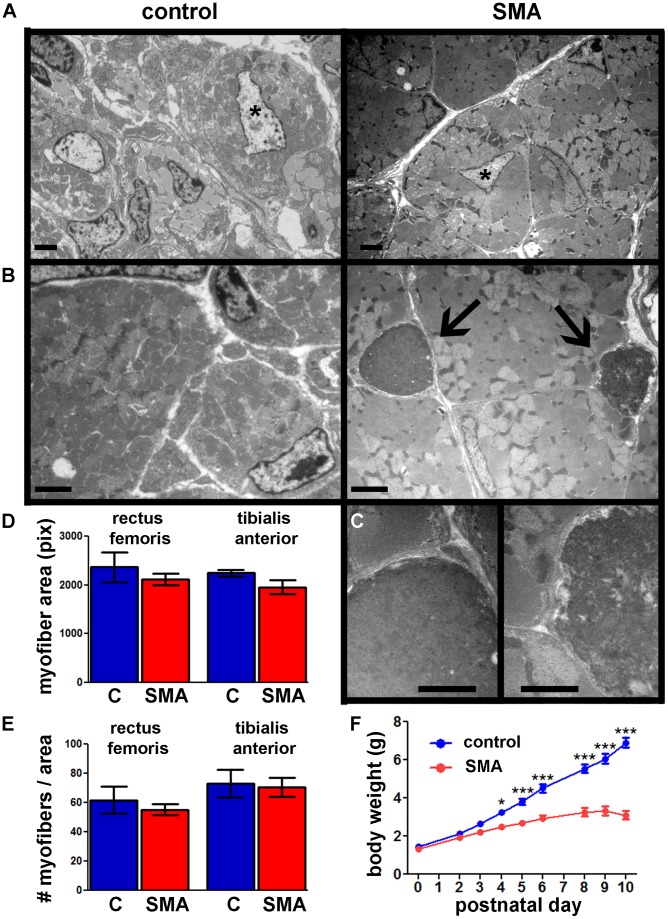
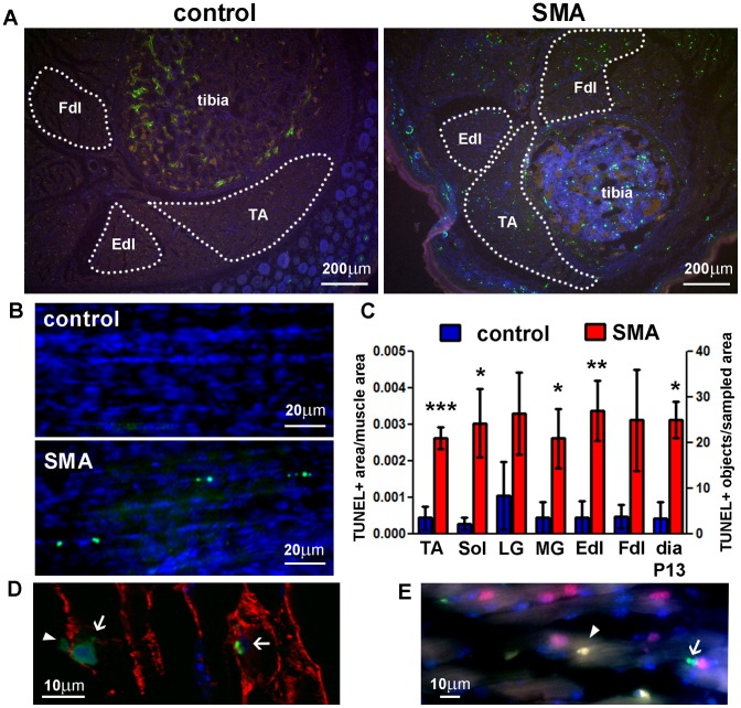
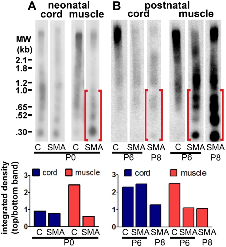
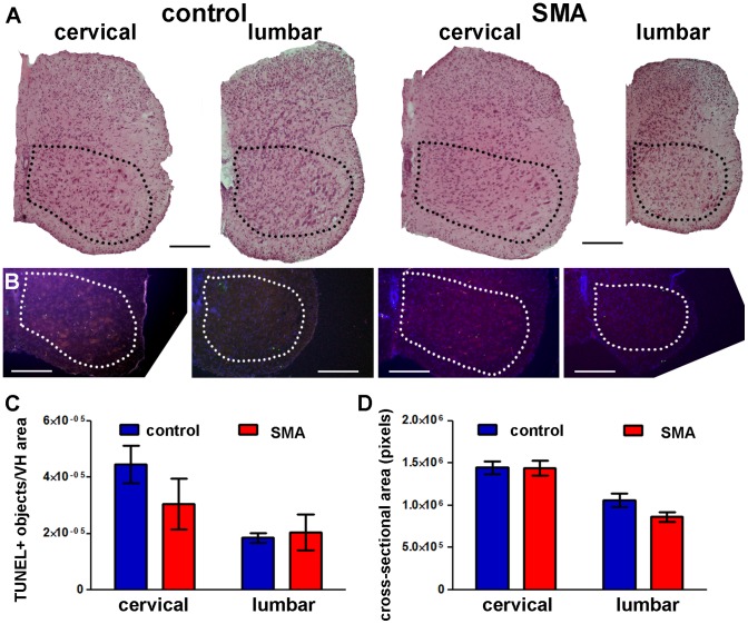
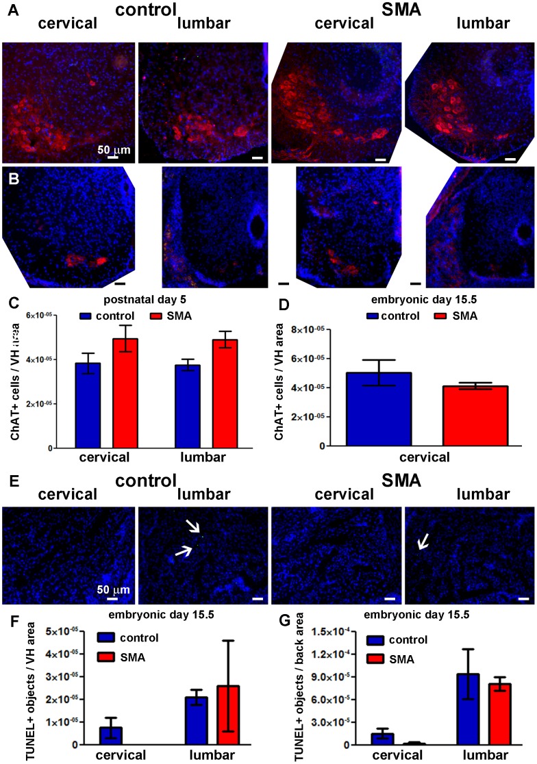
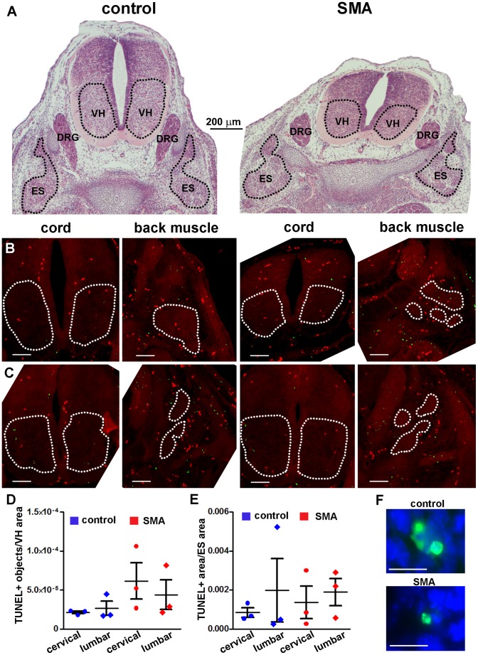
Spinal Muscular Atrophy (SMA) is a hereditary childhood disease that causes paralysis by progressive degeneration of skeletal muscles and spinal motor neurons. SMA is associated with reduced levels of full-length Survival of Motor Neuron (SMN) protein, due to mutations in the Survival of Motor Neuron 1 gene. The mechanisms by which lack of SMN causes SMA pathology are not known, making it very difficult to develop effective therapies. We investigated whether DNA damage is a perinatal pathological event in SMA, and whether DNA damage and cell death first occur in skeletal muscle or spinal cord of SMA mice. We used a mouse model of severe SMA to ascertain the extent of cell death and DNA damage throughout the body of prenatal and newborn mice. SMA mice at birth (postnatal day 0) exhibited internucleosomal fragmentation in genomic DNA from hindlimb skeletal muscle, but not in genomic DNA from spinal cord. SMA mice at postnatal day 5, compared with littermate controls, exhibited increased apoptotic cell death profiles in skeletal muscle, by hematoxylin and eosin, terminal deoxynucleotidyl transferase dUTP nick end labeling, and electron microscopy. SMA mice had no increased cell death, no loss of choline acetyl transferase (ChAT)-positive motor neurons, and no overt pathology in the ventral horn of the spinal cord. At embryonic days 13 and 15.5, SMA mice did not exhibit statistically significant increases in cell death profiles in spinal cord or skeletal muscle. Motor neuron numbers in the ventral horn, as identified by ChAT immunoreactivity, were comparable in SMA mice and control littermates at embryonic day 15.5 and postnatal day 5. These observations demonstrate that in SMA, disease in skeletal muscle emerges before pathology in spinal cord, including loss of motor neurons. Overall, this work identifies DNA damage and cell death in skeletal muscle as therapeutic targets for SMA.


Conflict of interest statement

Competing Interests: The authors have declared that no competing interests exist.

Figures

Figure 1
Figure 1. Skeletal muscle atrophy and apoptosis in SMA mice.
H&E staining was performed on transverse sections of the lower hindlimb at P5 (A), and whole body at P4 (B – lumbar spinal cord and back muscles, C – masseter, D – forelimb muscles, E – lumbar back muscles). Representative skeletal muscle images from control (left column) and SMA mice (right column) are shown at the same scale. Arrows and insets indicate apoptotic profiles (condensed, fragmented nuclei). Dotted lines in A delineate areas quantified. Some muscle groups were not quantified in their entirety because they were not intact or were not identifiable on all sections. TA – tibialis anterior muscle, Fdl/Edl – flexor/extensor digitorum longus muscle, Sol – soleus muscle, LG – gastrocnemius lateralis muscle, MG – gastrocnemius medialis muscle.
Figure 2
Figure 2. SMA mouse skeletal muscles exhibit apoptotic cell death in satellite cells, but normal myofiber size.
A, B, C– transmission electron microscopy images of TA muscles from SMA mice (right) and control littermates (left) at P6. A. Central nuclei (asterisk) are infrequent, but can be seen in both control and SMA muscles. Scale bar = 2 μm. B. Apoptotic satellite cell nuclei (arrows) were observed in SMA muscles, but not control muscles. Adjacent myofibers appear normal. Scale bar = 2 μm. C. Magnified images of the two apoptotic cells shown in the panel above. Cell membranes are distinct from the adjacent myofiber membranes, indicating that these dying cells are satellite cells. The apoptotic profile shown at right is at a more advanced stage of apoptosis compared to the cell at left. Nuclear condensation, membrane involution, and membrane fragmentation can be seen. Scale bar = 1 μm. D, E. Myofiber area (D) and myofiber number per muscle area (E) measurements in TA and RF muscles from SMA mice (red) and control littermates (blue) at P5 (Mean ± SE; n = 3). C = control. There were no statistically significant differences (t-test). F. Body weights of SMA mice and control littermates (Mean ± SE; n = 4–5). Statistical significance between SMA and control (two-way repeated measures ANOVA, Bonferroni test): * p<0.05, ** p<0.01, *** p<0.001.
Figure 3
Figure 3. Cell death in SMA mouse skeletal muscle at postnatal days 5 and 13.
A, B. TUNEL (green) was performed on transverse sections of the lower hindlimb at P5 (A) and whole diaphragms at P13 (B). Hoechst (blue) was used to stain cell nuclei. The red channel was used to exclude autofluorescent cells (e.g. red blood cells) from analysis. Dotted lines delineate the muscle areas quantified. C. TUNEL-positive counts in lower hindlimb skeletal muscles and diaphragm (Mean ± SE; n = 5–6 for hindlimb, n = 2 for diaphragm). TA – tibialis anterior muscle, Fdl/Edl – flexor/extensor digitorum longus muscle, Sol – soleus muscle, LG – gastrocnemius lateralis muscle, MG – gastrocnemius medialis muscle, dia – diaphragm. Statistical significance between SMA and control for each muscle group (one-tailed t-test): * p<0.05, ** p<0.01, *** p<0.001. Micrographs of additional hindlimb muscle groups are shown in Figure S1. TUNEL-positive objects per muscle area are shown for diaphragm; TUNEL-positive area per muscle area is shown for all other muscles. D. Confocal immunofluorescent image of hindlimb skeletal muscle at P5; green – TUNEL, red – laminin (myofiber cell membrane marker), blue – nuclei. E. Conventional immunofluorescent image of hindlimb skeletal muscle at P5; green – TUNEL, red – Pax7 (satellite cell marker), blue – nuclei. Arrows – myotube nuclei, arrowheads – satellite cell nuclei.
Figure 4
Figure 4. Internucleosomal fragmentation of DNA emerges in skeletal muscle before spinal cord in SMA mice.
Whole genomic DNA from spinal cord and hind limb muscle (postnatal days P0–P8) was separated by gel electrophoresis. DNA breaks were end-labeled with DIG-dUTP by TdT and detected using a DIG-based Southern blot. A DIG-conjugated molecular weight ladder was run on the same gel (size marked on the left). Brackets show areas of lower molecular weight DNA in skeletal muscle, indicating DNA fragmentation. Substantial differences in internucleosomal fragmentation of DNA were not observed between control and SMA spinal cord until postnatal day 8. Bar graphs under each blot represent integrated density measurements of the top band in each lane divided by the bottom band in each lane. A. Southern blot of spinal cord and skeletal muscle DNA from SMA and control (“C”) littermates at postnatal day 0. B. Southern blot of spinal cord and skeletal muscle DNA from SMA and control (“C”) littermates at postnatal day 6 and a separate SMA mouse at postnatal day 8.
Figure 5
Figure 5. Cell death in P5 SMA mouse spinal cord is not increased significantly.
A. Representative images of H&E-stained transverse sections at P5 showing cervical and lumbar levels of spinal cord from control (left) and SMA (right) littermates. B. TUNEL (green) was performed on transverse whole body sections at P5. Hoechst (blue) was used to stain cell nuclei. A separate channel (red) was used to identify anatomical landmarks and exclude autofluorescent signal (e.g. red blood cells). A and B: dotted lines delineate the ventral horn (VH) areas analyzed; all scale bars = 200 μm. C. TUNEL-positive counts in the VH of spinal cord at lumbar and cervical levels (Mean ± SE; n = 4–6). D. Cross-sectional area of spinal cord at lumbar and cervical levels (Mean ± SE; n = 3–6). There were no statistically significant differences between SMA and control groups (one-tailed t-test).
Figure 6
Figure 6. No significant motor neuron loss in spinal cord at E15.5 or P5 in SMA mice.
A, B, C. TUNEL (green), ChAT immunostaining (red), and nuclear staining (blue) were performed on transverse whole body sections at E15.5 (B) and isolated spinal cord sections at P5 (A). Representative images of lumbar and cervical spinal cord are shown for the control (left) and SMA (right) mouse groups. Scale bar = 50 μm in all panels. C, D. ChAT-positive motor neuron counts in the VH of spinal cord at lumbar and cervical levels at P5 (C; Mean ± SE; n = 4–5) and cervical level at E15.5 (D; Mean ± SD; n = 2 (SMA), n = 4 (control)). E. TUNEL (green) and nuclear staining (blue) were performed on transverse whole body sections at E15.5. Representative images of lumbar and cervical ES back muscles are shown for the control (left) and SMA (right) littermates. Scale bar = 50 μm in all panels. Arrows denote TUNEL-positive nuclei. F, G. TUNEL-positive structure counts in spinal cord VH (F) and ES muscle (G) at lumbar and cervical levels at E15.5 (Mean ± SD; n = 2 (SMA), n = 4 (control)). There were no statistically significant differences between SMA and control groups (two-tailed t-test).
Figure 7
Figure 7. Cell death in E13 SMA mouse skeletal muscle or spinal cord is not increased significantly.
A. Representative images of H&E-stained transverse sections of the whole body at E13 showing lumbar levels of spinal cord back from control (left) and SMA (right) littermates. Images are shown at the same scale. The areas delineated as ventral horn (VH) of spinal cord and erector spinae muscle group (ES) were analyzed. B, C. TUNEL (green) was performed on transverse whole body sections at E13. A separate channel (red) was used to identify anatomical landmarks and exclude autofluorescent signal (e.g. red blood cells). The total red signal was subtracted from the total green signal to show TUNEL-specific signal only. Representative images of lumbar (B) and cervical (C) spinal cord and back muscles are shown for the control (left) and SMA (right) mouse groups. Scale bar = 100 μm. D. TUNEL-positive structure counts in the VH of spinal cord at lumbar and cervical levels (individual counts and Mean ± SE; n = 3). E. TUNEL-positive structure counts in ES muscle at lumbar and cervical levels (individual counts and Mean ± SE; n = 3). There were no statistically significant differences between SMA and control groups (two-tailed t-test). F. Representative high-magnification images identifying TUNEL-positive structures (green) in E13 control and SMA mouse skeletal muscle as apoptotic nuclei. Hoechst (blue) was used to stain cell nuclei. Scale bar = 10 μm.
Figure 8
Figure 8. Expression of DNA damage signaling genes in P2 SMA skeletal muscle is not increased significantly.
A commercially available gene array was used to assay skeletal (back) muscle from SMA mice and control littermates (n = 3). There were no statistically significant changes in the expression of 84 genes (see Table S1 for a complete list of genes). Each circle represents a single gene assayed. The black line denotes a fold-change of 1. Red lines denote a fold-change of 3.

Similar articles

Cited by

References

    1. Roberts DF, Chavez J, Court SD (1970) The genetic component in child mortality. Arch Dis Child 45: 33–38. - PMC - PubMed
    1. MacLeod MJ, Taylor JE, Lunt PW, Mathew CG, Robb SA (1999) Prenatal onset spinal muscular atrophy. Eur J Paediatr Neurol 3: 65–72. - PubMed
    1. Crawford TO, Pardo CA (1996) The neurobiology of childhood spinal muscular atrophy. Neurobiol Dis 3: 97–110. - PubMed
    1. Thomas NH, Dubowitz V (1994) The natural history of type I (severe) spinal muscular atrophy. Neuromuscul Disord 4: 497–502. - PubMed
    1. Dubowitz V (1964) Infantile muscular atrophy. A prospective study with particular reference to a slowly progressive variety. Brain 87: 707–718. - PubMed

Publication types