

Intrinsic susceptibility MRI identifies tumors with ALKF1174L mutation in genetically-engineered murine models of high-risk neuroblastoma
- PMID: 24667968
- PMCID: PMC3965493
- DOI: 10.1371/journal.pone.0092886
Intrinsic susceptibility MRI identifies tumors with ALKF1174L mutation in genetically-engineered murine models of high-risk neuroblastoma
Abstract
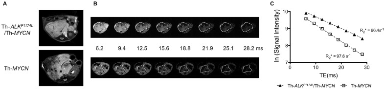
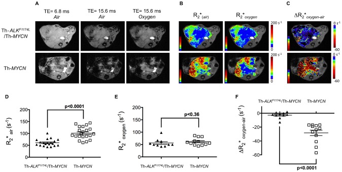
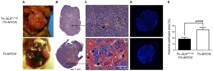
The early identification of children presenting ALK(F1174L)-mutated neuroblastoma, which are associated with resistance to the promising ALK inhibitor crizotinib and a marked poorer prognosis, has become a clinical priority. In comparing the radiology of the novel Th-ALK(F1174L)/Th-MYCN and the well-established Th-MYCN genetically-engineered murine models of neuroblastoma using MRI, we have identified a marked ALK(F1174L)-driven vascular phenotype. We demonstrate that quantitation of the transverse relaxation rate R2* (s(-1)) using intrinsic susceptibility-MRI under baseline conditions and during hyperoxia, can robustly discriminate this differential vascular phenotype, and identify MYCN-driven tumors harboring the ALK(F1174L) mutation with high specificity and selectivity. Intrinsic susceptibility-MRI could thus potentially provide a non-invasive and clinically-exploitable method to help identifying children with MYCN-driven neuroblastoma harboring the ALK(F1174L) mutation at the time of diagnosis.
Conflict of interest statement
Figures
Similar articles
-
The ALK(F1174L) mutation potentiates the oncogenic activity of MYCN in neuroblastoma.Cancer Cell. 2012 Jul 10;22(1):117-30. doi: 10.1016/j.ccr.2012.06.001. Cancer Cell. 2012. PMID: 22789543 Free PMC article.
-
Proliferation and Survival of Embryonic Sympathetic Neuroblasts by MYCN and Activated ALK Signaling.J Neurosci. 2016 Oct 5;36(40):10425-10439. doi: 10.1523/JNEUROSCI.0183-16.2016. J Neurosci. 2016. PMID: 27707976 Free PMC article.
-
Molecular rationale for the use of PI3K/AKT/mTOR pathway inhibitors in combination with crizotinib in ALK-mutated neuroblastoma.Oncotarget. 2014 Sep 30;5(18):8737-49. doi: 10.18632/oncotarget.2372. Oncotarget. 2014. PMID: 25228590 Free PMC article.
-
Neuroblastoma models for insights into tumorigenesis and new therapies.Expert Opin Drug Discov. 2015 Jan;10(1):53-62. doi: 10.1517/17460441.2015.974544. Epub 2014 Oct 25. Expert Opin Drug Discov. 2015. PMID: 25345447 Review.
-
Emerging importance of ALK in neuroblastoma.Semin Cancer Biol. 2011 Oct;21(4):267-75. doi: 10.1016/j.semcancer.2011.09.005. Epub 2011 Sep 16. Semin Cancer Biol. 2011. PMID: 21945349 Free PMC article. Review.
Cited by
-
The BET Protein Inhibitor JQ1 Decreases Hypoxia and Improves the Therapeutic Benefit of Anti-PD-1 in a High-Risk Neuroblastoma Mouse Model.Cells. 2022 Sep 6;11(18):2783. doi: 10.3390/cells11182783. Cells. 2022. PMID: 36139358 Free PMC article.
-
Pre-clinical imaging of transgenic mouse models of neuroblastoma using a dedicated 3-element solenoid coil on a clinical 3T platform.Br J Cancer. 2017 Sep 5;117(6):791-800. doi: 10.1038/bjc.2017.251. Epub 2017 Aug 8. Br J Cancer. 2017. PMID: 28787429 Free PMC article.
-
In vivo quantification of cerebral r2*-response to graded hyperoxia at 3 tesla.J Clin Imaging Sci. 2015 Jan 30;5:1. doi: 10.4103/2156-7514.150439. eCollection 2015. J Clin Imaging Sci. 2015. PMID: 25806136 Free PMC article.
-
Cyclin-Dependent Kinase Inhibitor AT7519 as a Potential Drug for MYCN-Dependent Neuroblastoma.Clin Cancer Res. 2015 Nov 15;21(22):5100-9. doi: 10.1158/1078-0432.CCR-15-0313. Epub 2015 Jul 22. Clin Cancer Res. 2015. PMID: 26202950 Free PMC article.
-
Response of GEM models of neuroblastoma to cabozantinib assessed by multiparametric magnetic resonance imaging.Neoplasia. 2025 Jul;65:101170. doi: 10.1016/j.neo.2025.101170. Epub 2025 May 12. Neoplasia. 2025. PMID: 40359728 Free PMC article.
References
-
- Meitar D, Crawford SE, Rademaker AW, Cohn SL (1996) Tumor angiogenesis correlates with metastatic disease, N-myc amplification, and poor outcome in human neuroblastoma. J Clin Oncol 14: 405–414. - PubMed
-
- De Brouwer S, De Preter K, Kumps C, Zabrocki P, Porcu M, et al. (2010) Meta-analysis of neuroblastomas reveals a skewed ALK mutation spectrum in tumors with MYCN amplification. Clin Cancer Res 16: 4353–4362. - PubMed
Publication types
MeSH terms
Substances
Grants and funding
LinkOut - more resources
Full Text Sources
Other Literature Sources
Medical
Molecular Biology Databases
