Review
. 2014 Apr 16;33(8):823-41.
doi: 10.1002/embj.201387442. Epub 2014 Mar 25.

Role of receptor binding specificity in influenza A virus transmission and pathogenesis

Affiliations
Review

Role of receptor binding specificity in influenza A virus transmission and pathogenesis

Miranda de Graaf et al. EMBO J. .

Erratum in

  • EMBO J. 2014 Jul 17;33(14):1614

Abstract

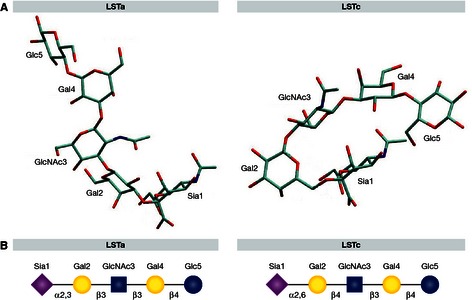
The recent emergence of a novel avian A/H7N9 influenza virus in poultry and humans in China, as well as laboratory studies on adaptation and transmission of avian A/H5N1 influenza viruses, has shed new light on influenza virus adaptation to mammals. One of the biological traits required for animal influenza viruses to cross the species barrier that received considerable attention in animal model studies, in vitro assays, and structural analyses is receptor binding specificity. Sialylated glycans present on the apical surface of host cells can function as receptors for the influenza virus hemagglutinin (HA) protein. Avian and human influenza viruses typically have a different sialic acid (SA)-binding preference and only few amino acid changes in the HA protein can cause a switch from avian to human receptor specificity. Recent experiments using glycan arrays, virus histochemistry, animal models, and structural analyses of HA have added a wealth of knowledge on receptor binding specificity. Here, we review recent data on the interaction between influenza virus HA and SA receptors of the host, and the impact on virus host range, pathogenesis, and transmission. Remaining challenges and future research priorities are also discussed.


Figures

Figure 1
Figure 1. Cartoon representation of the hemagglutinin structure of an H5N1 influenza A virus (protein database code 4KDO, representing a mutant version of A/Vietnam/1203/04)
The human receptor analog, LSTc (pink) is docked into the receptor binding site; the vestigial esterase subdomain and fusion peptide are depicted in yellow and orange, respectively. The amino acid substitutions described by Herfst et al are shown in red, and the mutations of Imai et al are shown in blue. Substitution Gln‐226‐Leu was described by both groups (Herfst et al, 2012; Imai et al, 2012).
Figure 2
Figure 2. N‐glycans, O‐glycans, and glycolipids
Examples of three types of N‐glycans are shown; complex with multiple N‐acetyllactosamine repeats (LN), oligomannose, and hybrid (with the common core boxed). A glycolipid and two types of O‐glycans are also shown; core 1 (in box) and core 2 (in box) O‐glycans. Monosaccharides are depicted using the following symbolic representations: fucose (red triangle), galactose (yellow circle), N‐acetyl glucosamine (blue square), N‐acetyl galactosamine (yellow square), glucosamine (blue circle), mannose (green circle), sialic acid (purple diamond).
Figure 3
Figure 3. Structures of Neu5Aca and Neu5Gc
Figure 4
Figure 4
Overall distribution of α2,6‐sialic acid (SA; red stars) and α2,3‐SA based on Maackia amurensis agglutinin (MAA)‐I (blue stars) and MAAII staining (yellow stars) in the epithelium of the respiratory tract (nasal cavity, trachea, bronchus, bronchiole, and alveoli) of humans (Nicholls et al, 2007a), ferrets (Xu et al, 2010a), pigs (Nelli et al, 2010), and chicken, and the small and large intestine of chickens (Costa et al, 2012). The size of the star shape corresponds with the relative receptor abundance. Receptor distribution may vary between studies, possibly due to the lectins used, experimental methods, or real variation in receptor distribution between animals. Lectin staining was not performed for the human trachea, nasal cavity of pigs and nasal cavity and bronchioles of ferrets in the cited studies. No MAA‐I staining was performed for the chicken tissues. Figure based on Herfst et al, 2012.
Figure 5
Figure 5
Cartoon representation of a human influenza hemagglutinin (database code 2YP4) receptor binding site with the 130‐loop, 190‐helix, and 220‐loop and the conserved residues, 98‐Tyr, 153‐Trp, 183‐His, and 195‐Tyr in complex with the human receptor analog LSTc in cis conformation (A). Amino acid alignment of the 130‐loop, 190‐helix, and 220‐loop of human (Hu), avian (Av), pig (Pi) influenza viruses, and an airborne transmissible H5N1 virus (Tr) (B). The sequences are derived from the following virus strains: A/Teal/NL/10/2000 (CY060178), A/Memphis/7/2001 (CY020149), A/SC/1/1918 (F116575), A/Mallard/SW/50/2000 (CY060308), A/HK/01/1968 (CY112249), A/Mallard/NL/5/1999 (CY064950), A/NL/056H1/1960 (CY077786), A/Indo/5/2005 (CY116646), airborne transmissible A/Indo/05/2005 (CY116686), A/Mallard/12/2000 (GU053030), A/NL/219/2003 (AY338459) and A/Shanghai/02/2013 (KF021597).
Figure 6
Figure 6. Human and avian receptor analogs in free conformation (Figure adapted from Xu et al, 2009)
Avian receptor analog LSTa is shown in trans conformation and the human receptor analog LSTc in cis conformation (A). Symbolic representation of the LSTa and LSTc structures (B), with the monosaccharides sialic acid (purple diamond), galactose (yellow circle), N‐acetylglucosamine (blue square) and glucose (blue circle).

References

    1. Ablan S, Rawat SS, Blumenthal R, Puri A (2001) Entry of influenza virus into a glycosphingolipid‐deficient mouse skin fibroblast cell line. Arch Virol 146: 2227–2238 - PubMed
    1. Aich U, Beckley N, Shriver Z, Raman R, Viswanathan K, Hobbie S, Sasisekharan R (2011) Glycomics‐based analysis of chicken red blood cells provides insight into the selectivity of the viral agglutination assay. FEBS J 278: 1699–1712 - PMC - PubMed
    1. Almond JW (1977) A single gene determines the host range of influenza virus. Nature 270: 617–618 - PubMed
    1. Bateman AC, Karamanska R, Busch MG, Dell A, Olsen CW, Haslam SM (2010) Glycan analysis and influenza A virus infection of primary swine respiratory epithelial cells: the importance of NeuAc{alpha}2‐6 glycans. J Biol Chem 285: 34016–34026 - PMC - PubMed
    1. Baum LG, Paulson JC (1991) The N2 neuraminidase of human influenza virus has acquired a substrate specificity complementary to the hemagglutinin receptor specificity. Virology 180: 10–15 - PubMed

Publication types