. 2014 Sep;60(3):1044-53.
doi: 10.1002/hep.27137. Epub 2014 May 9.

Alcohol dehydrogenase III exacerbates liver fibrosis by enhancing stellate cell activation and suppressing natural killer cells in mice

Affiliations

Alcohol dehydrogenase III exacerbates liver fibrosis by enhancing stellate cell activation and suppressing natural killer cells in mice

Hyon-Seung Yi et al. Hepatology. 2014 Sep.

Abstract

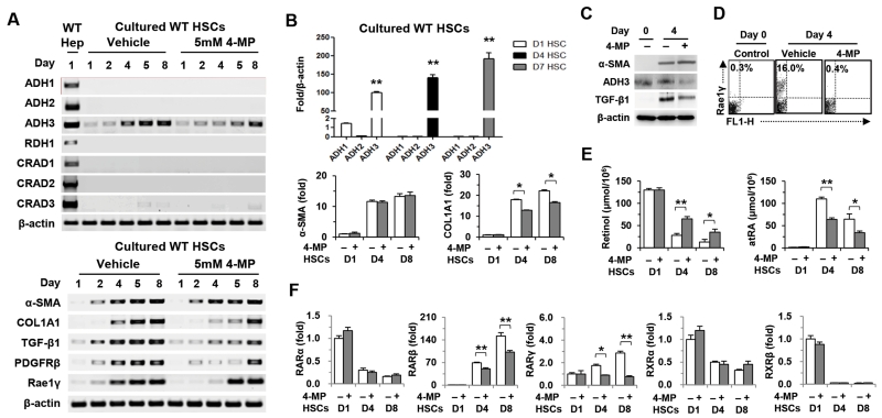
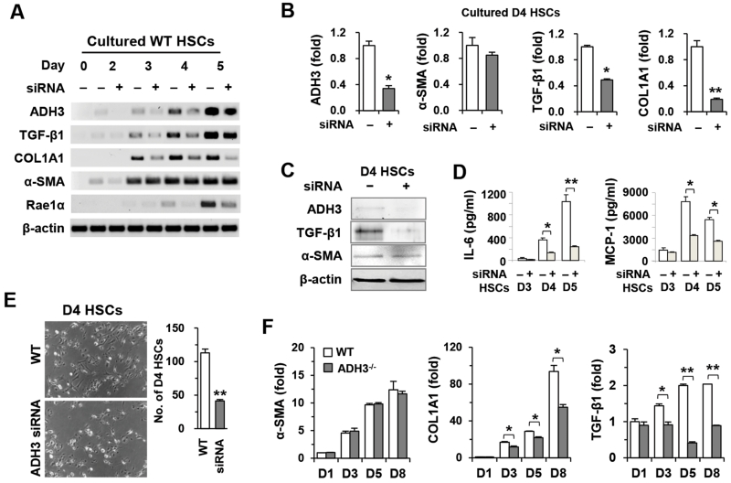
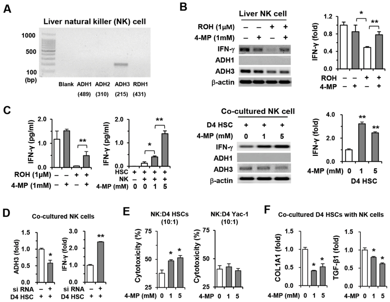
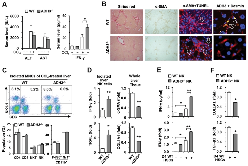
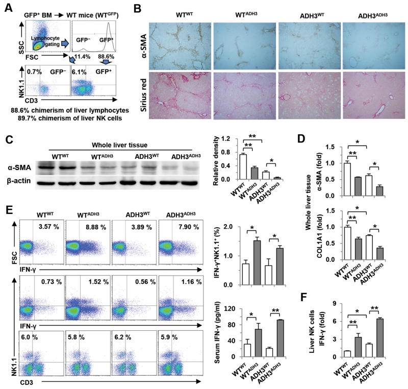
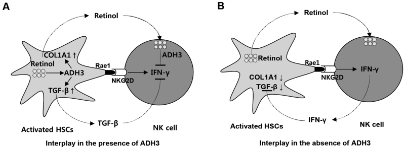
The important roles of retinols and their metabolites have recently been emphasized in the interactions between hepatic stellate cells (HSCs) and natural killer (NK) cells. Nevertheless, the expression and role of retinol metabolizing enzyme in both cell types have yet to be clarified. Thus, we investigated the expression of retinol metabolizing enzyme and its role in liver fibrosis. Among several retinol metabolizing enzymes, only alcohol dehydrogenase (ADH) 3 expression was detected in isolated HSCs and NK cells, whereas hepatocytes express all of them. In vitro treatment with 4-methylpyrazole (4-MP), a broad ADH inhibitor, or depletion of the ADH3 gene down-regulated collagen and transforming growth factor-β1 (TGF-β1) gene expression, but did not affect α-smooth muscle actin gene expression in cultured HSCs. Additionally, in vitro, treatments with retinol suppressed NK cell activities, whereas inhibition of ADH3 enhanced interferon-γ (IFN-γ) production and cytotoxicity of NK cells against HSCs. In vivo, genetic depletion of the ADH3 gene ameliorated bile duct ligation- and carbon tetrachloride-induced liver fibrosis, in which a higher number of apoptotic HSCs and an enhanced activation of NK cells were detected. Freshly isolated HSCs from ADH3-deficient mice showed reduced expression of collagen and TGF-β1, but enhanced expression of IFN-γ was detected in NK cells from these mice compared with those of control mice. Using reciprocal bone marrow transplantation of wild-type and ADH3-deficient mice, we demonstrated that ADH3 deficiency in both HSCs and NK cells contributed to the suppressed liver fibrosis.

Conclusion: ADH3 plays important roles in promoting liver fibrosis by enhancing HSC activation and inhibiting NK cell activity, and could be used as a potential therapeutic target for the treatment of liver fibrosis.


Figures

Fig. 1
Fig. 1
4-MP treatment inhibits activation of HSCs. (A) RT-PCR analyses of wild type (WT) hepatocytes and WT HSCs treated with 4-MP. (B) Real-time PCR analyses of ADH3, α-SMA and COL1A1 in WT HSCs. (C) Western blotting for α-SMA, ADH3 and TGF-β1 in cultured HSCs. (D) Flow cytometry for Rae1 expression in cultured HSCs. (E) Intracellular concentration of retinol and atRA from cultured HSCs. (F) Real-time PCR analyses on retinoic acid receptors (RARs) and retinoic X receptors (RXRs) of 4-MP-treated WT HSCs. Data are expressed as the mean ± SEM. *P < 0.05, **P < 0.01 in comparison with the corresponding controls.
Fig. 2
Fig. 2
Suppression of the ADH3 gene inhibits the activation and proliferation of HSCs. WT and ADH3−/− HSCs were isolated and cultured. (A,B) RT- and real-time PCR analyses of HSCs after the addition of ADH3 siRNA. (C) Western blot analyses of 4-day cultured HSCs (D4 HSCs) after ADH3 siRNA. (D) Supernatant levels of IL-6 and MCP-1 after ADH3 siRNA in HSCs. (E) Cultured D4 HSCs of WT mice with ADH3 siRNA and their numbers (original magnification ×100). (F) Cultured HSCs of WT and ADH3−/− mice were subjected to real-time PCR analyses. Data are expressed as the mean ± SEM. *P < 0.05, **P < 0.01 in comparison with the corresponding controls.
Fig. 3
Fig. 3
Retinol suppresses IFN-γ production and cytotoxicity of NK cells against HSCs through ADH3. Freshly isolated NK cells from WT mice were used for in vitro experiments. (A) Expression of retinol metabolizing enzymes was analyzed using liver NK cells. (B) NK cells were treated with retinol or co-cultured with D4 HSCs for 3 hours in the presence or absence of 4-MP and then subjected to RT- and real-time PCR analyses. (C) IFN-γ levels of supernatants were measured. (D) Liver NK cells treated with ADH3 siRNA or scramble siRNA were co-cultured with D4 HSCs and subjected to real-time PCR analyses. (E) Liver NK cells were used as effector cells for a cytotoxicity assay against D4 HSCs and Yac-1 cells in the presence or absence of 4-MP. (F) Co-cultured D4 HSCs with NK cells were subjected to real-time PCR analyses. Data are expressed as the mean ± SEM. *P < 0.05, **P < 0.01 in comparison with the corresponding controls.
Fig. 4
Fig. 4
Ablation of the ADH3 gene inhibits liver fibrosis by up-regulation of IFN-γ production of NK cells and down-regulation of the activation of HSCs. WT and AD3−/− mice were injected with CCl4 for 2 weeks. (A) Levels of ALT, AST and IFN-γ were measured in collected sera. (B) Liver sections were stained with Sirius red and α-SMA antibody (original magnification, ×100), co-stained with α-SMA antibody (red) and TUNEL (green) or with ADH3 (green) and desmin (red) (original magnification, ×800, ×1200). Nuclei were stained with DAPI (blue) (C) Liver MNCs were subjected to flow cytometry. (D) Isolated liver NK cells and whole liver tissues were subjected to real-time PCR analyses. (E) IFN-γ production and its expression were analyzed by co-culturing NK cells of WT and ADH3−/− mice with D4 WT HSCs. (F) Co-cultured D4 WT HSCs with NK cells of WT and ADH3−/− mice were subjected to real-time PCR analyses. Data are expressed as the mean ± SEM. *P < 0.05, **P < 0.01 in comparison with the corresponding controls.
Fig. 5
Fig. 5
ADH3-deficient bone marrow transplantation ameliorates liver fibrosis. (A) At week 8 after the transplantation of GFP-producing WT bone marrow into WT mice. Chimerism of NK cells was assessed by flow cytometry. (B) After reciprocal bone marrow transplantation between WT and ADH3-deficient mice, mice were injected with CCl4 for 2 weeks. Liver sections were stained with α-SMA antibody and Sirius red (original magnification, ×100). (C, D) Liver tissues were removed and used for Western blotting and real-time PCR analyses. (E) IFN-γ production of liver NK cells was analyzed by flow cytometry, and serum levels of IFN-γ were measured. (F) IFN-γ expression in isolated liver NK cells was analyzed by real-time PCR analyses. Data are expressed as the mean ± SEM. *P < 0.05, **P < 0.01 in comparison with the corresponding controls.
Fig. 6
Fig. 6
Cell type-specific roles of ADH3 in liver fibrosis. During liver injury, retinols are metabolized by ADH3 in HSCs and NK cells, which affects the interaction of HSCs and NK cells in 2 ways. (A) In the presence of ADH3 in HSCs and NK cells, ADH3 induces activation of HSCs by expressing COL1A1 or activating latent TGF-β. In the meantime, Rae1 expression in activated HSCs recruits liver NK cells, which is followed by the suppression of IFN-γ production of NK cells by activated TGF-β. In addition, released retinols from activated HSCs are metabolized by the ADH3 of NK cells, resulting in the inhibition of IFN-γ production in NK cells. (B) In the absence of ADH3 in HSCs and NK cells, the expression of COL1A1 and TGF-β was significantly reduced in HSCs, whereas activated NK cells by HSCs produce more IFN-γ, resulting in the inhibition of HSC activation. Therefore, ADH3 is a positive regulator in the activation of HSCs but a negative regulator in IFN-γ production of NK cells.

References

    1. Friedman SL. Hepatic stellate cells: protean, multifunctional, and enigmatic cells of the liver. Physiol Rev. 2008;88:125–172. - PMC - PubMed
    1. Radaeva S, Wang L, Radaev S, Jeong WI, Park O, Gao B. Retinoic acid signaling sensitizes hepatic stellate cells to NK cell killing via upregulation of NK cell activating ligand RAE1. Am J Physiol Gastrointest Liver Physiol. 2007;293:G809–816. - PubMed
    1. Pares X, Farres J, Kedishvili N, Duester G. Medium- and short-chain dehydrogenase/reductase gene and protein families : Medium-chain and short-chain dehydrogenases/reductases in retinoid metabolism. Cell Mol Life Sci. 2008;65:3936–3949. - PMC - PubMed
    1. Duester G, Mic FA, Molotkov A. Cytosolic retinoid dehydrogenases govern ubiquitous metabolism of retinol to retinaldehyde followed by tissue-specific metabolism to retinoic acid. Chem Biol Interact. 2003;143-144:201–210. - PubMed
    1. Crabb DW, Matsumoto M, Chang D, You M. Overview of the role of alcohol dehydrogenase and aldehyde dehydrogenase and their variants in the genesis of alcohol-related pathology. Proc Nutr Soc. 2004;63:49–63. - PubMed

Publication types

Substances