. 2014 May 2;289(18):12823-34.
doi: 10.1074/jbc.M114.565473. Epub 2014 Mar 25.

A microRNA processing defect in smokers' macrophages is linked to SUMOylation of the endonuclease DICER

Affiliations

A microRNA processing defect in smokers' macrophages is linked to SUMOylation of the endonuclease DICER

Thomas J Gross et al. J Biol Chem. .

Abstract

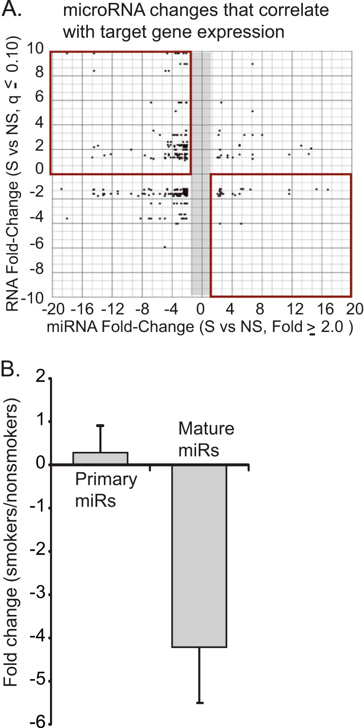
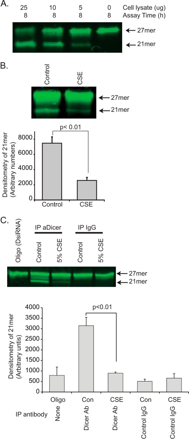
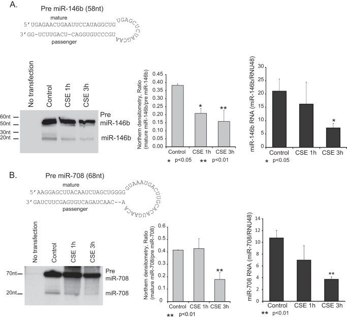
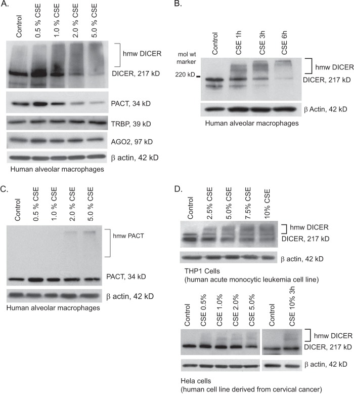
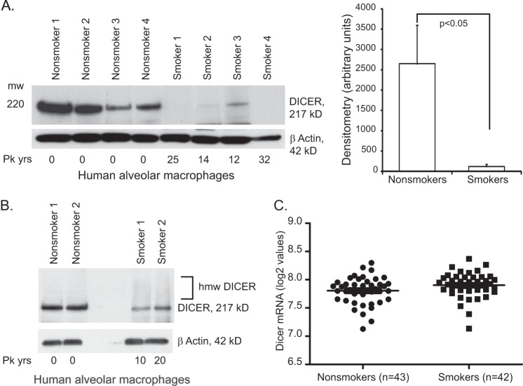
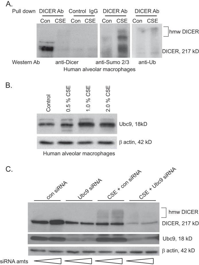
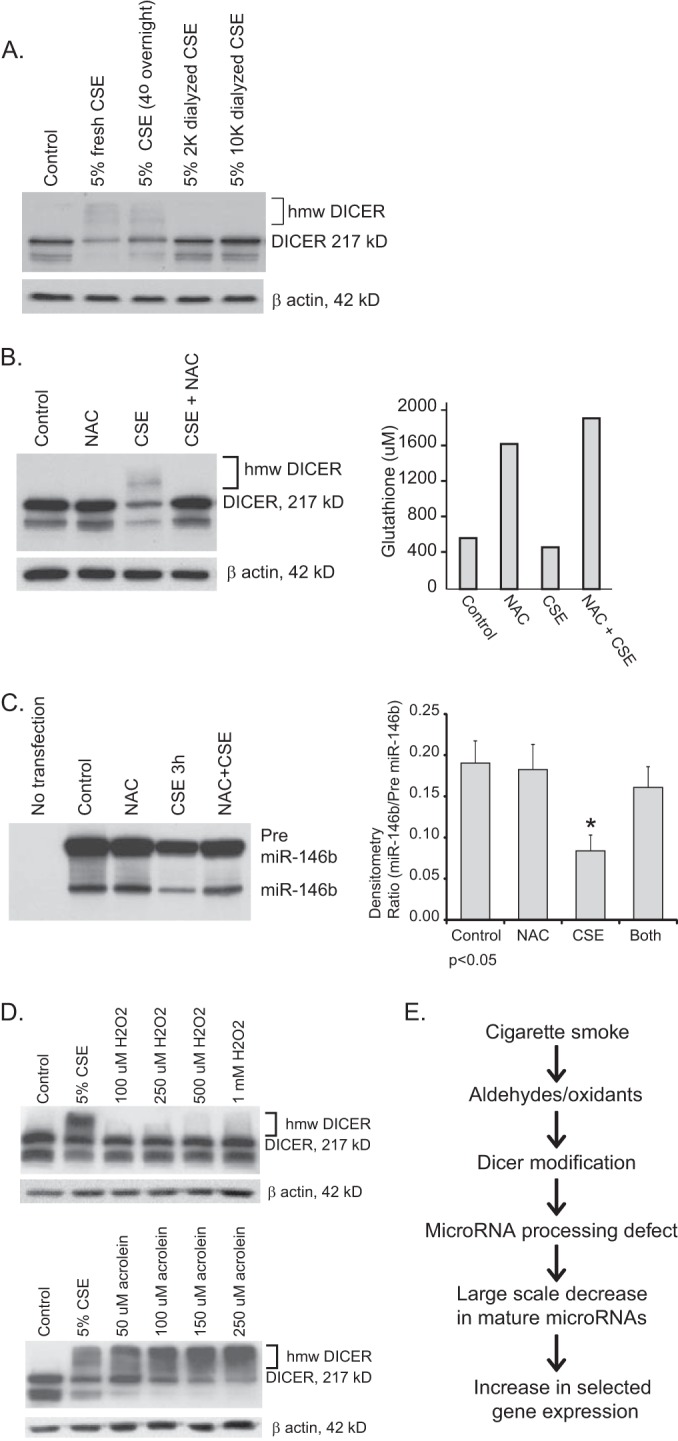
Despite the fact that alveolar macrophages play an important role in smoking-related disease, little is known about what regulates their pathophysiologic phenotype. Evaluating smoker macrophages, we found significant down-regulation of multiple microRNAs (miRNAs). This work investigates the hypothesis that cigarette smoke alters mature miRNA expression in lung macrophages by inhibiting processing of primary miRNA transcripts. Studies on smoker alveolar macrophages showed a defect in miRNA maturation. Studies on the miRNA biogenesis machinery led us to focus on the cytosolic RNA endonuclease, DICER. DICER cleaves the stem-loop structure from pre-miRNAs, allowing them to dissociate into their mature 20-22-nucleotide single-stranded form. DICER activity assays confirmed impaired DICER activity following cigarette smoke exposure. Further protein studies demonstrated a decreased expression of the native 217-kDa form of DICER and an accumulation of high molecular weight forms with cigarette smoke exposure. This molecular mass shift was shown to contain SUMO moieties and could be blocked by silencing RNA directed at the primary SUMOylating ligase, Ubc9. In determining the cigarette smoke components responsible for changes in DICER, we found that N-acetylcysteine, an antioxidant and anti-aldehyde, protected DICER protein and activity from cigarette smoke extract. This massive down-regulation of miRNAs (driven in part by alterations in DICER) may be an important regulator of the disease-promoting macrophage phenotype found in the lungs of smokers.

Keywords: Chronic Obstructive Pulmonary Disease (COPD); Cigarette Smoke; DICER; Epigenetics; Macrophage; MicroRNA (miRNA); SUMOylation; Small Ubiquitin-like Modifier (SUMO).


Figures

FIGURE 1.
FIGURE 1.
In vivo cigarette smoke exposure alters miRNA profiles in alveolar macrophages. A, differentially expressed miRNAs in smokers' alveolar macrophages are down-regulated and link to up-regulated mRNAs. Alveolar macrophages were isolated from four active smokers (>10 pack years) and four never smokers. An analysis of mRNA expression was performed using Human Exon Array 1.0 ST arrays from Affymetrix. miRNA expression was analyzed using TaqMan Low Density Arrays version 2.0 (ABI). Altered miRNAs (>2-fold change) were analyzed for potential mRNA targets using TargetScan and TarBase. The miRNA target information was compared with significantly altered mRNAs (q ≤ 0.10). The q value (false discovery rate) for the mRNAs was calculated using the Partek GS implementation of the step-up method. Gene IDs common to both lists (miRNA targets and mRNAs) were plotted. Each dot represents a gene/miRNA pair as predicted by Target Scan and/or TarBase. The top right quadrant is of particular interest because it identifies down-regulated miRNAs that link to up-regulated mRNAs. B, miRNAs that are down-regulated in smokers' alveolar macrophages have intact transcriptional profiles. A panel of down-regulated miRNAs (miR-708, miR-200a, miR-210, miR-187, miR-149, miR-429, miR-146b-3p, and miR-200c) was tested in RNA isolated from smoker and never smoker alveolar macrophages. Each miRNA was tested using primers specific for the mature miRNA and primers specific for the primary miRNA transcript (the stem loop structure with accompanying 5′- and 3′-ends). The data are a composite of results from four smokers and four never smokers with significance determined by Student's t test. Error bars, S.E.
FIGURE 2.
FIGURE 2.
Cigarette smoke exposure decreases dsRNA endonuclease activity. To test the effect of cigarette smoke on DICER activity, we used an in vitro assay of endonuclease cleavage of a fluorescent 27-mer target. A, to validate the assay, varying amounts of whole cell lysate protein (THP-1 cells) were incubated with the FITC-27-mer. Shown is a gel demonstrating a dose-dependent cleavage of the 27-mer target dsRNA. B, whole cell lysates from control THP-1 cells and CSE-exposed THP-1 cells (5% CSE for 6 h) were incubated with the FITC-27-mer. Shown is a fluorescent image of the DNA gel and densitometry from three separate experiments. Significance was determined using Student's t test. Error bars, S.E. C, DICER was immunoprecipitated (IP) from 500 μg of THP-1 protein (control and CSE-exposed, 5% for 6 h). 20% of the resulting protein-coated beads (pulled down with an anti-DICER antibody or a nonspecific IgG) were mixed with the FITC-27-mer to assay for DICER-specific ribonuclease activity. Shown is the fluorescent image of a DNA gel showing both the original 27-mer and the cleaved 21-mer product. Densitometry is from three separate experiments.
FIGURE 3.
FIGURE 3.
Cigarette smoke exposure decreases pre-miRNA processing activity. Two highly CSE regulated miRNAs were chosen for an in vivo pre-miRNA processing assay. A and B, transfection with either pre-miR-146b (A) or pre-miR-708 (B) and processing of the respective miRNAs were evaluated by Northern blot and qRT-PCR. HeLa cells were transfected with a plasmid expressing pre-miRNAs under control of the constitutive H1 promoter. 24 h after transfection, cells were exposed to 1 or 3 h of CSE, and RNA was isolated. Northern analysis was performed to demonstrate the relative degree of pre-miRNA processing. qRT-PCR to compare pre-miRNA to mature miRNA levels was also performed. All assays were done in triplicate, and statistical analysis for both the densitometry data and qRT-PCR was performed (Student's t test). Error bars, S.E.
FIGURE 4.
FIGURE 4.
DICER protein is modified by cigarette smoke exposure. A, human alveolar macrophages freshly isolated from nonsmokers were exposed to varying concentrations of CSE for 6 h (0.5–5.0%). Whole cell lysates were obtained, and Western analysis was performed for DICER, PACT, TRBP, and AGO2. Equal loading of the gel was determined by staining for β-actin. B, normal human alveolar macrophages were exposed to CSE (2%) for varying times (1–6 h). Whole cell lysates were obtained, and Western analysis performed for DICER. C, normal human alveolar macrophages were exposed to CSE at varying concentrations, and whole cell lysates were run on an SDS-7.5% polyacrylamide gel to optimize transfer of high molecular weight (hmw) proteins. PACT protein levels were determined by Western analysis. D, THP-1 cells were exposed to varying concentrations of CSE for 6 h; HeLa cells were exposed to CSE for 3 and 6 h. Whole cell lysates were obtained, and Western analysis was performed for DICER.
FIGURE 5.
FIGURE 5.
Smoking alters alveolar macrophage DICER protein and not DICER mRNA. A, human alveolar macrophages were obtained from active smokers and never smokers. Whole cell lysates were obtained immediately (baseline sample), and Western analysis was performed for DICER. Shown on the right is densitometry. Significance was determined using Student's t test. B, human alveolar macrophages were obtained from active smokers and never smokers. Fresh whole cell lysates were immediately analyzed by Western blotting and probed for DICER. The gel was optimized to maximize visualization of the high molecular weight bands (5% gel with a long transfer). C, total RNA was obtained from freshly isolated human alveolar macrophages (43 nonsmoker subjects and 42 smoker subjects). The mRNA levels for DICER were analyzed using an Affymetrix array (Gene Chip Human Exon Array 1.0 ST arrays). Shown are the log 2 values for DICER, as determined by Partek analysis of the raw data. There is no difference in DICER mRNA levels between smokers and nonsmokers. Error bars, S.E.
FIGURE 6.
FIGURE 6.
Cigarette smoke exposure increases conjugation of SUMO chains to the protein DICER. A, alveolar macrophages from nonsmokers were exposed to 2% CSE in vitro for 6 h. Whole cell lysates were isolated, and 500 μg of protein was used for immunoprecipitation using an anti-DICER antibody. The resulting samples were divided into thirds, and Western analysis was performed for DICER, SUMO 2/3, and ubiquitin. As a control, 500 μg of whole cell lysate protein was also subjected to immunoprecipitation with a nonspecific IgG. B, human alveolar macrophages were exposed to varying concentrations of CSE. Western analysis was performed for Ubc9. C, HeLa cells were incubated with Ubc9-specific siRNA or a control siRNA. The cells were then exposed to 5% CSE for 6 h, and whole cell protein was obtained. Western analysis was performed for DICER, Ubc9 (verifying knockdown), and β-actin.
FIGURE 7.
FIGURE 7.
DICER modification by cigarette smoke is inhibited by N-acetylcysteine. A, to explore the chemical properties of the DICER-active component of cigarette smoke, CSE was dialyzed overnight in tubing with cut-offs of 2 or 10 kDa. THP-1 cells were then exposed to freshly prepared CSE, CSE that sat in the refrigerator overnight (control for dialysis), or the dialyzed CSE for 6 h. Western analysis for DICER was performed on whole cell lysates. B, THP-1 cells were exposed to CSE alone (2%) or CSE in the presence of NAC (2 mm) for 6 h. Duplicate assays were set up. One set of cells was utilized for glutathione assays, and the other set of cells were lysed, and whole cell protein was obtained for Western analysis for DICER. C, HeLa cells were transfected with a plasmid that expresses pre-miRNA-146b under control of the constitutive H1 promoter. 24 h following transfection, cells were exposed to CSE (10%) with and without added NAC (2 mm) for 3 h. RNA was isolated, and Northern analysis for miRNA-146b was performed. Shown is a representative blot and densitometry from three separate experiments. Error bars, S.E. D, THP-1 cells were exposed to varying concentrations of H2O2 or acrolein for 6 h. Whole cell lysates were obtained, and Western analysis for DICER was performed. E, this diagram represents an overview of the conclusions supported by the study data.

References

    1. Martin T. R., Raghu G., Maunder R. J., Springmeyer S. C. (1985) The effects of chronic bronchitis and chronic air-flow obstruction on lung cell populations recovered by bronchoalveolar lavage. Am. Rev. Respir. Dis. 132, 254–260 - PubMed
    1. Janoff A., Scherer J. (1968) Mediators of inflammation in leukocyte lysosomes. IX. Elastinolytic activity in granules of human polymorphonuclear leukocytes. J. Exp. Med. 128, 1137–1155 - PMC - PubMed
    1. Shapiro S. D., Ingenito E. P. (2005) The pathogenesis of chronic obstructive pulmonary disease: advances in the past 100 years. Am. J. Respir. Cell Mol. Biol. 32, 367–372 - PubMed
    1. Tetley T. D. (2002) Macrophages and the pathogenesis of COPD. Chest 121, 156S–159S - PubMed
    1. Shaykhiev R., Krause A., Salit J., Strulovici-Barel Y., Harvey B. G., O'Connor T. P., Crystal R. G. (2009) Smoking-dependent reprogramming of alveolar macrophage polarization: implication for pathogenesis of chronic obstructive pulmonary disease. J. Immunol. 183, 2867–2883 - PMC - PubMed

Publication types

MeSH terms