. 2014 May 9;289(19):13615-26.
doi: 10.1074/jbc.M114.550418. Epub 2014 Mar 25.

Z-disc-associated, alternatively spliced, PDZ motif-containing protein (ZASP) mutations in the actin-binding domain cause disruption of skeletal muscle actin filaments in myofibrillar myopathy

Affiliations

Z-disc-associated, alternatively spliced, PDZ motif-containing protein (ZASP) mutations in the actin-binding domain cause disruption of skeletal muscle actin filaments in myofibrillar myopathy

Xiaoyan Lin et al. J Biol Chem. .

Abstract

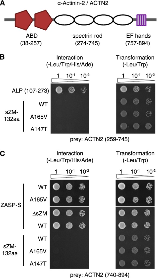
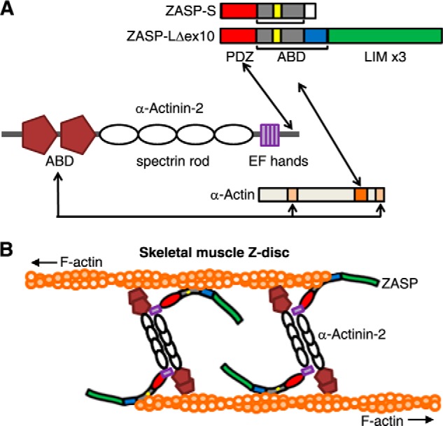
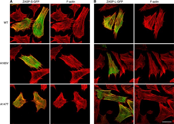
The core of skeletal muscle Z-discs consists of actin filaments from adjacent sarcomeres that are cross-linked by α-actinin homodimers. Z-disc-associated, alternatively spliced, PDZ motif-containing protein (ZASP)/Cypher interacts with α-actinin, myotilin, and other Z-disc proteins via the PDZ domain. However, these interactions are not sufficient to maintain the Z-disc structure. We show that ZASP directly interacts with skeletal actin filaments. The actin-binding domain is between the modular PDZ and LIM domains. This ZASP region is alternatively spliced so that each isoform has unique actin-binding domains. All ZASP isoforms contain the exon 6-encoded ZASP-like motif that is mutated in zaspopathy, a myofibrillar myopathy (MFM), whereas the exon 8-11 junction-encoded peptide is exclusive to the postnatal long ZASP isoform (ZASP-LΔex10). MFM is characterized by disruption of skeletal muscle Z-discs and accumulation of myofibrillar degradation products. Wild-type and mutant ZASP interact with α-actin, α-actinin, and myotilin. Expression of mutant, but not wild-type, ZASP leads to Z-disc disruption and F-actin accumulation in mouse skeletal muscle, as in MFM. Mutations in the actin-binding domain of ZASP-LΔex10, but not other isoforms, cause disruption of the actin cytoskeleton in muscle cells. These isoform-specific mutation effects highlight the essential role of the ZASP-LΔex10 isoform in F-actin organization. Our results show that MFM-associated ZASP mutations in the actin-binding domain have deleterious effects on the core structure of the Z-discs in skeletal muscle.

Keywords: Actin; Actinin; Muscular Dystrophy; Myofibrillar Myopathy; Myotilin; Protein Complex; Protein-Protein Interaction; Skeletal Muscle; Z-disc; ZASP.


Figures

FIGURE 1.
FIGURE 1.
Characterization of ZASP-skeletal muscle α-actin 1 (ACTA1) interaction in yeast cells. Representative images of Y2H pairwise assays demonstrating the interaction of different regions of ZASP and ACTA1 are shown. Positive interactions were determined by yeast growth on the media deficient in HIS3 and ADE2. The transformation efficiency was uniform for all constructs (right panels). Sequential 10-fold yeast dilutions are shown. Inset, the human LDB3/ZASP genomic structure and the three skeletal muscle-specific ZASP splice isoforms: the pre-natal long isoform (L), the post-natal long isoform (LΔex10), and the short isoform (S). The known domains of ZASP are highlighted. The location of ZASP A147T and A165V mutations in the sZM domain is indicated. The peptide sZM-132aa, wild-type or A165V, was used as bait in aY2H screen. A, Y2H pairwise assays show interactions between the indicated ZASP regions and the peptide encoding amino acids 247–377 in the C terminus of ACTA1. WT and mutant (A165V and A147T) sZM-132aa interact with the ACTA1 peptide. ZASP exon 6-encoded peptide, which contains the sZM domain, interacts with the C terminus of ACTA1. Yeast cells cotransformed with empty bait vector and the ACTA1 peptide prey show no growth on the interaction media (−). ZASP-S interacts with the ACTA1 peptide, and deletion of the sZM domain (ΔsZM) abolishes the interaction. The ZASP peptide encoded by exons 8–11Δex10, corresponding to ZASP-LΔex10, interacts with the ACTA1 peptide, and inclusion of exon 10, corresponding to ZASP-L, abolishes the interaction of this ZASP region with the C terminus of ACTA1. B and C, Y2H pairwise assays show the interaction between the indicated ACTA1 regions and ZASP sZM-132aa (B) and exon 8–11Δex10 (C) baits. The ZASP baits interact with amino acids 247–377 of ACTA1, corresponding to the clones identified in our Y2H screen. ACTA1 peptides encoding overlapping amino acid residues within the C terminus were tested against the ZASP baits. Deletion of ACTA1 amino acids 287–303 and 304–325 abolished the interaction with ZASP peptides encoded by exons 8–11Δex10 and the sZM-132aa (wild-type and A165V and A147T mutants), respectively.
FIGURE 2.
FIGURE 2.
Characterization of ZASP-skeletal muscle α-actin 1 (ACTA1) interaction in mammalian cells and in vitro with purified proteins. A, co-IP (IP) assay showing the interaction between the ZASP isoforms and ACTA1 in wild-type mouse vastus muscle lysates. B and C, co-IP assays showing the interaction between the indicated ZASP isoforms and ACTA1 in non-muscle (COS7/HEK293) cells. The proteins were detected with anti-FLAG and anti-HA antibodies. D, purified GST-tagged ZASP-LΔex10 (WT, A165V, and A147T), but not GST alone, binds to biotinylated G-actin (G-actin) in a slot blot overlay assay. Biotin-G-actin was detected with HRP-streptavidin. GST and GST-ZASP were detected by immunoblotting with GST antibody (anti-GST). Arp2/3 and BSA served as positive and negative controls for G-actin binding. E, high-speed cosedimentation assay of purified GST or the indicated GST-ZASP-LΔex10 proteins and F-actin. Representative colloidal blue-stained gel images demonstrate a shift of the indicated GST-ZASP proteins from the supernatant (S) to the pellet (P) fraction in the presence of F-actin, whereas GST remained in the supernatant. Nonlinear regression analysis of the ZASP-actin interaction is shown in the right panel (triplicate assays, data represent mean ± S.D.).
FIGURE 3.
FIGURE 3.
Characterization of the interaction between ZASP and α-actinin-2 (ACTN2) in yeast cells. A, diagram of the ACTN2 domain structure. ACTN2 has two N-terminal actin-binding domains (ABD), an internal spectrin rod domain, and the C-terminal EF-hands. ACTN2 amino acids encoding these domains are numbered as in the human sequence and shown in parentheses below each domain. B, Y2H pairwise assays show that the internal ZM region of ALP (107–273) interacts with the spectrin rod domain of ACTN2. In the same assay, WT and mutant (A165V and A147T) sZM-132aa do not interact with the spectrin rod domain of ACTN2. C, Y2H pairwise assays show that ZASP-S (WT and A165V) interacts with the C terminus of ACTN2. This interaction persists after deletion of the sZM domain (ΔsZM). Neither the wild-type nor mutant ZASP-sZM-132aa baits interact with the C terminus of ACTN2. Transformation efficiency was uniform for all constructs (right panel). Sequential 10-fold yeast dilutions are shown.
FIGURE 4.
FIGURE 4.
A model of interactions between ZASP, α-actinin-2, and skeletal muscle α-actin. A, the actin-binding domains (ABD) of α-actinin-2 bind to amino acids 86–117 and 350–375 of α-actin (light orange) (21). The PDZ domain of ZASP (red) interacts with the C terminus of α-actinin-2 (11, 12, 14). The actin-binding domain of ZASP containing the sZM-132aa (yellow/gray) and exon 8–11Δex10-encoded peptide (blue) binds to the C terminus of α-actin, and amino acids 287–325 of α-actin (dark orange) are required for this interaction in yeast cells (this study). The ZASP A165V and A147T mutations do not interrupt these interactions. B, schematic of the skeletal muscle Z-disc core structure showing skeletal actin filaments (F-actin) from adjacent sarcomeres cross-linked by antiparallel homodimers of α-actinin-2. The predicted position of ZASP-LΔex10 between α-actinin-2 and skeletal α-actin filaments is shown. The domain structures of the proteins are as shown in A.
FIGURE 5.
FIGURE 5.
Effects of ZASP-LΔex10 mutants on actin stress fibers in C2C12 mouse myoblasts. C2C12 cells were transfected with WT or mutant (A165V and A147T) ZASP-LΔex10-GFP for 48 h. The morphology of actin stress fibers and microtubules in transfected cells was examined with rhodamine-phalloidin and α-tubulin-1 antibody. WT and mutant ZASP isoforms (green) colocalize with phalloidin-stained actin stress fibers (red). The organization of F-actin and microtubules (blue) is normal in the cells expressing WT protein. There is a disruption of F-actin in the cells transfected with mutant ZASP proteins, whereas the organization of microtubules in these cells is normal. Scale bar = 20 μm.
FIGURE 6.
FIGURE 6.
Effects of ZASP-S and ZASP-L mutants on actin stress fibers in C2C12 mouse myoblasts. C2C12 cells were transfected with either WT or mutant (A165V and A147T) versions of ZASP-S-GFP (A) or ZASP-L-GFP (B) for 48 h. The morphology of actin stress fibers was examined with rhodamine-phalloidin. WT and mutant proteins colocalize with F-actin. The organization of F-actin is normal in the cells expressing the WT and mutant ZASP-S and ZASP-L isoforms. Scale bar = 20 μm (A and B).
FIGURE 7.
FIGURE 7.
Effects of ZASP-LΔex10-A165V on actin filaments in skeletal muscle. A–C, longitudinal sections of mouse TA muscles electroporated with either WT or mutant (A165V) versions of ZASP-LΔex10-GFP. D, cross-section of the vastus lateralis muscle of a patient. A and B, immunostaining of mouse TA muscle sections with α-actinin-2 antibody shows colocalization of ZASP-WT (green) with α-actinin-2 (red) at the Z-discs 1 week (A) and 4 weeks (B) after electroporation. A, ZASP-A165V (green) localized to the Z-discs, and there is a loss of α-actinin-2 from the Z-discs 1 week after electroporation. B, at 4 weeks, ZASP-A165V localizes to the Z-discs, whereas there is a generalized loss of α-actinin-2 from the Z-discs in the muscle fibers. We also observed the GFP fluorescence of ZASP-A165V in the sarcoplasm between the Z-discs. Relative fluorescence intensity distributions of ZASP-WT and ZASP-A165V and α-actinin-2 across the white lines (40 μm) in the merged images are shown in the right panels. C, phalloidin staining of mouse TA muscle fibers showing that ZASP-A165V (green) and F-actin (red) colocalize in sarcoplasmic accumulations 4 weeks after electroporation. D, immunostaining with ZASP antibody and phalloidin shows colocalization of ZASP (green) and F-actin (red) accumulations in the sarcoplasm of the muscle fibers of a patient. Scale bar = 10 μm (A and B) and 25 μm (C and D).
FIGURE 8.
FIGURE 8.
Effects of ZASP-LΔex10-A165V on myotilin in mouse skeletal muscle. A, diagram of skeletal muscle Z-disc showing interaction of myotilin with ZASP, skeletal muscle α-actin, and α-actinin-2. The domain structures of the proteins are same as those shown in Fig. 4. B, GST pulldown assay showing the interaction between wild-type and mutant (A165V and A147T) GST-ZASP-LΔex10 proteins with myotilin in wild-type mouse vastus muscle lysates. C, longitudinal sections of mouse tibialis anterior muscles electroporated with WT and mutant (A165V) versions of ZASP-LΔex10-GFP. Immunostaining with myotilin antibody shows localization of myotilin to the Z-discs in muscle fibers. ZASP-WT colocalizes with myotilin at the Z-discs of transfected muscle fibers. In contrast, there is a loss of myotilin from the Z-discs of the muscle fibers expressing ZASP-A165V 4 weeks after electroporation. Focal accumulation of myotilin and ZASP-A165V are seen in the sarcoplasm of the muscle fiber with evidence for colocalization of the two proteins in some areas. Scale bars = 10 μm.
FIGURE 9.
FIGURE 9.
Ultrastructure of mouse skeletal muscle. The architecture of the Z-discs (arrow) and the sarcomeres appears normal in mouse tibialis anterior muscle electroporated with ZASP-LΔex10-WT-GFP. In contrast, there is disruption of the Z-discs, loss of the adjacent actin filament pattern, and accumulation of cellular debris, including clusters of membranous organelles, in the muscle expressing ZASP-LΔex10-A165V-GFP. Scale bar = 500 nm.

Similar articles

Cited by

References

    1. Luther P. K. (1991) Three-dimensional reconstruction of a simple Z-band in fish muscle. J. Cell Biol. 113, 1043–1055 - PMC - PubMed
    1. Luther P. K. (2000) Three-dimensional structure of a vertebrate muscle Z-band: implications for titin and α-actinin binding. J. Struct. Biol. 129, 1–16 - PubMed
    1. Sanger J. M., Sanger J. W. (2008) The dynamic Z bands of striated muscle cells. Sci. Signal. 1, pe37. - PubMed
    1. Frank D., Kuhn C., Katus H. A., Frey N. (2006) The sarcomeric Z-disc: a nodal point in signalling and disease. J. Mol. Med. 84, 446–468 - PubMed
    1. Nakano S., Engel A. G., Waclawik A. J., Emslie-Smith A. M., Busis N. A. (1996) Myofibrillar myopathy with abnormal foci of desmin positivity: I: light and electron microscopy analysis of 10 cases. J. Neuropathol. Exp. Neurol. 55, 549–562 - PubMed

Publication types

MeSH terms

Supplementary concepts

LinkOut - more resources