Isolated ureteric endometriosis presenting as a ureteric tumor
- PMID: 24669135
- PMCID: PMC3963356
- DOI: 10.4103/0974-7796.127035
Isolated ureteric endometriosis presenting as a ureteric tumor
Abstract
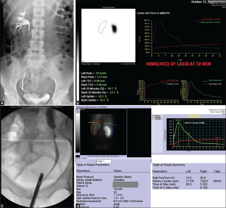
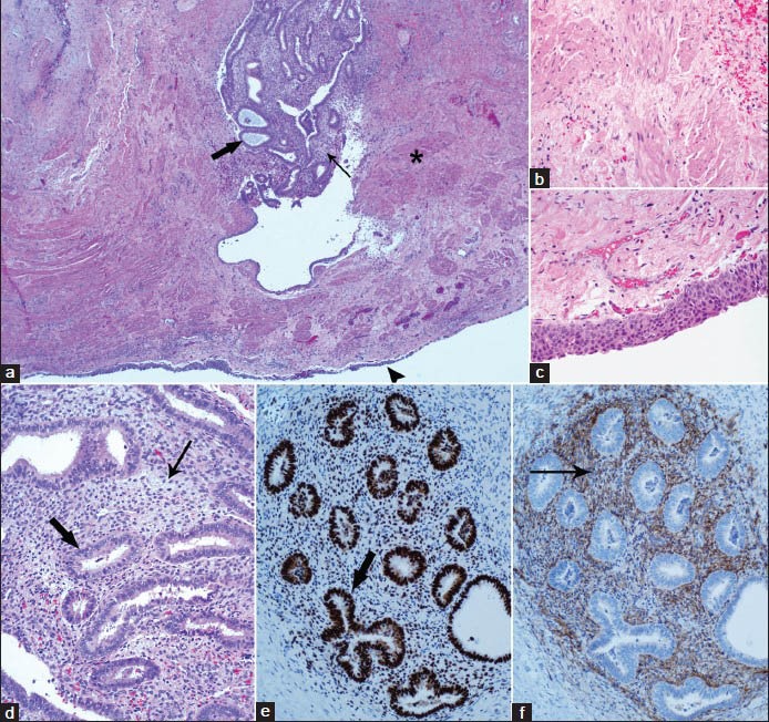
A 32 year old lady presented with recurrent left flank pain for 4 weeks and chronic lower back pain. CT without contrast showed no stones and mild left hydronephrosis. CT of the spine suggested an inflammatory process at L5-S1 vertebra. The diagnosis was supported by a bone scan. Incidentally, the scan showed nonfunctioning left kidney. Diuretic renography confirmed poor perfusion and no excretion. A retrograde study showed narrowing of the ureter at the pelvic brim. Ureteroscopy showed a papillary mass in the lumen of the ureter from which multiple cold cup biopsies were taken. The pathology however was not conclusive. A robotic nephroureterectomy was carried out. Definitive pathology showed intrinsic endometriosis of the ureter. We conclude that endometriosis should be considered in the differential diagnosis of unexplained ureteric obstruction and ureteric lumen filling defects in young women.
Keywords: Endometriosis; tumor; ureter.
Conflict of interest statement
Figures
Similar articles
-
Unilateral Ureteric Endometriosis at the Pelvic Brim Resulting in Complete Loss of Renal Function.Case Rep Obstet Gynecol. 2019 Oct 13;2019:9194615. doi: 10.1155/2019/9194615. eCollection 2019. Case Rep Obstet Gynecol. 2019. PMID: 31737388 Free PMC article.
-
[Mixed type ureteral endometriosis : a case report and a review of the Japanese literature].Hinyokika Kiyo. 2012 Jul;58(7):329-33. Hinyokika Kiyo. 2012. PMID: 22895128 Review. Japanese.
-
Management of ureteric endometriosis associated with hydronephrosis: An Australian case series of 13 patients.BMC Res Notes. 2010 Feb 25;3:45. doi: 10.1186/1756-0500-3-45. BMC Res Notes. 2010. PMID: 20184728 Free PMC article.
-
Medical treatment of ureteral obstruction associated with ovarian remnants and/or endometriosis: report of three cases and review of the literature.J Minim Invasive Gynecol. 2015 Mar-Apr;22(3):462-8. doi: 10.1016/j.jmig.2014.12.153. Epub 2014 Dec 20. J Minim Invasive Gynecol. 2015. PMID: 25533869 Review.
-
Intrinsic unilateral ureteral endometriosis: A rare case report.Int J Surg Case Rep. 2023 Mar;104:107966. doi: 10.1016/j.ijscr.2023.107966. Epub 2023 Mar 4. Int J Surg Case Rep. 2023. PMID: 36889152 Free PMC article.
Cited by
-
Unilateral ureteral endometriosis masquerading as a ureteral tumor and resulting in complete loss of renal function: A case report.J Int Med Res. 2025 Feb;53(2):3000605251322252. doi: 10.1177/03000605251322252. J Int Med Res. 2025. PMID: 40019105 Free PMC article.
-
Extravertebral low back pain: a scoping review.BMC Musculoskelet Disord. 2024 May 7;25(1):363. doi: 10.1186/s12891-024-07435-9. BMC Musculoskelet Disord. 2024. PMID: 38714994 Free PMC article.
References
-
- Abrao MS, Dias JA, Jr, Bellelis P, Podgaec S, Bautzer CR, Gromatsky C. Endometriosis of the ureter and bladder are not associated diseases. Fertil Steril. 2009;91:1662–7. - PubMed
-
- Carmignani L, Vercellini P, Spinelli M, Fontana E, Frontino G, Fedele L. Pelvic endometriosis and hydroureteronephrosis. Fertil Steril. 2010;93:1741–4. - PubMed
-
- Clement PB. Kurman RJ. Blaustein's Pathology of the Female Genital Tract. 3rd ed. Vol. 17. New York: Springer-Verlag; 1987. Endometriosis, lesions of the secondary mullerian system, and pelvic mesothelial proliferations; pp. 516–59.
Publication types
LinkOut - more resources
Full Text Sources
Other Literature Sources