Review
. 2014 Mar 3:6:13.
doi: 10.12703/P6-13. eCollection 2014.

The M1 and M2 paradigm of macrophage activation: time for reassessment

Affiliations
Review

The M1 and M2 paradigm of macrophage activation: time for reassessment

Fernando O Martinez et al. F1000Prime Rep. .

Abstract

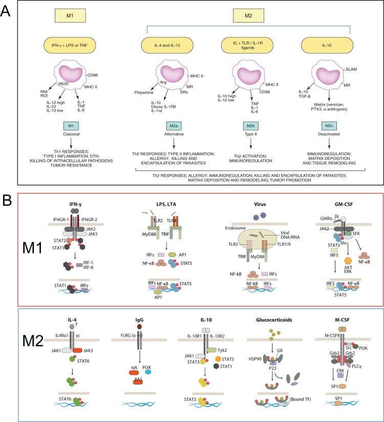
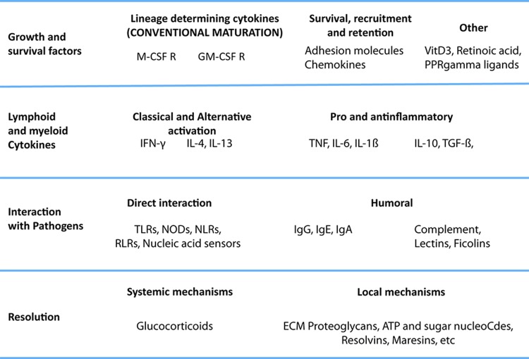
Macrophages are endowed with a variety of receptors for lineage-determining growth factors, T helper (Th) cell cytokines, and B cell, host, and microbial products. In tissues, macrophages mature and are activated in a dynamic response to combinations of these stimuli to acquire specialized functional phenotypes. As for the lymphocyte system, a dichotomy has been proposed for macrophage activation: classic vs. alternative, also M1 and M2, respectively. In view of recent research about macrophage functions and the increasing number of immune-relevant ligands, a revision of the model is needed. Here, we assess how cytokines and pathogen signals influence their functional phenotypes and the evidence for M1 and M2 functions and revisit a paradigm initially based on the role of a restricted set of selected ligands in the immune response.


Figures

Figure 1.
Figure 1.. The M1/M2 paradigm, origin, and molecular basis
(A) Mantovani and colleagues [11] proposed an M1-M2 macrophage model, in which M1 included interferon-gamma (IFN-γ) + lipopolysaccharide (LPS) or tumor necrosis factor (TNF) and M2 was subdivided to accommodate similarities and differences between interleukin-4 (IL-4) (M2a), immune complex + Toll-like receptor (TLR) ligands (M2b), and IL-10 and glucocorticoids (M2c). Diagram reproduced with the permission of Elsevier. (B) The signaling behind the effects of M1 and M2 stimuli in macrophages has gained clarity in recent years. Here, we highlight receptors and key signaling mediators in common and distinct pathways, explained in the text. The diagram includes granulocyte macrophage colony-stimulating factor (GM-CSF) and macrophage colony-stimulating factor (M-CSF) as M1 and M2 stimuli.
Figure 2.
Figure 2.. A multipolar view of the macrophage activation paradigm from an immunological perspective
The cytokines and stimuli that we call M1 and M2 play different roles in the development, maturation, and activation of macrophages. Integration of these isolated stimuli is necessary to represent the complex changes that macrophages undergo during full activation. Lessons from basic immunology suggest that many of the mechanisms that affect innate and acquired immunity are underappreciated (for example, humoral pattern recognition receptors, inflammatory cytokines other than interferon-gamma (IFN-γ) and tumor necrosis factor (TNF)). Here, we focus on the levels of activation imposed by the immune system rather than proposing a new classification of macrophage phenotypes, which is currently under discussion internationally. We propose that the stimuli governing macrophage activation should be organized according to their role in the immune response, rather than in groups that overlook their individual differences, and highlight the fact that complex combinations should be assessed to understand the full repertoire of macrophages. We identify at least four levels: growth and survival factors, interaction with lymphoid and myeloid cytokines, interaction with pathogens, and resolution. In the first level, in addition to prototypic maturation signals, we add stimuli known to promote survival of monocytes. A second level is interaction with cytokines. Here, we place interleukin-4 (IL-4), and IFN-γ and other cytokines produced by lymphoid and myeloid cells. However, the contribution of non-hematopoietic cells cannot be ignored. The next level is that of interaction with pathogens directly or through humoral recognition receptors, such as lectins, ficolins, and the B cell-derived immunoglobulins. A final level of resolution we classify as systemic (e.g. glucocorticoids) or local, such as ATP, resolvins, and other mediators with general anti-inflammatory properties. The combinations of stimuli commonly associated with extreme phenotypes of cells are IFN-γ + lipopolysaccharide (LPS) or TNF, immune complexes + Myd88, and granulocyte macrophage colony-stimulating factor (GM-CSF) + IL-4, which in monocytes induce a dendritic cell-like phenotype. However, there are many other possible combinations that are not considered special cases. We plan to extend the potential of macrophages, taking into consideration other functionally relevant combinations.

References

    1. Mosmann TR, Cherwinski H, Bond MW, Giedlin MA, Coffman RL. Two types of murine helper T cell clone. I. Definition according to profiles of lymphokine activities and secreted proteins. J Immunol. 1986;136:2348–57. - PubMed
    1. Coffman RL. Origins of the T(H)1-T(H)2 model: a personal perspective. Nat Immunol. 2006;7:539–41. doi: 10.1038/ni0606-539. - DOI - PubMed
    2. http://f1000.com/prime/718270380

    1. Mackaness GB. Cellular resistance to infection. J Exp Med. 1962;116:381–406. doi: 10.1084/jem.116.3.381. - DOI - PubMed
    1. Nathan CF, Murray HW, Wiebe ME, Rubin BY. Identification of interferon-gamma as the lymphokine that activates human macrophage oxidative metabolism and antimicrobial activity. J Exp Med. 1983;158:670–89. doi: 10.1084/jem.158.3.670. - DOI - PMC - PubMed
    1. Pace JL, Russell SW, Schreiber RD, Altman A, Katz DH. Macrophage activation: priming activity from a T-cell hybridoma is attributable to interferon-gamma. Proc Natl Acad Sci USA. 1983;80:3782–6. doi: 10.1073/pnas.80.12.3782. - DOI - PMC - PubMed