Primary labial tuberculosis: a rare presentation
- PMID: 24669346
- PMCID: PMC3952285
- DOI: 10.4103/2141-9248.126623
Primary labial tuberculosis: a rare presentation
Abstract
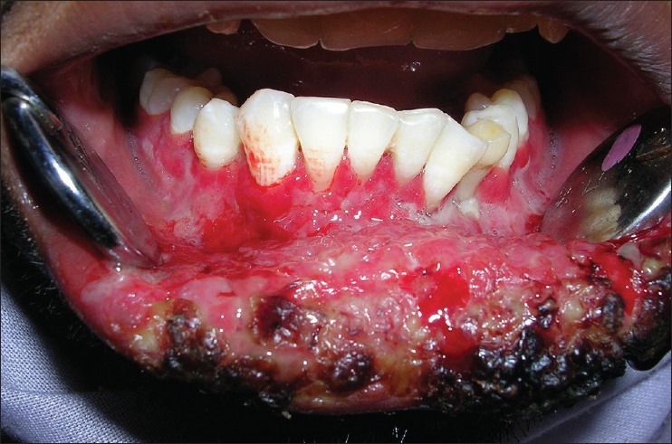
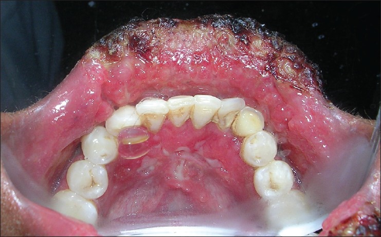
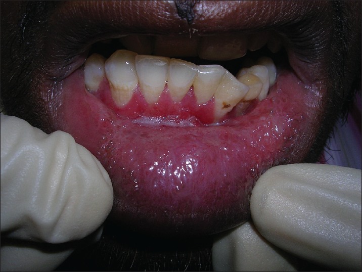
Tuberculosis is one of the oldest scorches of mankind that has not left this world even today. The disease is more common in the developing countries. Oral tuberculosis has been considered in 0.1-5% of all tuberculous infections. Mostly, the oral tuberculous lesions are secondary to pulmonary tuberculosis, but rarely primary lesions may occur. Primary lesions occur due to direct inoculation of the microorganism into the oral mucosa and mainly seen in the young individuals. Tongue is the most common oral site involved. Of all the sites involved, labial involvement is extremely rare. This case report intends to throw light on one such unique case, where a young male patient presented with a primary tubercular lesion of the lip. The lesion resolved immediately after anti tubercular therapy.
Keywords: Granulomatosis; Labial; Oral; Tuberculosis.
Conflict of interest statement
Figures
Similar articles
-
Primary Tuberculosis of Buccal and Labial Mucosa: Literature Review and a Rare Case Report of a Public Health Menace.Case Rep Dent. 2023 Oct 5;2023:6543595. doi: 10.1155/2023/6543595. eCollection 2023. Case Rep Dent. 2023. PMID: 37842328 Free PMC article.
-
Tubercular Osteomyelitis of Pubis with Labial Abscess: A Rare Presentation of a Common Disease.J Orthop Case Rep. 2021 Dec;11(12):22-25. doi: 10.13107/jocr.2021.v11.i12.2550. J Orthop Case Rep. 2021. PMID: 35415143 Free PMC article.
-
Isolated primary cold abscess of the sternum: a case report.J Med Case Rep. 2019 Aug 25;13(1):267. doi: 10.1186/s13256-019-2210-9. J Med Case Rep. 2019. PMID: 31445516 Free PMC article.
-
Orofacial tubercular lesions.Indian J Tuberc. 2014 Oct;61(4):325-30. Indian J Tuberc. 2014. PMID: 25675696 Review.
-
Primary lingual tuberculosis: a case report.J Laryngol Otol. 1998 Jan;112(1):86-7. doi: 10.1017/s0022215100139982. J Laryngol Otol. 1998. PMID: 9538456 Review.
Cited by
-
[Tuberculous cheilitis revealing pulmonary tuberculosis].Pan Afr Med J. 2016 Jun 30;24:176. doi: 10.11604/pamj.2016.24.176.9862. eCollection 2016. Pan Afr Med J. 2016. PMID: 27795773 Free PMC article. French.
-
A buccal mucosa ulcer as the first sign of tuberculosis.J Oral Maxillofac Pathol. 2022 Jul-Sep;26(3):399-403. doi: 10.4103/jomfp.jomfp_443_21. Epub 2022 Oct 17. J Oral Maxillofac Pathol. 2022. PMID: 36588851 Free PMC article.
-
Non-infectious granulomatous disorders of the upper lip: clinicopathological analysis of 11 patients.BMC Oral Health. 2022 May 11;22(1):173. doi: 10.1186/s12903-022-02189-z. BMC Oral Health. 2022. PMID: 35545768 Free PMC article.
-
Oral manifestation of tuberculosis: a case-report.Braz J Infect Dis. 2016 Mar-Apr;20(2):210-3. doi: 10.1016/j.bjid.2015.12.001. Epub 2015 Dec 31. Braz J Infect Dis. 2016. PMID: 26748230 Free PMC article.
-
Primary Tuberculosis of Buccal and Labial Mucosa: Literature Review and a Rare Case Report of a Public Health Menace.Case Rep Dent. 2023 Oct 5;2023:6543595. doi: 10.1155/2023/6543595. eCollection 2023. Case Rep Dent. 2023. PMID: 37842328 Free PMC article.
References
-
- Kakisi OK, Kechagia AS, Kakisis IK, Rafailidis PI, Falagas ME. Tuberculosis of the oral cavity: A systematic review. Eur J Oral Sci. 2010;118:103–9. - PubMed
-
- Rafailidis PI, Avramopoulos I, Sapkas G, Falagas ME. Multidrug-resistant tuberculous spondylodiscitis: Need for aggressive management and drug susceptibility testing of Mycobacterium tuberculosis isolates. J Infect. 2006;52:e35–7. - PubMed
-
- Miziara ID. Tuberculosis affecting the oral cavity in Brazilian HIV-infected patients. Oral Surg Oral Med Oral Pathol Oral Radiol Endod. 2005;100:179–82. - PubMed
-
- Mignogna MD, Muzio LL, Favia G, Ruoppo E, Sammartino G, Zarrelli C, et al. Oral tuberculosis: A clinical evaluation of 42 cases. Oral Dis. 2000;6:25–30. - PubMed
-
- Iype EM, Ramdas K, Pandey M, Jayasree K, Thomas G, Sebastian P, et al. Primary tuberculosis of the tongue: Report of three cases. Br J Oral Maxillofac Surg. 2001;39:402–3. - PubMed
Publication types
LinkOut - more resources
Full Text Sources
Other Literature Sources