. 2014 Aug 1;23(15):1809-19.
doi: 10.1089/scd.2013.0618. Epub 2014 May 20.

Extracellular vesicles released from mesenchymal stromal cells modulate miRNA in renal tubular cells and inhibit ATP depletion injury

Affiliations

Extracellular vesicles released from mesenchymal stromal cells modulate miRNA in renal tubular cells and inhibit ATP depletion injury

Rafael S Lindoso et al. Stem Cells Dev. .

Abstract

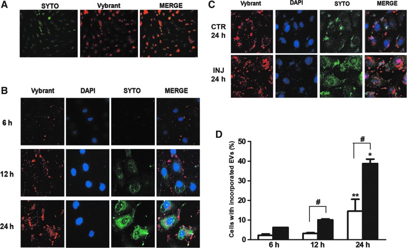
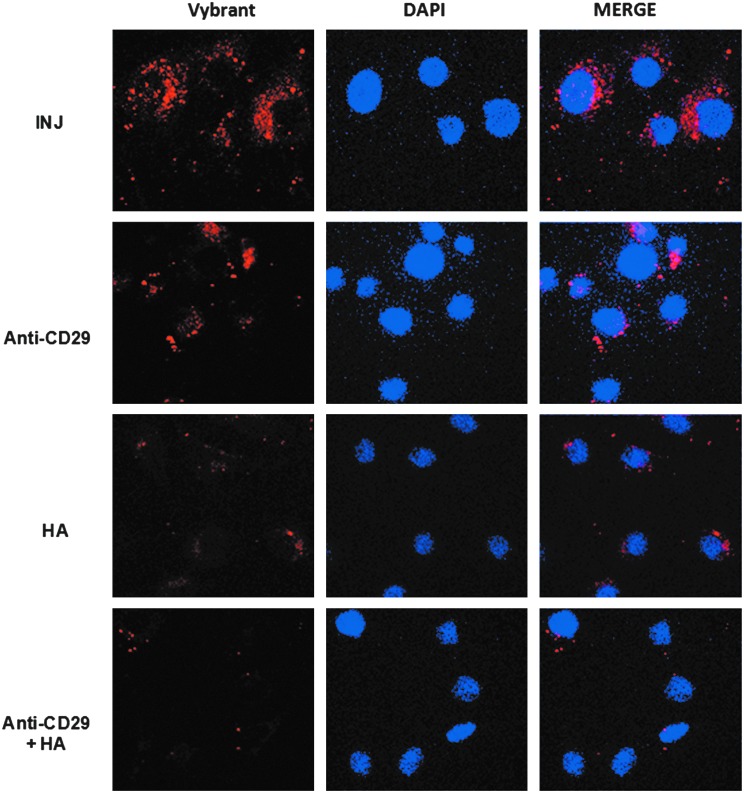
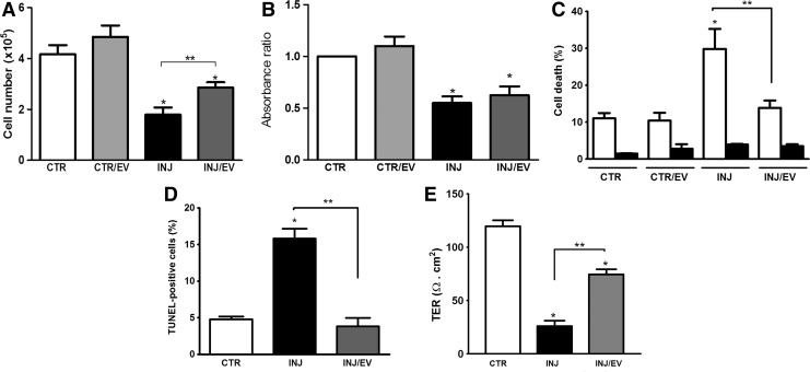
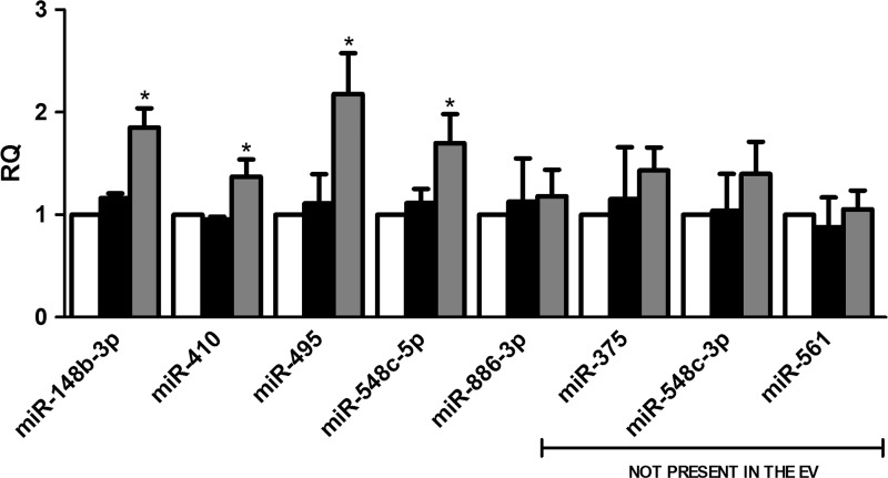
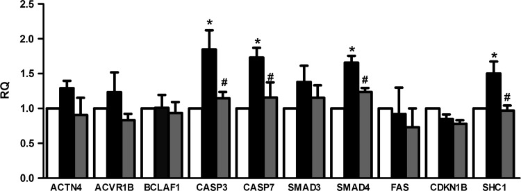
The mechanisms involved in renal repair by mesenchymal stromal cells (MSCs) are not entirely elucidated. The paracrine secretion of bioactive molecules has been implicated in the protective effects. Besides soluble mediators, MSCs have been shown to release extracellular vesicles (EVs), involved in renal repair process for different injury models. EVs have been shown to mediate communication between cells through the transference of several molecules, like protein, bioactive lipids, mRNA, and microRNAs (miRNAs). The miRNAs are noncoding RNAs that posttranscriptionally modulate gene expression and are involved in the regulation of several cellular processes, including those related to repair. The aim of the present study was to investigate the role of MSC-EVs in the modulation of miRNAs inside renal proximal tubular epithelial cells (PTECs) in an in vitro model of ischemia-reperfusion injury induced by ATP depletion. In this model we evaluated whether changes in miRNA expression were dependent on direct miRNA transfer or on transcription induction by MSC-EVs. The obtained results showed an enhanced incorporation of MSC-EVs in injured PTECs with protection from cell death. This biological effect was associated with EV-mediated miRNA transfer and with transcriptional modulation of miRNAs expressed by injured PTECs. Prediction of miRNA targets showed that miRNAs modulated in PTECs are involved in process of renal recovery with downregulation of coding-mRNAs associated with apoptosis, cytoskeleton reorganization, and hypoxia, such as CASP3 and 7, SHC1 and SMAD4. In conclusion, these results indicate that MSC-EVs may transfer and modulate the expression of several miRNAs involved in the repair and recovery process in PTECs.


Figures

<b>FIG. 1.</b>
FIG. 1.
Incorporation of MSC-EVs and RNA transfer in proximal tubular epithelial cells (PTECs). (A) MSCs were double-stained in red (with Vybrant Dil, 15-min incubation) and green (with Syto-RNA, 30-min incubation). Original magnification: ×200. Labeled MSCs released double-labeled EVs (see “Materials and Methods” section). (B) Double-labeled MSC-EVs were incubated for 6, 12, and 24 h with PTECs in normal condition and after ATP depletion injury. The first column of panels from the left shows the internalization of MSC-EV membranes. The second column of panels is the nuclei of PTECs stained with DAPI (blue). The third column of panels shows the distribution of Syto-RNA carried by MSC-EVs inside PTECs. The fourth column of panels shows a merge between the two previous panels. These experiments were realized in normal culture condition. (C) MSC-EV incorporation in PTECs after 24 h of incubation in normal culture condition and after ATP depletion injury. The panel description is the same as indicated above. Three experiments were performed with similar results using MSC-EVs derived from different MSCs. Original magnification: ×630. (D) FACS analysis of Vybrant Dil-labeled MSC-EV incorporation rate by PTECs. White bars represent the experiments realized in normal control conditions and black bars represent incorporation rate by PTECs after ATP depletion injury. Statistical analysis was performed by ANOVA with Newman-Keuls multicomparison test: *,**statistical difference between the 6- and 24-h experimental conditions; #statistical difference between normal and injury conditions, in the same incubation time (P<0.05; n=4). EV, extracellular vesicle; MSCs, mesenchymal stromal cells; FACS, fluorescence activated cell sorting.
<b>FIG. 2.</b>
FIG. 2.
Blockage of MSC-EV incorporation by PTECs. MSC-EVs stained with Vybrant Dil (red) were incubated for 24 h with PTECs submitted to ATP depletion injury. To block CD29 and CD44 integrins the MSC-EVs were previously incubated with anti-CD29 antibody, hialuronic acid (HA), or both simultaneously as indicated in the panels. Left panels indicate internalization of MSC-EV membrane. INJ indicated cells submitted to injury without any blockage. Middle panels show the nuclei of PTECs stained with DAPI (blue). Right panels show a merge of the two previous images. Three experiments were performed with similar results using MSC-EVs derived from different MSCs. Original magnification: ×630.
<b>FIG. 3.</b>
FIG. 3.
MSC-EVs promoted protection but not proliferation in PTECs after injury. After ATP depletion injury, MSC-EVs were incubated with PTECs for 24 h. (A) Number of viable cells by counting with Trypan blue staining. (B) Proliferation was performed by an ELISA for Brdu incorporation. (C) Cell death analysis by Muse Annexin V & Dead Cell Assay. Black bars indicate cell death rate by early apoptosis and white bars represent late apoptosis. (D) Apoptosis was also evaluated by TUNEL and expressed as percentage of positive cells (500 cells were counted in random fields using a fluorescent microscopy at a magnification of ×200). (E) Effect of MSC-EVs on TER of PTECs. TER was measured in all groups before submitted to the different conditions and no significant difference was observed (not shown). Final measures were performed 24 h after the cell incubation with antimycin A. Each group is indicated in the abscissa; in the control group (CTR) the cells were not submitted to injury. CTR/EV represents PTECs that were incubated with MSC-EVs; INJ indicates the PTECs submitted to injury, while INJ/EV is the group submitted to injury and then incubated with MSC-EVs. Statistical analysis was performed by ANOVA with Newman-Keuls multicomparison test: *statistical difference related to the control group; **statistical difference between injured group and injured group treated with MSC-EVs (P<0.05; n=5). ANOVA, analysis of variance; TER, transespithelial resistance.
<b>FIG. 4.</b>
FIG. 4.
Characterization of miRNAs transferred or upregulated by MSC-EVs. PTECs were first incubated with actinomycin D. After transcription blockage, the cells were submitted to injury and treated or not with MSC-EVs. White bars indicate control group maintained in normal condition. Black bars represent PTECs submitted to injury and gray bars represent the cells treated with MSC-EVs after injury. Three experiments were performed in triplicate. Analysis of upregulated miRNAs was performed by quantitative real-time polymerase chain reaction. The abscissa indicates the miRNAs evaluated. Data are expressed as relative quantification (RQ), normalized to RNU48. Statistical analysis was performed by ANOVA with Dunnett multicomparison test: *statistical difference to the injured group (P<0.05; n=6).
<b>FIG. 5.</b>
FIG. 5.
Changes in the expression of predicted miRNA targets modulated by MSC-EVs. Evaluation of the gene expression of miRNA targets predicted by GO analysis related to hypoxia, cytoskeleton reorganization, and apoptosis processes. The changes in gene expression were performed by quantitative real-time polymerase chain reaction. The analysis was performed in all three conditions: control (white bars), injury (black bars), and injury treated with MSC-EVs (gray bars). Data are expressed as RQ, normalized to GAPDH. The abscissa indicated the evaluated genes. Statistical analysis was performed by ANOVA with Dunnett multicomparison test: *statistical difference to the control group; #statistical difference to the injured group (P<0.05; n=4).

References

    1. Bussolati B, Hauser PV, Carvalhosa R. and Camussi G. (2009). Contribution of stem cells to kidney repair. Curr Stem Cell Res Ther 4:2–8 - PubMed
    1. Asanuma H, Meldrum DR. and Meldrum KK. (2010). Therapeutic applications of mesenchymal stem cells to repair kidney injury. J Urol 184:26–33 - PubMed
    1. Camussi G, Deregibus MC. and Tetta C. (2010). Paracrine/endocrine mechanism of stem cells on kidney repair: role of microvesicle-mediated transfer of genetic information. Curr Opin Nephrol Hypertens 19:7–12 - PubMed
    1. Bruno S, Grange C, Deregibus MC, Calogero RA, Saviozzi S, Collino F, Morando L, Busca A, Falda M, et al. (2009). Mesenchymal stem cell-derived microvesicles protect against acute tubular injury. J Am Soc Nephrol 20:1053–1067 - PMC - PubMed
    1. Ratajczak J, Miekus K, Kucia M, Zhang J, Reca R, Dvorak P. and Ratajczak MZ. (2006). Embryonic stem cells-derived microvesicles reprogram hematopoietic progenitors: evidence for horizontal transfer of mRNA and protein delivery. Leukemia 20:847–856 - PubMed

Publication types