Cone beam CT evaluation of the presence of anatomic accessory canals in the jaws
- PMID: 24670010
- PMCID: PMC4082258
- DOI: 10.1259/dmfr.20130259
Cone beam CT evaluation of the presence of anatomic accessory canals in the jaws
Abstract
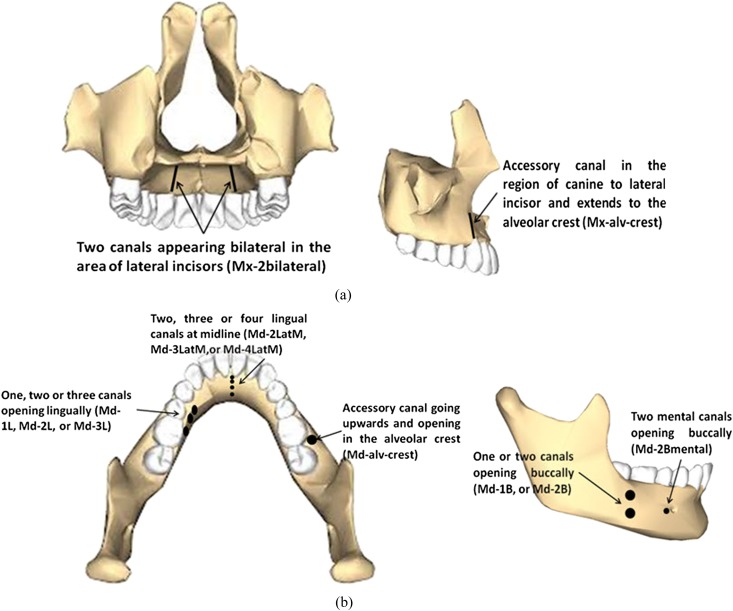
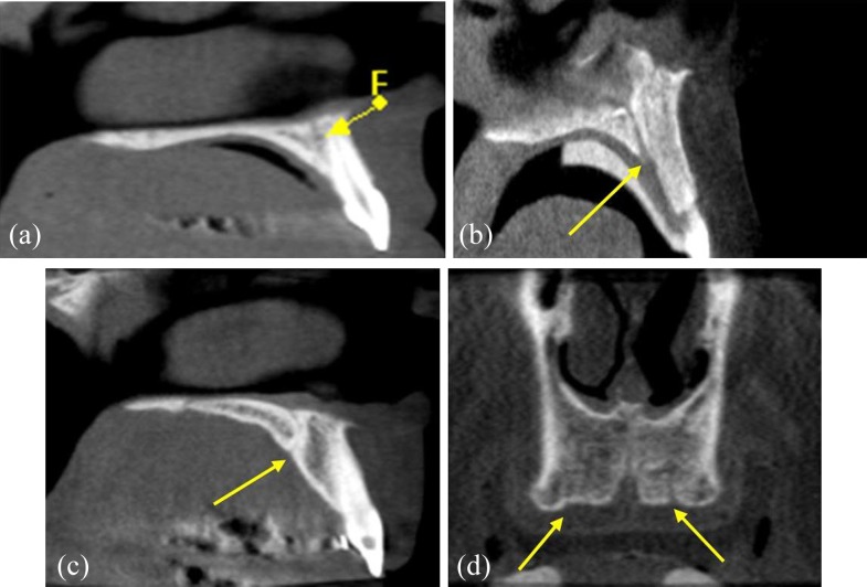
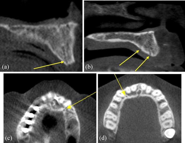
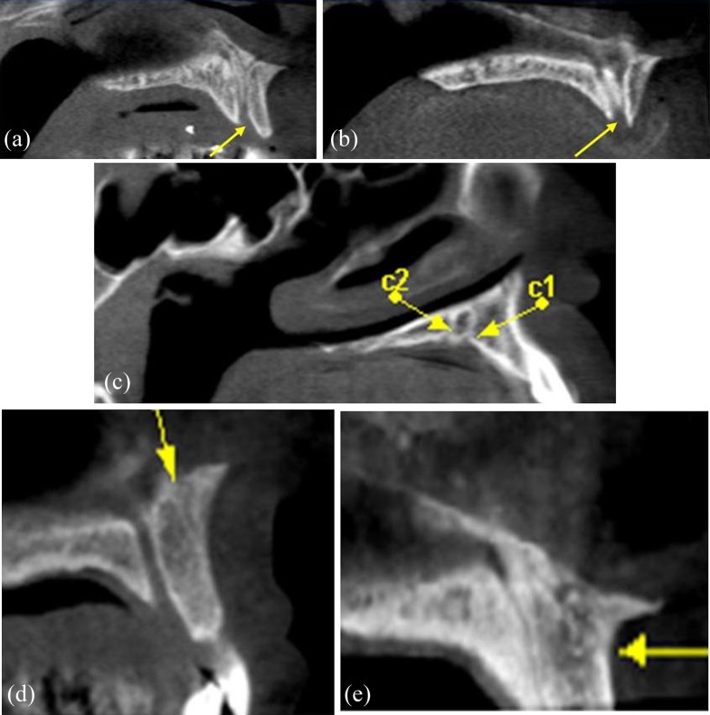
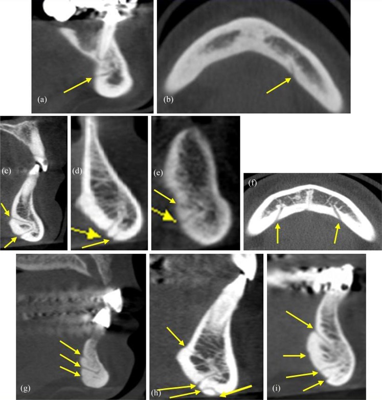
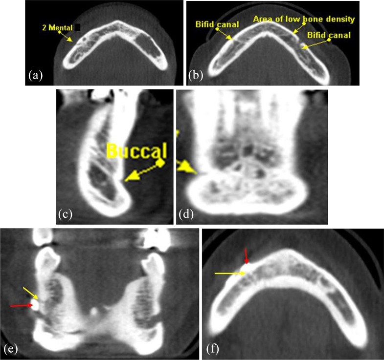
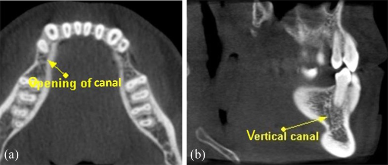
Objectives: To assess the prevalence, location and anatomical course of accessory canals of the jaws using cone beam CT.
Methods: A retrospective analysis of 4200 successive cone beam CT scans, for patients of both genders and ages ranging from 7 to 88 years, was performed. They were exposed at the School of Dentistry, University of Michigan, Ann Arbor, MI. After applying the exclusion criteria (the presence of severe ridge resorption, pre-existing implants, a previously reported history of craniofacial malformations or syndromes, a previous history of trauma or surgery, inadequate image quality and subsequent scans from the same individuals), 4051 scans were ultimately included in this study.
Results: Of the 4051 scans (2306 females and 1745 males) that qualified for inclusion in this study, accessory canals were identified in 1737 cases (42.9%; 1004 females and 733 males). 532 scans were in the maxilla (13.1%; 296 females and 236 males) and 1205 in the mandible (29.8%; 708 females and 497 males).
Conclusions: A network of accessory canals bringing into communication the inner and outer cortical plates of the jaws was identified. In light of these findings, clinicians should carefully assess for the presence of accessory canals prior to any surgical intervention to decrease the risk for complications.
Keywords: CBCT; accessory canals; incisive canal; interforaminal; lingual vascular canals.
Figures
Similar articles
-
Cone beam computed tomography analysis of accessory canals of the canalis sinuosus: A prevalent but often overlooked anatomical variation in the anterior maxilla.J Prosthet Dent. 2021 Oct;126(4):560-568. doi: 10.1016/j.prosdent.2020.05.028. Epub 2020 Sep 29. J Prosthet Dent. 2021. PMID: 33004226
-
An anatomical variant: evaluation of accessory canals of the canalis sinuosus using cone beam computed tomography.Folia Morphol (Warsz). 2018;77(3):551-557. doi: 10.5603/FM.a2018.0003. Epub 2018 Jan 18. Folia Morphol (Warsz). 2018. PMID: 29345719 Clinical Trial.
-
Assessment of bone channels other than the nasopalatine canal in the anterior maxilla using limited cone beam computed tomography.Surg Radiol Anat. 2013 Nov;35(9):783-90. doi: 10.1007/s00276-013-1110-8. Epub 2013 Mar 29. Surg Radiol Anat. 2013. PMID: 23539212
-
Evaluation of root canal morphology of human primary molars by using CBCT and comprehensive review of the literature.Acta Odontol Scand. 2016;74(4):250-8. doi: 10.3109/00016357.2015.1104721. Epub 2015 Nov 2. Acta Odontol Scand. 2016. PMID: 26523502 Review.
-
Clinical anatomy and surgical significance of the lingual foramina and their canals.Clin Anat. 2017 Mar;30(2):194-204. doi: 10.1002/ca.22824. Epub 2017 Feb 9. Clin Anat. 2017. PMID: 28033685 Review.
Cited by
-
[Importance of cone beam computed tomography in the recognition of the trajectory and anatomical variants of the mandibular canal. A review of the literature].Rev Cient Odontol (Lima). 2021 Mar 11;9(1):e046. doi: 10.21142/2523-2754-0901-2021-046. eCollection 2021 Jan-Mar. Rev Cient Odontol (Lima). 2021. PMID: 38464412 Free PMC article. Review. Spanish.
-
Determination of presence and morphometry of lingual foramina and canals in Chilean mandibles using cone-beam CT images.Surg Radiol Anat. 2018 Dec;40(12):1405-1410. doi: 10.1007/s00276-018-2080-7. Epub 2018 Aug 18. Surg Radiol Anat. 2018. PMID: 30121723
-
Advocacy of diagnostic criteria for maxillary incisive canal cysts based on alteration of normal maxillary incisive canals according to aging in Japanese populations.Head Face Med. 2019 Oct 22;15(1):25. doi: 10.1186/s13005-019-0209-5. Head Face Med. 2019. PMID: 31640721 Free PMC article.
-
Cone beam computerized tomography evaluation of incisive canal and anterior maxillary bone thickness for placement of immediate implants.J Indian Prosthodont Soc. 2018 Oct-Dec;18(4):356-363. doi: 10.4103/jips.jips_167_18. J Indian Prosthodont Soc. 2018. PMID: 30449964 Free PMC article.
-
Systematic review and meta-analysis of lingual foramina anatomy and surgical-related aspects on cone-beam computed tomography: a PROSPERO-registered study.Oral Radiol. 2022 Jan;38(1):1-16. doi: 10.1007/s11282-021-00516-8. Epub 2021 Feb 20. Oral Radiol. 2022. PMID: 33609258
References
-
- Romanos GE, Greenstein G. The incisive canal. Considerations during implant placement: case report and literature review. Int J Oral Maxillofac Implants 2009; 24: 740–5. - PubMed
MeSH terms
LinkOut - more resources
Full Text Sources
Other Literature Sources
