. 2014 Nov;7(6):1340-53.
doi: 10.1038/mi.2014.21. Epub 2014 Mar 26.

Targeted colonic claudin-2 expression renders resistance to epithelial injury, induces immune suppression, and protects from colitis

Affiliations

Targeted colonic claudin-2 expression renders resistance to epithelial injury, induces immune suppression, and protects from colitis

R Ahmad et al. Mucosal Immunol. 2014 Nov.

Abstract

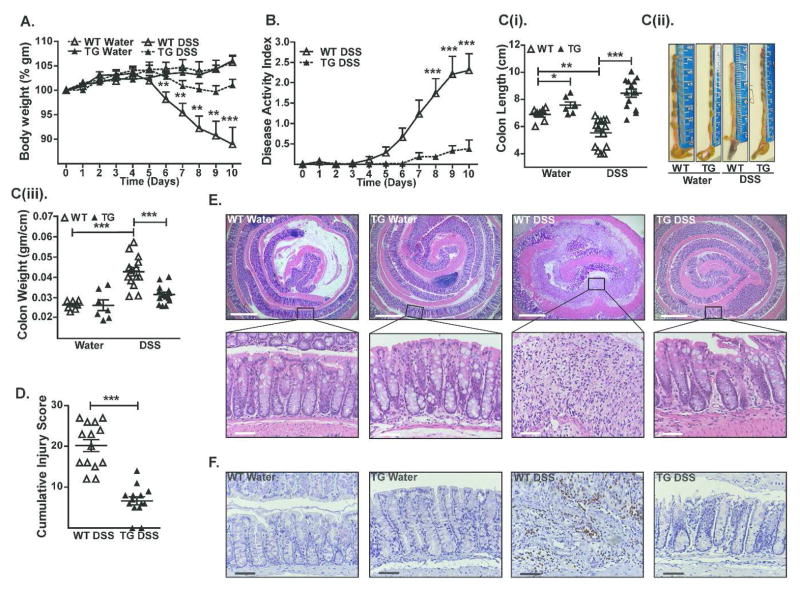
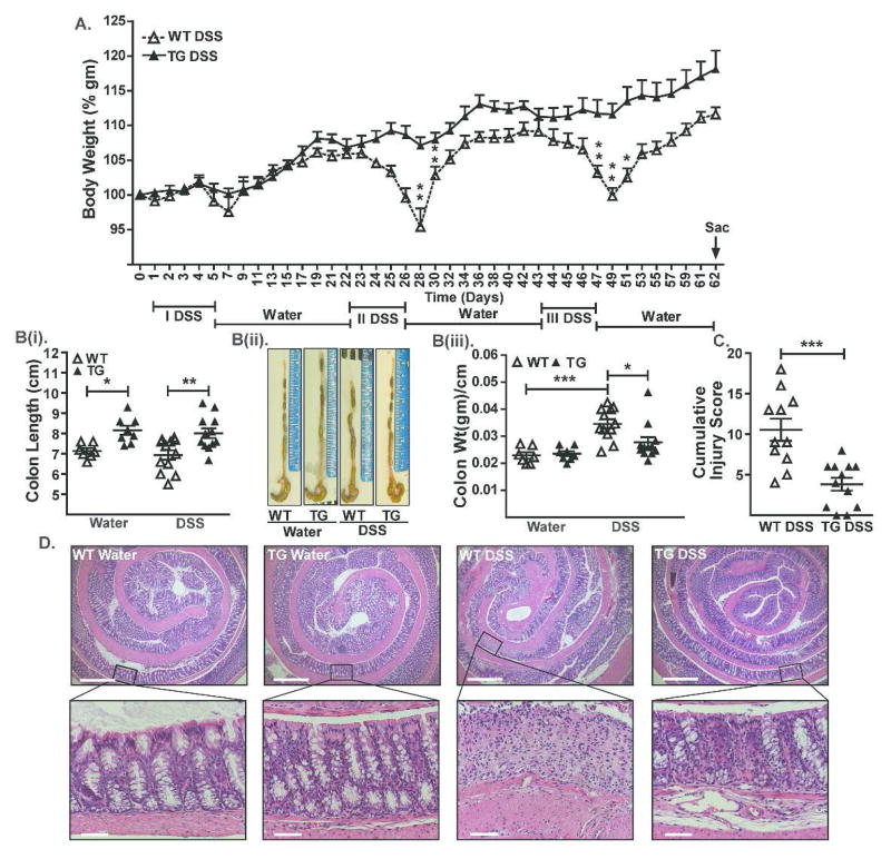
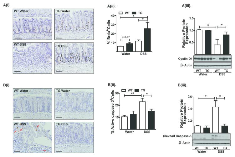
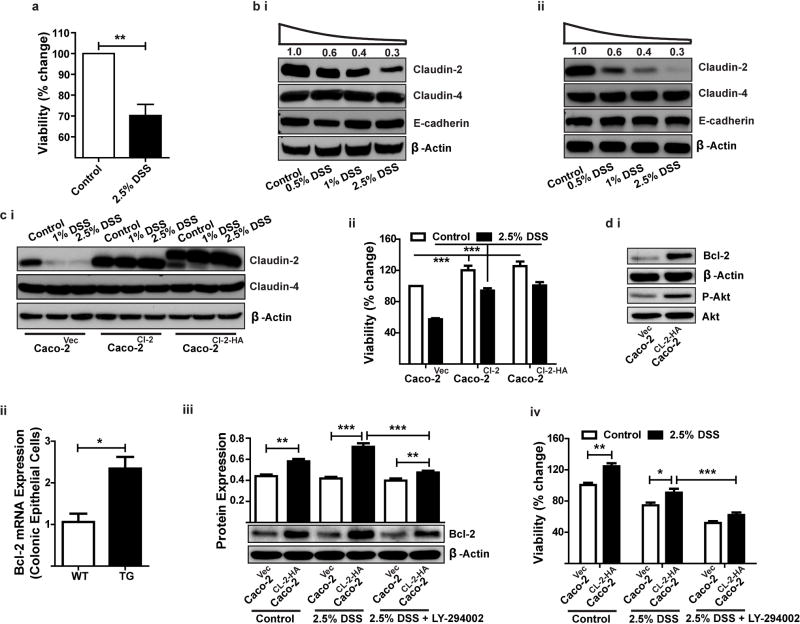
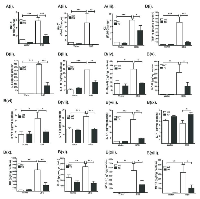
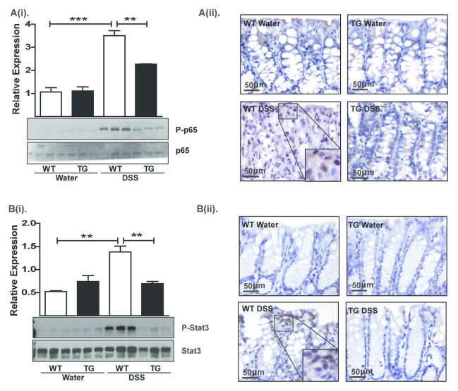
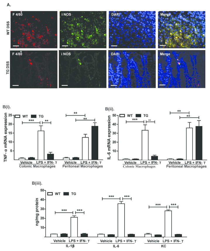
Expression of claudin-2, a tight junction protein, is highly upregulated during inflammatory bowel disease (IBD) and, due to its association with epithelial permeability, has been postulated to promote inflammation. Notably, claudin-2 has also been implicated in the regulation of intestinal epithelial proliferation. However, precise role of claudin-2 in regulating colonic homeostasis remains unclear. Here, we demonstrate, using Villin-Claudin-2 transgenic mice, that increased colonic claudin-2 expression augments mucosal permeability as well as colon and crypt length. Most notably, despite leaky colon, Cl-2TG mice were significantly protected against experimental colitis. Importantly, claudin-2 expression increased colonocyte proliferation and provided protection against colitis-induced colonocyte death in a PI-3Kinase/Bcl-2-dependent manner. However, Cl-2TG mice also demonstrated marked suppression of colitis-induced increases in immune activation and associated signaling, suggesting immune tolerance. Accordingly, colons from naive Cl-2TG mice harbored significantly increased numbers of regulatory (CD4(+)Foxp3(+)) T cells than WT littermates. Furthermore, macrophages isolated from Cl-2TG mouse colon exhibited immune anergy. Importantly, these immunosuppressive changes were associated with increased synthesis of the immunoregulatory cytokine TGF-β by colonic epithelial cells in Cl-2TG mice compared with WT littermates. Taken together, our findings reveal a critical albeit complex role of claudin-2 in intestinal homeostasis by regulating epithelial permeability, inflammation and proliferation and suggest novel therapeutic opportunities.


Figures

Figure 1
Figure 1. Villin-Claudin-2 transgenic mice exhibit increased colon length and permeability
A(i). Immunoblot analysis using mouse colon lysates and antigen-specific antibodies, and A(ii). Quantitative analysis of protein expression relative to the expression in WT mice; (B). Immunohistochemical analysis of endogenous and exogenous colonic claudin-2 expression; C(i). Colon length (N# 10, **p<0.01), and C(ii). Crypt height; (D). BrdU-positive colonocytes in Cl-2TG versus WT mice. Cytokeratin-positive cells were isolated from the colons and subjected to FACS-analysis following staining with anti-BrdU antibody; (E). Transepithelial resistance (TER) (N# 5, *p<0.05); F(i). Transmucosal permeability using Ussing chamber (N#5, *p<0.05, **p<0.01); and F(ii). Transmucosal permeability using intrarectal delivery of FITC-dextran (4kDa) (N#5, *p<0.05).
Figure 2
Figure 2. Cl-2TG mice are protected from DSS-induced acute colitis
To induce colitis, mice received DSS (4% w/v) in the drinking water (10 days). (A). Weight loss during the course of DSS administration; (B). Disease activity index (DAI) changes among DSS-treated groups; C(i). Colon length (cm) in control and DSS-treated mice, C(ii). Representative colon images, and C(iii). Colon weight/cm; (D). Cumulative injury scores. Control mice did not show inflammation and associated injury; (E). Representative H&E staining of the colonic tissues from control and DSS-treated mice; (F). Representative photomicrographs demonstrating immunostaining to determine colonic myeloperoxidase (MPO) activity from control and DSS-treated mice. Values are presented as mean ± sem. *p<0.05, **p<0.01, ***p<0.001. Scale bars=500 or 50 μm.
Figure 3
Figure 3. Cl-2TG mice are protected from chronic colitis
Mice were subjected to three cycles of DSS (4% w/v) drinking water (5 days/cycle) followed by regular drinking water (16 days/cycle). (A). Weight loss during the treatment course. Body weight did not differ significantly among control groups; B(i). Mean colon length (cm) in control and DSS-treated mice, B(ii) representative images, and B(iii). Colon weight/cm of colon length; (C). Cumulative injury score. Control mice did not show inflammation and associated injury; (D). Representative H&E staining of colonic tissues from control and DSS-treated mice. Values are presented as mean ± sem. *p<0.05, **p<0.01, ***p<0.001. Scale bars=500 or 50 μm.
Figure 4
Figure 4. Claudin-2 modulates colonic epithelial cell homeostasis
A(i). Representative images showing BrdU-positive cells in control and DSS-treated animals. Please note that crypts in DSS-treated WT mice are BrdU-negative but that adjoining infiltrating immune cells are positive, A(ii) FACS-analysis of colonic epithelial cells co-immunostained for BrdU and cleaved-caspase-3, and A(iii). Immunoblot analysis to determine cyclin-D1 expression; B(i) Representative images showing cleaved caspase-3-positive cells in control and DSS-treated animals, B(ii). FACS analysis using colonic epithelial cells co-immunostained for BrdU and cleaved-caspase-3, and B(iii). Immunoblot analysis to determine cleaved caspase-3 expression in control and DSS-treated mice: N# 3–6. Values are mean+sem. *p<0.05, **p<0.01. Scale bars=500 μm.
Figure 5
Figure 5. In Caco-2 cells, claudin-2 overexpression protects against DSS-dependent decrease in cell viability
(A). Effect of DSS treatment on cell viability; B(i–ii). Immunoblot analysis to determine expression of claudin-2, claudin-4 and E-cadherin in Caco-2 (i) or HT-29 (ii) cells exposed to different concentrations of DSS for 24 hours; C(i). Immunoblot analysis demonstrating stable overexpression of an untagged claudin-2 or an HA-tagged claudin-2 cDNA construct in Caco-2 cells; C(ii). Effect of claudin-2 expression on cell viability in DSS-treated Caco-2 cells; D(i) Immunoblot analysis using total cell lysate from unchallenged control and Caco-2Cl-2HA cells; D(ii) qRT-PCR analysis to determine Bcl-2 expression in colonic epithelial cells isolated from Cl-2TG mice and WT-littermates (n#4/group); D(iii) Effect of DSS-treatment upon Bcl-2 expression in control and Caco-2Cl-2HA cells with or without pre-treatment with LY-294002; D(iv) Effect of DSS-treatment upon cell viability in control and Caco-2Cl-2HA cells with or without pre-treatment with LY-294002. Data is presented as mean+sem from at least three independent experiments. **p<0.01 and ***p<0.001.
Figure 6
Figure 6. Expression of inflammatory cytokines/chemokines is sharply down-regulated in DSS-treated Cl-2TG mice
Mice were subjected to DSS colitis as described in the legend to Fig. 2. (A). Cytokine/chemokine levels as determined by quantitative real-time RT-PCR; and (B). Cytokine/chemokine levels as determined by Luminex analysis (N#5/group). Values are presented as mean ± sem. *p<0.05, **p<0.01, ***p<0.001.
Figure 7
Figure 7. The DSS-induced activation of NF-κB and STAT3-signaling is attenuated in Cl-2TG mice while expression of the immunoregulatory cytokine TGF-β is significantly upregulated
Samples from mice subjected to DSS colitis were used. A(i). Immunoblot analysis using anti-phospho-p65, p65 antibodies and representative densitometric analyses, A(ii). Immunohistochemical analysis to determine cellular localization of p65 protein; B(i). Immunoblot analysis using anti-phospho-STAT3 and –STAT3 antibodies and representative densitometric analyses, B(ii). Immunohistochemical analysis to determine the tissue distribution and cellular localization of phospho-STAT3; n#3–6, *p<0.05, **p<0.01, ***p<0.001. Scale bar=500 μm.
Figure 8
Figure 8. Colonic macrophages in Cl-2TG mice exhibit inflammatory anergy
(A). Representative images demonstrating macrophage (F4/80) infiltration in the colonic mucosa in control and DSS-treated WT and Cl-2TG mice. F4/80+ cells were co-immunostained with anti-iNOS antibody; B(i–ii). Colonic or peritoneal macrophages were isolated from naïve WT and Cl-2TG mice and were subjected to immune activation using LPS and IFN-γ for 24 hours. mRNA expression of TNF-α and IL-6 was determined using qRT-PCR; B(iii). Supernatant from in vitro activated macrophages isolated from Cl-2TG and WT-mice was subjected to ELISA analysis using antigen-specific antibody. Values are presented as mean ± sem. **p<0.01, ***p<0.001. Scale bars=500 μm.
Figure 9
Figure 9. Regulatory T-cells and expression of the immunoregulatory cytokine TGF-β are significantly upregulated in the colon of naïve Cl-2TG mice
Samples from unchallenged Cl-2TG mice and WT-littermates were used. (A). Genomic DNA isolated from the colon was subjected to PCR using primers based on universal bacterial sequence; B(i–ii). FACS-analysis using antigen-specific antibody was performed using cells isolated from the lamina propria of naïve Cl-2TG mice and WT-littermates; C(i). Immunoblot analysis to determine TGF-β expression and representative densitometric analysis. Total tissue lysate from control and DSS-treated mice was used. C(ii). TGF-β expression analysis using mRNA isolated from specific cell populations isolated from the colon of unchallenged Cl-2TG mice and WT-littermates. n#3–6, *p<0.05 and **p<0.01.

References

    1. Hollander D. Intestinal permeability, leaky gut, and intestinal disorders. Current gastroenterology reports. 1999;1(5):410–416. - PubMed
    1. Arrieta MC, Madsen K, Doyle J, Meddings J. Reducing small intestinal permeability attenuates colitis in the IL10 gene-deficient mouse. Gut. 2009;58(1):41–48. - PMC - PubMed
    1. Tsukita S, Furuse M. Occludin and claudins in tight-junction strands: leading or supporting players? Trends in cell biology. 1999;9(7):268–273. - PubMed
    1. Heller F, Florian P, Bojarski C, Richter J, Christ M, Hillenbrand B, et al. Interleukin-13 is the key effector Th2 cytokine in ulcerative colitis that affects epithelial tight junctions, apoptosis, and cell restitution. Gastroenterology. 2005;129(2):550–564. - PubMed
    1. Zeissig S, Burgel N, Gunzel D, Richter J, Mankertz J, Wahnschaffe U, et al. Changes in expression and distribution of claudin 2, 5 and 8 lead to discontinuous tight junctions and barrier dysfunction in active Crohn’s disease. Gut. 2007;56(1):61–72. - PMC - PubMed

Publication types