. 2014 Mar 26;6(229):229ra40.
doi: 10.1126/scitranslmed.3007696.

Defective neutrophil recruitment in leukocyte adhesion deficiency type I disease causes local IL-17-driven inflammatory bone loss

Affiliations

Defective neutrophil recruitment in leukocyte adhesion deficiency type I disease causes local IL-17-driven inflammatory bone loss

Niki M Moutsopoulos et al. Sci Transl Med. .

Abstract

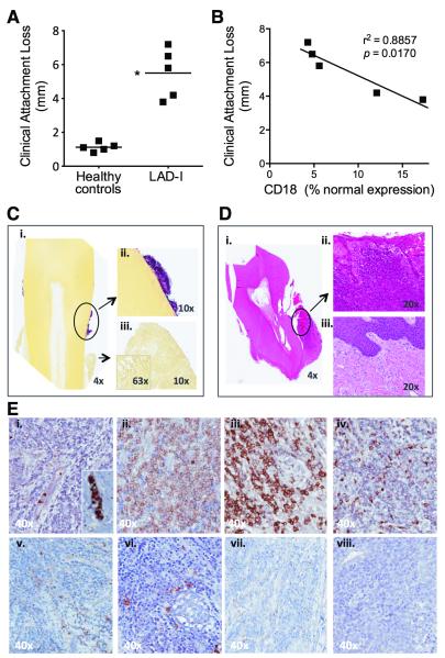
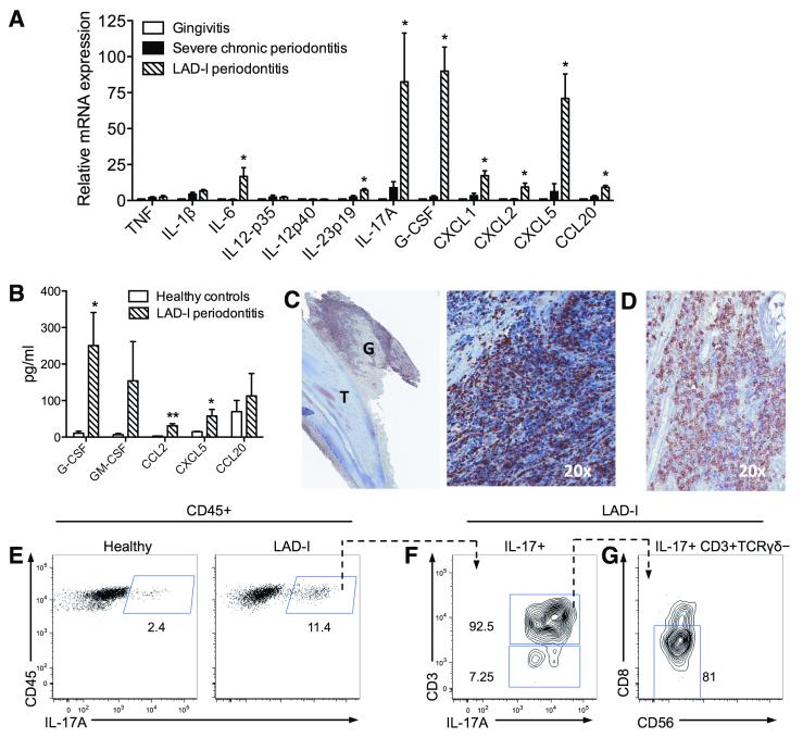
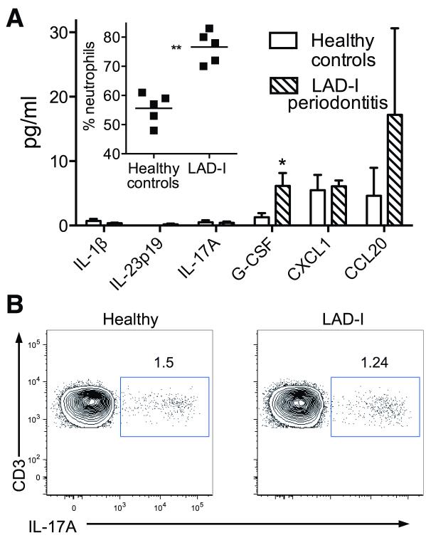
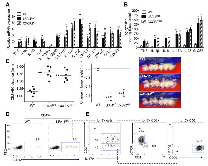
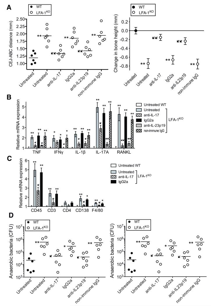
Leukocyte adhesion deficiency type I (LAD-I), a disease syndrome associated with frequent microbial infections, is caused by mutations on the CD18 subunit of β₂ integrins. LAD-I is invariably associated with severe periodontal bone loss, which historically has been attributed to the lack of neutrophil surveillance of the periodontal infection. We provide an alternative mechanism by showing that the cytokine interleukin-17 (IL-17) plays a major role in the oral pathology of LAD-I. Defective neutrophil recruitment in LAD-I patients or in LFA-1 (CD11a/CD18)-deficient mice--which exhibit the LAD-I periodontal phenotype--was associated with excessive production of predominantly T cell-derived IL-17 in the periodontal tissue, although innate lymphoid cells also contributed to pathological IL-17 elevation in the LFA-1-deficient mice. Local treatment with antibodies to IL-17 or IL-23 in LFA-1-deficient mice not only blocked inflammatory periodontal bone loss but also caused a reduction in the total bacterial burden, suggesting that the IL-17-driven pathogenesis of LAD-I periodontitis leads to dysbiosis. Therefore, our findings support an IL-17-targeted therapy for periodontitis in LAD-I patients.


Figures

Fig. 1
Fig. 1. Clinical and histological profile of LAD-I periodontitis
(A) Clinical attachment loss (marker of bone loss) was measured on all teeth of five 11-13 year-old LAD-I patients and healthy controls. Shown are mean measurements per individual (*P < 0.01; unpaired t test). (B) Correlation between clinical attachment loss and CD18 expression on peripheral neutrophils of LAD-I patients (Pearson correlation coefficient r2=0.8857; P=0.0170). CD18 expression on neutrophils evaluated by flow cytometry and expressed as percent of normal. (C) Gram staining (Brown and Hoops) on an extracted tooth and surrounding soft tissues: (i) Blue/pink positive staining on the tooth encircled and shown in higher magnification (ii); Soft tissues with no visible gram staining (iii). (D) H&E of extracted tooth and surrounding soft tissues: (i) Soft tissue surrounding the entire tooth indicating total loss of bone support. Encircled soft tissue in higher magnification (ii) revealing dense inflammatory infiltrate in the lesion compared to healthy gingival tissue from a control subject (iii). (E) Immunohistochemistry for major cell populations in the lesion: (i) neutrophils, inset showing positive cells within a vessel; (ii) T cells; (iii) plasma cells; (iv) macrophages; (v) γδ T cells; (vi) mast cells; (vii) DC; (viii) NK cells. C, D, and E representative of four patients and multiple extracted teeth. All original magnifications indicated.
Fig. 2
Fig. 2. IL-17 signature in LAD-I periodontitis
(A) Quantitative real-time PCR analysis of indicated cytokine mRNA expression levels in the lesions of LAD-I periodontitis compared to those from severe chronic periodontitis (severe inflammatory bone loss) or gingivitis (gingival inflammation without associated bone loss); results were normalized to HPRT mRNA and presented as fold induction relative to gingivitis, assigned an average value of 1. Data are means ± SEM (n=4 per group). *P < 0.05 vs. both chronic periodontitis and gingivitis (one-way ANOVA). (B) The indicated cytokines/chemokines were measured in gingival crevicular fluid from healthy control subjects and LAD-I patients, using multiplex luminex assays. Data are means ± SEM (n=5 per group) *P < 0.05 and **P < 0.01; unpaired t test. (C) Immunohistochemistry for IL-17A in LAD-I gingiva [G] surrounding an extracted tooth [T] (left). Numerous IL-17A+ cells are seen throughout the lesion, shown in larger magnification (right). (D) Immunohistochemistry for CD3 in IL-17A+ cell regions. C and D are serial sections, representative of 4 patients and multiple tooth sites. (E-G) Characterization of IL-17-producing cells in LAD-I periodontitis. (E) Flow cytometry after intracellular staining for IL-17A in isolated CD45+ gingival cells, from healthy (left) or LAD-I (right) subjects, stimulated with PMA and ionomycin. Plots F and G show further characterization of IL-17+ populations in LAD-I: (F) IL-17A versus CD3 staining gated on CD45+IL-17+ cells; (G) CD56 versus CD8 staining gated on CD45+IL-17+CD3+TCRγδ cells. Representative of two separate experiments with paired LAD-I versus healthy control comparisons.
Fig. 3
Fig. 3. Systemic responses in LAD-I patients
(A) The indicated cytokines/chemokines were measured in blood plasma from healthy control subjects and LAD-I patients using multiplex luminex assays. The inset shows the percentage of neutrophils in the peripheral blood of the two groups (each point represents an individual). Data are means ± SEM (n = 5 per group). *P < 0.05 and **P < 0.01; unpaired t test. (B) Flow cytometry after intracellular staining for IL-17A in PBMC, from healthy (left) or LAD-I (right) subjects, stimulated with PMA and ionomycin. The PBMC were gated on CD45+CD3+ cells and the plots are representative of four separate experiments with paired LAD-I versus healthy control comparisons.
Fig. 4
Fig. 4. Cytokine profiles in periodontitis associated with defective neutrophil adhesion and/or recruitment
18-week-old WT control mice were compared with age-matched LFA-1KO and CXCR2KO mice for cytokine levels and bone loss. (A, B) Gingiva were dissected to assess the indicated cytokine responses at the mRNA (A) or protein (B) level. Cytokine mRNA expression levels were normalized against GAPDH mRNA and expressed as fold induction relative to the transcript levels of 18-week-old WT mice, which were assigned an average value of 1. (C) Measurement of periodontal bone heights (CEJ-ABC distance) in the indicated mouse groups (left), calculation of relative bone loss (middle), and representative images of maxillae (right). In the left panel, each symbol represents an individual mouse and small horizontal lines indicate the mean. In the middle panel, bone loss was calculated as bone height in WT control mice (0 baseline) minus bone height in experimental mice. Data are means ± SEM (n = 6 mice/group) from one of two independent experiments with similar results. *P< 0.05 and **P< 0.01 compared with WT controls (one-way ANOVA). (D, E) Flow cytometry of cell preparations isolated from mouse gingiva stimulated with PMA and ionomycin. (D) IL-17 staining in CD45+ cells from WT and LFA-1KO mice. (E) Further characterization of IL-17+ populations in LFA-1KO mice. Plots shown from left to right; staining for CD3 versus IL-17 gated on CD45+IL-17+ cells; TCRγδ versus CD4 gated on CD45+IL-17+CD3+ cells; and lineage staining (CD3CD19CD5NK1.1CD11cCD11bLy6GCD117) versus CD90 gated on CD45+IL-17+CD3 cells. Data are representative of three independent experiments.
Fig. 5
Fig. 5. Treatment of LFA-1KO mice with anti-IL-17 or anti-IL-23 inhibits bone loss and reduces the bacterial burden
LFA-1KO mice were either left untreated or were treated locally in the gingiva with anti-IL-17A mAb (or IgG2a isotype control) or anti-IL-23p19 polyclonal antibody (or non-immune IgG), three times weekly from the age of 4 to 18 weeks. (A) Periodontal bone heights (CEJ-ABC distance) in the indicated mouse groups (left) and bone loss (right), calculated as bone height in 18-week-old WT control mice (0 baseline) minus bone height in experimental mice. (B, C) Quantitative real-time PCR analysis of the indicated cytokine (B) and cell-specific marker (C) mRNA expression in the periodontal tissue of mice treated as shown; results were normalized to those of GAPDH mRNA and are presented as fold induction relative to the transcript levels of untreated WT controls, which were assigned an average value of 1. (D) Quantification of cultivatable oral anaerobic bacteria (left) and determination of total bacterial burden in the periodontal tissue of the indicated mouse groups by real-time PCR of the 16S rRNA gene (right). Data were pooled from two independent experiments with three mice per group in each experiment (i.e., total of six mice per group). In A (left) and D, each symbol represents an individual mouse and small horizontal lines indicate the mean. In A (right), B, and C data are means ± SEM (n = 6 mice/group). *P< 0.05 and **P< 0.01 compared with untreated WT control (one-way ANOVA). P< 0.05 and ↙↙P< 0.01 compared to untreated LFA-1KO (one-way ANOVA).

References

    1. Kolaczkowska E, Kubes P. Neutrophil recruitment and function in health and inflammation. Nat. Rev. Immunol. 2013;13:159–175. - PubMed
    1. Hajishengallis G, Chavakis T. Endogenous modulators of inflammatory cell recruitment. Trends Immunol. 2013;34:1–6. - PMC - PubMed
    1. von Vietinghoff S, Ley K. Homeostatic regulation of blood neutrophil counts. J. Immunol. 2008;181:5183–5188. - PMC - PubMed
    1. Bugl S, Wirths S, Muller MR, Radsak MP, Kopp HG. Current insights into neutrophil homeostasis. Ann. N. Y. Acad. Sci. 2012;1266:171–178. - PubMed
    1. Summers C, Rankin SM, Condliffe AM, Singh N, Peters AM, Chilvers ER. Neutrophil kinetics in health and disease. Trends Immunol. 2010;31:318–324. - PMC - PubMed

Publication types

MeSH terms

Substances

Supplementary concepts

LinkOut - more resources