. 2014 Jun;4(6):716-29.
doi: 10.1158/2159-8290.CD-13-0743. Epub 2014 Mar 26.

p38MAPK plays a crucial role in stromal-mediated tumorigenesis

Affiliations

p38MAPK plays a crucial role in stromal-mediated tumorigenesis

Elise Alspach et al. Cancer Discov. 2014 Jun.

Abstract

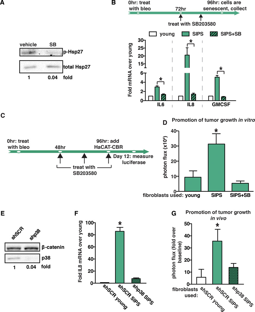
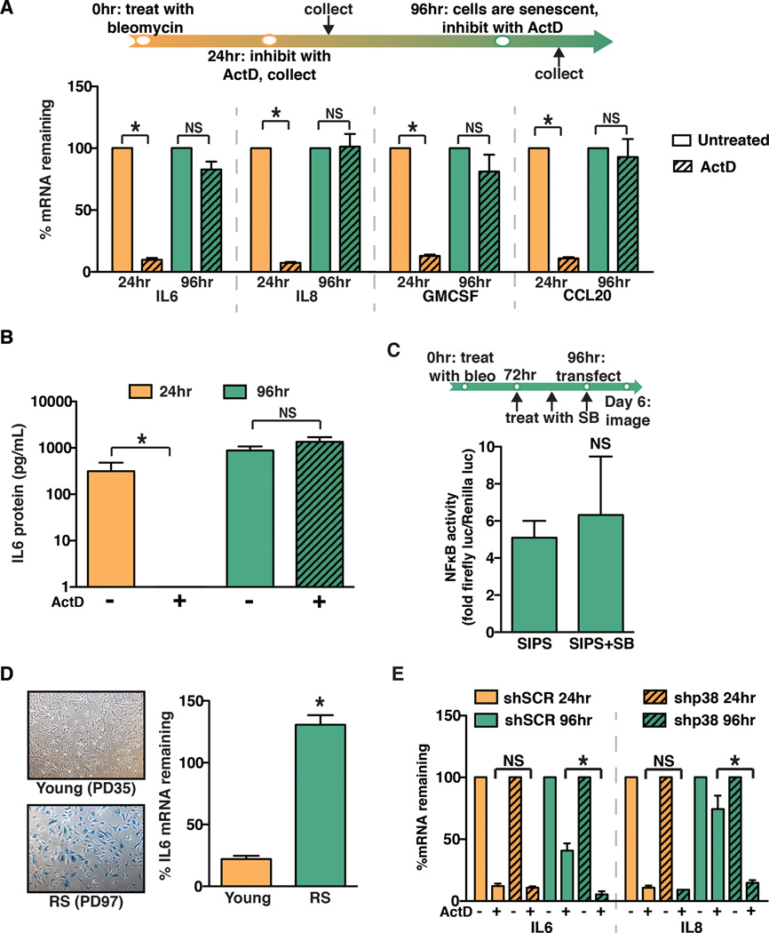
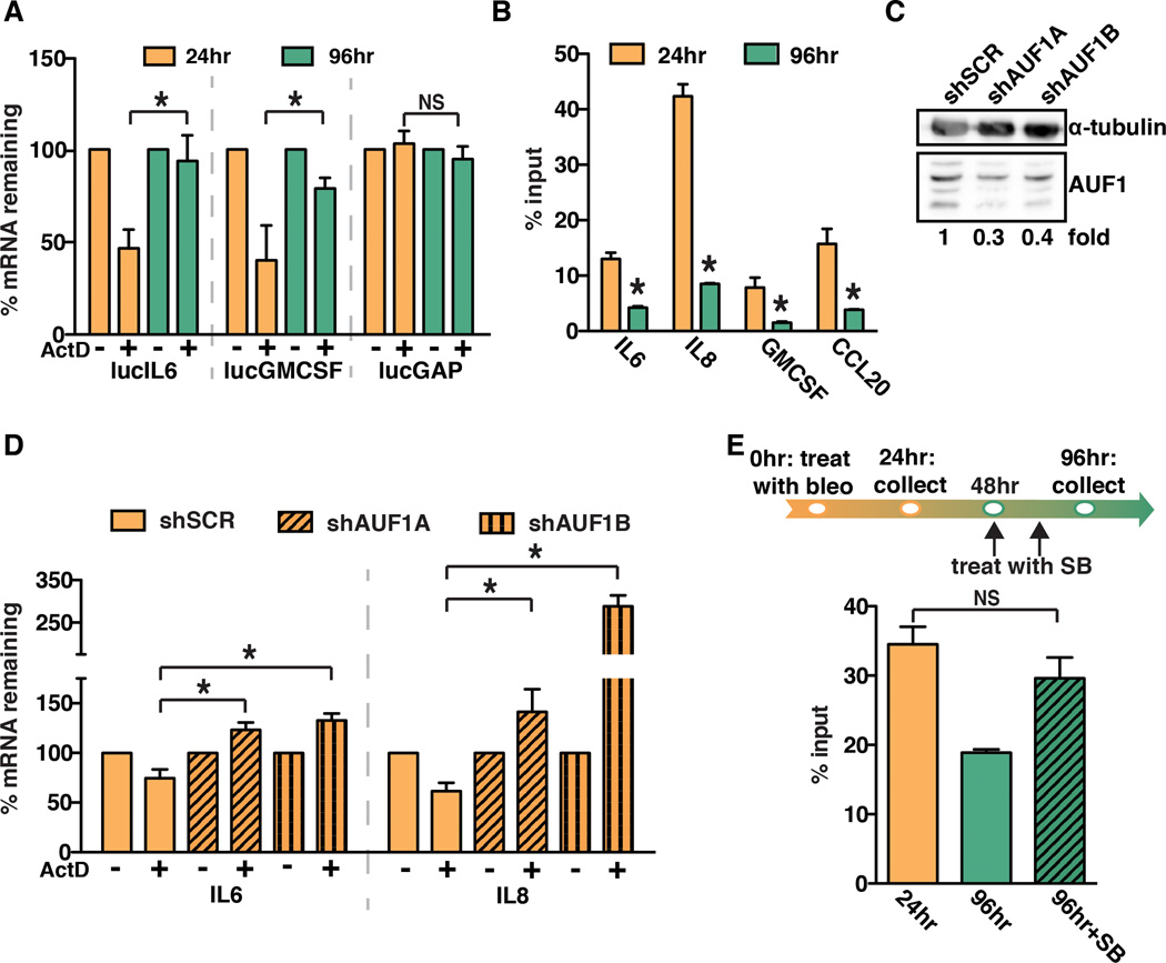
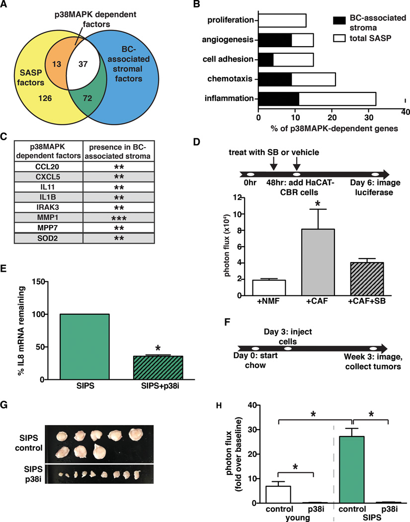
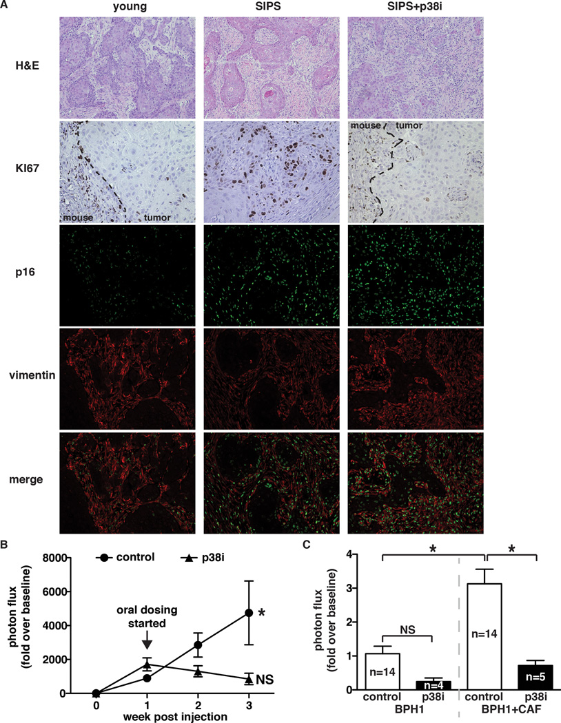
Neoplastic cells rely on the tumor microenvironment (TME) for survival and progression factors. Indeed, senescent and cancer-associated fibroblasts (CAF) express factors that promote tumorigenesis that are collectively referred to as the senescence-associated secretory phenotype (SASP). Despite their importance in tumorigenesis, the mechanisms that control TME-derived factor expression remain poorly understood. Here, we address a key unanswered question: how the SASP is sustained in senescent fibroblasts and CAFs. We find that the mitogen-activated protein kinase p38 (p38MAPK) controls AUF1 occupancy on SASP mRNAs and thus controls their stability. The importance of this regulatory mechanism is underscored by our findings that stromal-specific p38MAPK inhibition abrogates the tumor-promoting activities of CAFs and senescent fibroblasts. Our data suggest that targeting SASP mRNA stability through inhibition of p38MAPK will significantly aid the development of clinical strategies to target the TME.

Significance: The TME plays a key role in tumorigenesis. We demonstrate that p38MAPK governs a posttranscriptional mechanism that sustains the protumorigenic SASP. Inhibition of p38MAPK abrogates the tumor-promoting activities of CAFs and senescent fibroblasts. Thus, p38MAPK is a TME-specific Achilles' heel that may be exploited as a new therapeutic target.


Conflict of interest statement

Conflict of Interest: None

Figures

Fig. 1
Fig. 1. p38MAPK activity controls the pro-tumorigenic properties of the SASP
(A) Western blot analysis demonstrating that SB203580 treatment inhibits p38MAPK activity. As expected, SB203580 treatment significantly impacts phosphorylation of p38MAPK’s direct downstream target, Hsp27. Total Hsp27 was used as loading control. (B) Schematic of protocol to generate SIPS in BJ fibroblasts. Cells were treated with bleomycin for 24 hours. SB203580 (SB) treatment or vehicle control was initiated 48 hours after removal of bleomycin (bleo). 96 hours after bleomycin treatment, cells were collected for expression analysis of IL6, IL8, and GMCSF by qRT-PCR. SIPS: senescence induced premature senescence. Representative experiment, n=4. (C) Timeline of bleomycin (bleo) and SB203580 treatment of BJ fibroblasts in (d). SB203580 was replenished daily until co-culture with HaCAT-CBR cells was initiated. (D) Growth of human keratinocytes expressing click beetle red (HaCaT-CBR) measured 8 days following initiation of co-culture with indicated fibroblast populations. Representative experiment, n=3. (E) BJ fibroblasts were depleted of p38MAPK through the expression of shRNA (shp38) or control shRNA (shSCR). p38 depletion was verified by western blot analysis and β-catenin was used as a loading control. (F) Expression of IL8 was analyzed by qRT-PCR 96 hours following bleomycin treatment in p38MAPK-depleted (shp38) or control (shSCR) fibroblasts and represented relative to young fibroblasts expressing shSCR control. Representative experiment, n=3. (G) BJ fibroblasts expressing shp38 or shSCR were treated with bleomycin 72 hours prior to injection. Indicated fibroblast populations were admixed with preneoplastic epithelial cells expressing click beetle red (BPH1-CBR cells) and injected subcutaneously into the rear flanks of female NcR nude mice. Luciferase activity was measured using live, whole-animal imaging to monitor BPH1 cell growth relative to baseline signal. Data represents mean + SEM, n=8. Data represents mean + SD unless otherwise stated. * indicates p<0.05. SIPS: stress induced premature senescence.
Fig. 2
Fig. 2. p38MAPK post-transcriptionally regulates the SASP
(A) Schematic of protocol to generate SIPS in BJ fibroblasts. Cells were treated with bleomycin for 24 hours. Cells were subsequently treated with actinomycin D (ActD) for 24 hours. The ActD treatment was initiated 24 or 96 hours after the completion of bleomycin treatment. IL6, IL8, GMCSF, and CCL20 mRNA levels were analyzed by qRT-PCR. To account for changes in gene expression, levels mRNA in ActD-treated cells were normalized to the levels observed in untreated cells from the respective time points (% mRNA remaining). Representative experiment, n=3. (B) ELISA analysis of IL6 protein levels in conditioned media from cells treated as in (A). Representative experiment, n=4. (C) BJ fibroblasts were treated with bleomycin (bleo) for 24 hours and with SB203580 (SB) as indicated. 96 hours post bleomycin treatment cells were transiently transfected with an NFκB activity luciferase reporter. Luciferase activity was measured by live-cell imaging 48 hours post transfection. Representative experiment, n=2. (D) Young BJ fibroblasts (35 population doublings, PD) or replicatively senescent BJ fibroblasts (PD97) were stained for senescence-associated β-galactosidase to confirm senescent phenotype (left). Cells were treated with ActD and IL6 mRNA levels were analyzed by qRT-PCR. Representative experiment, n=3. (E) BJ fibroblasts expressing a control hairpin (shSCR) or shp38 were treated for 24 hours with ActD at 24 or 96 hours after the completion of bleomycin treatment. IL6 and IL8 mRNA levels were analyzed by qRT-PCR. Representative experiment, n=2. Data represent mean + SD. * indicates p<0.05. SIPS: stress-induced premature senescence.
Fig. 3
Fig. 3. AUF1 directly binds to SASP factor mRNA and modulates SASP factor stabilization
(A) BJ fibroblasts were stably transduced with luciferase constructs fused to the 3’ untranslated regions (UTR) of IL6, GMCSF, and GAPDH (lucIL6, lucGMCSF, and lucGAP). Cells were treated with ActD at 24 or 96 hours following bleomycin treatment. Luciferase mRNA levels were analyzed by qRT-PCR. Representative experiment, n=3. (B) RNA immunoprecipitation was performed for AUF1 using BJ fibroblast cell lysates collected 24 or 96 hours after bleomycin treatment. IL6, IL8, GMCSF, and CCL20 mRNA levels in immunoprecipitations were analyzed by qRT-PCR. Representative experiment, n=4. (C) BJ fibroblasts were transduced with shRNAs to deplete AUF1 (shAUF1A and shAUF1B) or a control shRNA (shSCR). Protein levels were analyzed by western blot analysis. Note: there are four AUF1 isoforms present and α-tubulin was used as a loading control. (D) 24 hours following bleomycin treatment, BJ fibroblasts expressing a control hairpin shSCR, shAUF1A, or shAUF1B were treated with ActD for 1 hour. IL6 and IL8 mRNA levels were analyzed by qRT-PCR. Representative experiment, n=2. (E) RNA immunoprecipitation for AUF1 was performed on BJ fibroblasts treated with bleomycin (bleo) and SB203580 (SB) as indicated. The level of IL8 mRNA in the AUF1 immunoprecipitation was measured by qRT-PCR. Representative experiment, n=3. Data represent mean + SD. * indicates p<0.05.
Fig. 4
Fig. 4. p38MAPK-dependent factors are expressed in the TME of breast cancer lesions
(A) RNA-seq analysis was performed on young fibroblasts, senescent fibroblasts, and senescent fibroblasts treated with SB203580. RNA-seq results were analyzed to determine the number of factors upregulated in response to senescence (SASP factors) and the number of p38MAPK-dependent factors. These results were also analyzed for overlap with the expression profiles of breast cancer (BC)-associated stroma. (B) GO processes analysis was performed on p38MAPK-dependent SASP factors. Results are presented as the percent of p38MAPK-depenedent genes assigned to the processes shown. Black regions of the bars represent the percent of p38MAPK-dependent SASP factors assigned to each process that are also expressed in BC-associated stroma. The significance threshold was set at p < 0.05. (C) p38MAPK-dependent SASP factors that are expressed in more than one BC-associated stroma data set. **indicates expression in 2 BC-associated stroma datasets, ***indicates expression in 3 BC-associated stroma datasets. (D) Tumor-educated human CAFs and their normal isogenic counterparts (NMF) were treated with SB203580 (SB) or vehicle as indicated and replenished daily until co-culture with HaCAT-CBR preneoplastic keratinocytes was initiated. Luciferase activity was measured using live-cell imaging 4 days following initiation of co-culture to monitor HaCaT cell growth. Representative experiment, n=2. (E) Senescent BJ fibroblasts in matrigel were injected subcutaneously into the rear flanks of nude mice fed either control or p38i chow. Cells were removed 10 days after injection and IL8 mRNA levels were measured using qRT-PCR. Representative experiment, n=4. (F, G, and H) Xenografts of BPH1-CBR cells co-injected with senescent BJ fibroblasts (SIPS) into female NcR nude mice. Control or the p38i compounded chow were performed as outlined in (F). Tumor are shown in (G). Tumor growth was analyzed by bioluminescence imaging (H). Data represent mean + SEM, n=8. Data represent mean + SD unless otherwise stated. * indicates p<0.05. SIPS: stress induced premature senescence.
Fig 5
Fig 5. p38MAPK inhibition is effective in both senescent fibroblast and CAF-driven tumors
(A) Tumors were removed at the endpoint of the experiment described in (Fig 4F) and stained for Ki67 (dashed line demarks the margin between the mouse and xenograft), p16, and vimentin. H&E images were captured with a 10× objective, all other images were captured with a 20× objective. Representative images, n=2. (B) Xenograft growth of BPH1-CBR cells co-injected with senescent BJ fibroblasts (SIPS) into female NcR nude mice. Tumors were allowed to grow for 1 week after injection, at which time mice were placed on control or p38i-compounded chow. Tumor growth was analyzed by bioluminescence imaging. Data represent mean + SEM, n=16. * indicates significance between 1 and 3 weeks post-injection in mice fed control chow. (C) Xenografts of BPH1-CBR cells co-injected with pCAFs into female NcR nude mice. Mice were fed control or p38i chow as outlined for the experiment in Fig 4F. Tumor growth was analyzed by bioluminescence imaging. Data represent mean + SEM, n is indicated for each sample. * indicates p<0.05. NS: not significant.

References

    1. Finak G, Bertos N, Pepin F, Sadekova S, Souleimanova M, Zhao H, et al. Stromal gene expression predicts clinical outcome in breast cancer. Nat Med. 2008;14:518–527. - PubMed
    1. Ma XJ, Dahiya S, Richardson E, Erlander M, Sgroi DC. Gene expression profiling of the tumor microenvironment during breast cancer progression. Breast Cancer Res. 2009;11:R7. - PMC - PubMed
    1. Karnoub AE, Dash AB, Vo AP, Sullivan A, Brooks MW, Bell GW, et al. Mesenchymal stem cells within tumour stroma promote breast cancer metastasis. Nature. 2007;449:557–563. - PubMed
    1. Olumi AF, Grossfeld GD, Hayward SW, Carroll PR, Tlsty TD, Cunha GR. Carcinoma-associated fibroblasts direct tumor progression of initiated human prostatic epithelium. Cancer research. 1999;59:5002–5011. - PMC - PubMed
    1. Dimri GP, Lee X, Basile G, Acosta M, Scott G, Roskelley C, et al. A biomarker that identifies senescent human cells in culture and in aging skin in vivo. Proceedings of the National Academy of Sciences of the United States of America. 1995;92:9363–9367. - PMC - PubMed

Publication types

MeSH terms