. 2014 May-Jun;6(3):728-39.
doi: 10.4161/mabs.28591. Epub 2014 Mar 26.

A novel tetravalent bispecific TandAb (CD30/CD16A) efficiently recruits NK cells for the lysis of CD30+ tumor cells

Affiliations

A novel tetravalent bispecific TandAb (CD30/CD16A) efficiently recruits NK cells for the lysis of CD30+ tumor cells

Uwe Reusch et al. MAbs. 2014 May-Jun.

Abstract

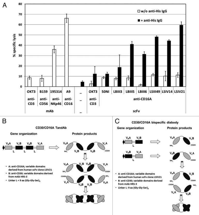
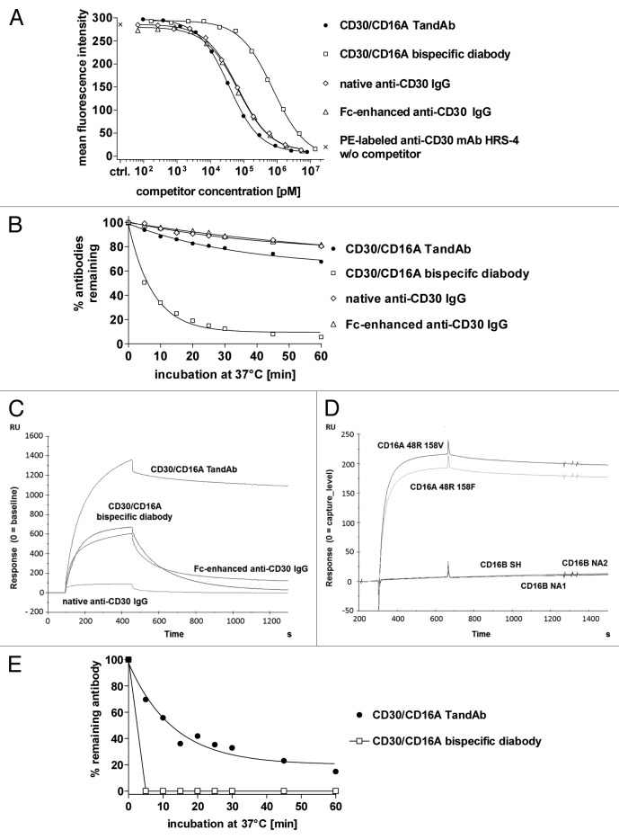
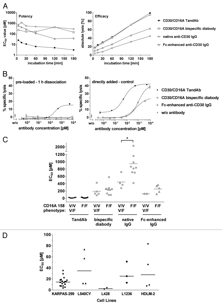
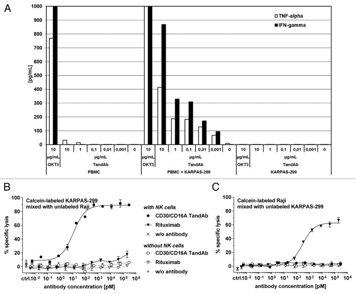
To improve recruitment and activation of natural killer (NK) cells to lyse tumor cells, we isolated a human anti-CD16A antibody with similar affinity for the CD16A 158F/V allotypes, but no binding to the CD16B isoform. Using CD16A-targeting Fv domains, we constructed a tetravalent bispecific CD30/CD16A tandem diabody (TandAb®) consisting solely of Fv domains. This TandAb has two binding sites for CD16A and two for CD30, the antigen identifying Hodgkin lymphoma cells. The binding and cytotoxicity of the TandAb were compared with antibodies with identical anti-CD30 domains: (1) a native IgG, (2) an IgG optimized for binding to Fc receptors, and (3) a bivalent bispecific CD30/CD16A diabody. Due to its CD16A-bivalency and reduced koff, the TandAb was retained longer on the surface of NK cells than the IgGs or the diabody. This contributed to the higher potency and efficacy of the TandAb relative to those of the other anti-CD30 antibodies. TandAb cytotoxicity was independent of the CD16A allotype, whereas the anti-CD30 IgGs were substantially less cytotoxic when NK cells with low affinity CD16A allotype were employed. TandAb activation of NK cells was strictly dependent on the presence of CD30(+) target cells. Therefore, the CD30/CD16A TandAb may represent a promising therapeutic for the treatment of Hodgkin's lymphoma; further, anti-CD16A TandAbs may function as potent immunotherapeutics that specifically recruit NK cells to destroy cancer cells.

Keywords: CD16A; FcγRIIIA; Hodgkin’s lymphoma; NK cells; bispecific antibodies.


Figures

None
Figure 1. Selection of anti-CD16A-specific scFv for activation of NK cells and construction of bispecific CD30/CD16A TandAb and diabody. (A) Induction of redirected cell lysis with affinity matured anti-CD16A scFvs. 1 × 10⁴ calcein-labeled FcγRII+ murine mastocytoma P-815 target cells were incubated in triplicate with freshly isolated and enriched human NK cells at an effector-to-target ratio of 10:1 in the presence of 1 µg/mL of the indicated scFv or IgGs with or without the addition of 1 µg/mL anti-His IgG1, clone 13/45/31–2, for 3 h. Percentage of specific target cell lysis was calculated from the measured fluorescence counts in the culture supernatant. Mean values and standard deviations from triplicates are plotted. (B and C) Schematic representation of the gene organization and domain order of the bispecific CD30/CD16A TandAb (B) and diabody (C). VHA and VLA represent the variable anti-CD16A domains that are derived from the human scFv clone LSIV21 isolated from Affimed's phage display library. VHB and VLB stand for the murine anti-CD30 Fv domains that are derived from the HRS-3 IgG. The HRS-3 hybridoma-derived light chain contains two mutations in framework 1: tyrosine at L23, instead of the conserved cysteine that forms a disulfide bond with CysL88, and asparagine at L20, which is a potential glycosylation site. The residues at L20 and L23 were restored to the original amino acids in the germline sequence. The linker peptides (L) are of 9 amino acids length and consist of three Gly-Gly-Ser repeats.
None
Figure 2. Binding of the TandAb, the diabody and IgGs to CD30 and CD16A. (A) Competition assay. 1x10⁶ KARPAS-299 cells were incubated with 3 µg/mL (~20 nM) PE-conjugated anti-CD30 HRS-4 IgG together with the indicated concentrations of recombinant bispecific antibodies or anti-CD30 IgGs for 45 min at 37 °C. After washing, the fluorescence of 1 × 10⁴ cells was measured with a flow cytometer, corrected for background staining with the corresponding isotype control and analyzed by nonlinear regression for calculation of IC50 and KD values. One out of two experiments is depicted. (B) Cell surface retention at 37 °C. Aliquots of KARPAS-299 cells were incubated with biotinylated CD30/CD16A TandAb and diabody or anti-CD30 IgG for 45 min on ice. After removing excess antibodies by washing, cells were incubated at 37 °C for the indicated periods of time. After repeated washing on ice, remaining antibodies were detected using Dylight488-conjugated streptavidin, and the fluorescence of 1 × 10⁴ cells was analyzed by flow cytometry. The mean fluorescence values from time-point 0 were set to 100% and the percentage of remaining antibodies was determined using nonlinear regression analysis. Data shown are representative of two independent experiments. (C) The CD16A 48R158V Fc-fusion protein was covalently immobilized on a CM5 chip. The different antibodies (CD30/CD16A TandAb and diabody, and anti-CD30 IgGs,) were injected with a flow rate of 10 µL/min for 360 s and the dissociation time was set to 600 s. Background signals in the control flow cell (without immobilized antigen) were subtracted from signals in the test flow cell. Chips were regenerated with 10 mM Glycine-HCL, pH 2.0. (D) The CD16A allotypes, 48R158V and 48R158F, and the CD16B allotypes, SH, NA1 and NA2, were expressed in HEK-293 cells as Fc-fusion proteins and directly immobilized on chips. TandAb was injected with a flow rate of 10 µL/min for 360 s. Background signals in the control flow cell (without immobilized antigen) were subtracted from signals in the test flow cell. Dissociation time was set to 600 s. Chips were regenerated with 10 mM Glycine-HCL, pH 2.0. (E) Cell surface retention assay. 1 × 10⁶ enriched human NK cells were stained with 1 µM of CD30/CD16A TandAb or diabody on ice, washed and incubated for indicated periods of time at 37 °C to allow dissociation. After washing, remaining antibodies were detected by anti-His IgG followed by FITC-conjugated goat anti-mouse IgG. The mean fluorescence values from time-point 0 were set to 100% and the percentage of remaining antibodies was plotted using nonlinear regression.
None
Figure 3. Comparison of CD30/CD16A TandAb and diabody, anti-CD30 IgG and Fc-enhanced anti-CD30 IgG in cytotoxicity assays. (A) Kinetics of antibody-mediated target cell lysis. 1 × 10⁴ calcein-labeled KARPAS-299 target cells were incubated for the indicated time periods with increasing concentrations of CD30/CD16A TandAb, CD30/CD16A diabody, anti-CD30 IgG and Fc-enhanced anti-CD30 IgG together with freshly isolated human NK cells at an E:T ratio of 5:1. Percent specific target cell lysis was calculated from the fluorescent calcein released into the cell culture supernatant from apoptotic target cells. EC50 values (potency) and maximal target cell lysis (efficacy) were determined by nonlinear regression for all antibodies and plotted. (B) Residual cytotoxicity of pre-opsonized effector cells to measure antibodies’ retentions. Freshly isolated human NK cells were incubated with increasing concentrations of CD30/CD16A TandAb, diabody and Fc-enhanced anti-CD30 IgG for 15 min at 37 °C, washed twice and then incubated for 1 h at 37 °C to allow dissociation of the bound antibodies. After an additional washing step, NK cells were used as effector cells in a 3 h cytotoxicity assay with calcein-labeled KARPAS-299 target cells at an E:T ratio of 4.4:1 (pre-loaded – 1 h dissociation). As a control, NK cells were pre-incubated in the absence of antibodies before they were used as effector cells together with added antibodies in the same cytotoxicity assay (directly added - control). Mean values and SD from duplicates of one out of two independent experiments are shown. (C) Potency of TandAb, diabody and IgG in relation to the CD16A 158 polymorphism. The EC50 values for TandAb, diabody and anti-CD30 IgG were determined in several independent 3 h cytotoxicity assays on KARPAS-299 target cells with NK cells as effector cells isolated from unrelated donors as described and plotted in the diagram together with the mean values shown as bars. The CD16A 158 phenotype of the NK cells was assessed by flow cytometry after staining of the NK cells with the CD16 158V-specific mAb MEM-154. NK cells were rated as CD16A 158F/F when the fluorescence signal was at background level. The asterisk (*) indicates statistical significance (P < 0.05). (D) Cytotoxic potency of the TandAb against a panel of five CD30+ cell lines. The EC50 values of the TandAb were determined in independent 3 h cytotoxicity assays on target CD30+ cells, with NK cells as effectors, isolated from independent donors, at a 1:5 ratio. Mean values for each cell line are shown as horizontal bars.
None
Figure 4. Specificity and safety in vitro. (A) Cytokine release in PBMC cultures in the presence of the CD30/CD16A TandAb. 5 × 10⁵ human PBMC with and without 1 × 10⁴ CD30+ KARPAS-299 cells, or 1 × 10⁴ KARPAS-299 cells alone were cultured in the presence of increasing concentrations of the CD30/CD16A TandAb (denoted by TandAb; at 0.001–10 µg/mL), 10 µg/mL OKT3, or without antibody (denoted by 0). The concentrations of secreted TNF and IFN-γ were quantified using multiplexing after 24 h incubation. Results from one representative experiment are shown. (B and C) Assessment of bystander cell killing in a cytotoxicity assay with mixed target cells. 1 × 10⁴ calcein-labeled CD20-/CD30+ KARPAS-299 target cells, combined with 1x10⁴ unlabeled CD20+/CD30- Raji target cells (B) or 1 × 10⁴ unlabeled KARPAS-299 combined with calcein-labeled Raji cells (C), were incubated for 3 h, together with increasing concentrations of the CD30/CD16A TandAb or rituximab (MabThera, Roche, Hertfordshire, UK), in the presence or absence of 5 × 10⁴ enriched human NK cells as effectors for 3 h in a cytotoxicity assay. The percentage of specific target cell lysis was calculated and the mean and SD from duplicates were plotted.

References

    1. Mandelboim O, Malik P, Davis DM, Jo CH, Boyson JE, Strominger JL. Human CD16 as a lysis receptor mediating direct natural killer cell cytotoxicity. Proc Natl Acad Sci U S A. 1999;96:5640–4. doi: 10.1073/pnas.96.10.5640. - DOI - PMC - PubMed
    1. Robertson MJ, Ritz J. Biology and clinical relevance of human natural killer cells. Blood. 1990;76:2421–38. - PubMed
    1. Cooper MA, Fehniger TA, Caligiuri MA. The biology of human natural killer-cell subsets. Trends Immunol. 2001;22:633–40. doi: 10.1016/S1471-4906(01)02060-9. - DOI - PubMed
    1. van de Winkel JG, Capel PJ. Human IgG Fc receptor heterogeneity: molecular aspects and clinical implications. Immunol Today. 1993;14:215–21. doi: 10.1016/0167-5699(93)90166-I. - DOI - PubMed
    1. van Spriel AB, van Ojik HH, van De Winkel JG. Immunotherapeutic perspective for bispecific antibodies. Immunol Today. 2000;21:391–7. doi: 10.1016/S0167-5699(00)01659-5. - DOI - PubMed

Publication types

MeSH terms