. 2014 Jun;55(6):1120-30.
doi: 10.1194/jlr.M047548. Epub 2014 Mar 26.

The macrophage LBP gene is an LXR target that promotes macrophage survival and atherosclerosis

Affiliations

The macrophage LBP gene is an LXR target that promotes macrophage survival and atherosclerosis

Tamer Sallam et al. J Lipid Res. 2014 Jun.

Abstract

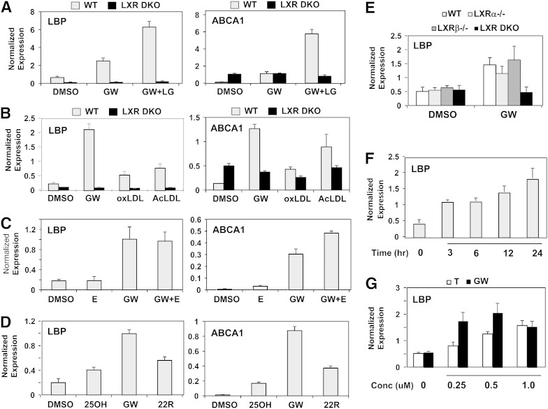
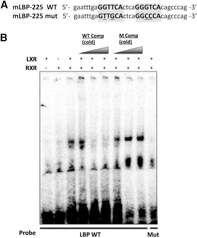
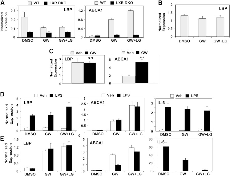
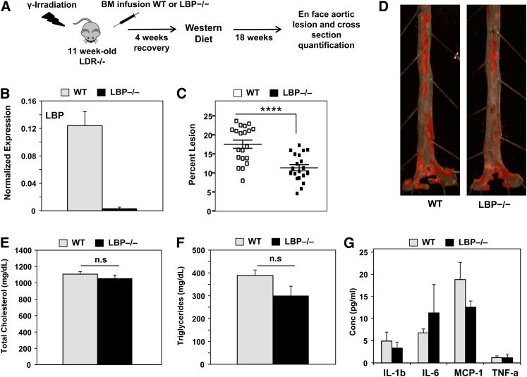
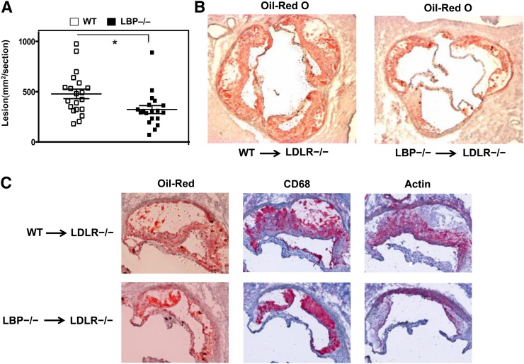
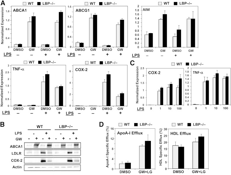
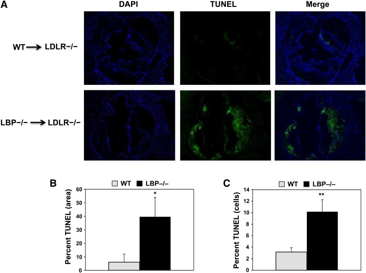
The liver X receptors (LXRs) are members of the nuclear receptor superfamily that regulate sterol metabolism and inflammation. We sought to identify previously unknown genes regulated by LXRs in macrophages and to determine their contribution to atherogenesis. Here we characterize a novel LXR target gene, the lipopolysaccharide binding protein (LBP) gene. Surprisingly, the ability of LXRs to control LBP expression is cell-type specific, occurring in macrophages but not liver. Treatment of macrophages with oxysterols or loading with modified LDL induces LBP in an LXR-dependent manner, suggesting a potential role for LBP in the cellular response to cholesterol overload. To investigate this further, we performed bone marrow transplant studies. After 18 weeks of Western diet feeding, atherosclerotic lesion burden was assessed revealing markedly smaller lesions in the LBP(-/-) recipients. Furthermore, loss of bone marrow LBP expression increased apoptosis in atherosclerotic lesions as determined by terminal deoxynucleotidyl transferase dUTP nick end labeling staining. Supporting in vitro studies with isolated macrophages showed that LBP expression does not affect cholesterol efflux but promotes the survival of macrophages in the setting of cholesterol loading. The LBP gene is a macrophage-specific LXR target that promotes foam cell survival and atherogenesis.

Keywords: atherogenesis; lipopolysaccharide binding protein; liver X receptor; nuclear receptor.


Figures

Fig. 1.
Fig. 1.
LXR regulates LBP expression in macrophages. A: Primary mouse peritoneal macrophages were treated with GW3965 (GW, 1 μM) and/or the RXR ligand LG268 (LG, 100 nM). Gene expression in this and all subsequent figures was analyzed by real-time PCR. Results are representative of three independent experiments. Values are mean ± SD. B: Primary bone marrow-derived macrophages were treated with GW3965 (GW, 1 μM), oxidized LDL (oxLDL, 50 μg/ml), or acetylated LDL (AcLDL, 50 μg/ml) for 60 h. Results are representative of four independent experiments. Values are mean ± SD. C: Primary peritoneal mouse macrophages were treated with GW3965 (GW, 1 μM) and/or the protein synthesis inhibitor emetine (E, 5 μg/ml). Results are representative of three independent experiments. Values are mean ± SD. D: Primary mouse peritoneal macrophages were treated with GW3965 (GW, 1 μM), endogenous ligand 22(R)-hydroxycholesterol (22R, 2.5 μM), or 25-hydroxycholesterol (25OH, 2.5 μM). E: Primary mouse peritoneal macrophages were treated with GW3965 (GW, 1 μM) overnight. Results are representative of two independent experiments. Values are mean ± SD. F: Primary mouse peritoneal macrophages were treated with GW3965 (GW, 0.5 μM). Results are representative of two independent experiments. Values are mean ± SD. G: Primary mouse peritoneal macrophages were treated with the LXR ligands GW3965 (GW, 0.5 μM) or T0901317 (T, 1 μM). Results are representative of three independent experiments. Values are mean ± SD. DKO, double knockout; Conc, concentration.
Fig. 2.
Fig. 2.
LXR/RXR heterodimers bind to a DR4 site on the LBP promoter. A: WT and mutant (mut) LXR DR4 binding sites 225 bp upstream of the LBP transcription start site. B: EMSA using labeled oligonucleotides (32-P) to either the WT or the mutant LXR DR4 sites, in vitro translated LXRα or RXRα protein. Competition (Comp) assays were performed by adding increasing concentrations of nonlabeled WT or mutant (M) LBP probe.
Fig. 3.
Fig. 3.
LBP regulation by LXRs and LPS is cell-type selective. A: Primary mouse hepatocytes were treated with GW3965 (GW, 1 μM) overnight. Results are representative of three independent experiments. Values are mean ± SD. B: Primary mouse hepatocytes were treated with GW3965 (GW, 1 μM) overnight. Results are representative of four independent experiments. Values are mean ± SD. C: LBP expression in livers of WT mice treated with 40 mg/kg/day GW3956 by oral gavage for 3 days (n = 5 per group). Values are mean ± SEM. D: Primary mouse hepatocytes were treated with GW3965 (GW, 1 μM) and/or the RXR ligand LG268 (LG, 50 nM) in the absence or presence of LPS stimulation (100 ng/ml). Results are representative of three independent experiments. Values are mean ± SD. E: Primary mouse peritoneal macrophages were treated with GW3965 (GW, 1 μM) and/or the RXR ligand LG268 (LG, 50 nM) in the absence or presence of LPS stimulation (100 ng/ml). Results are representative of three independent experiments. Values are mean ± SD. DKO, double knockout; Veh, vehicle.
Fig. 4.
Fig. 4.
Bone marrow expression of LBP promotes atherogenesis in LDLR-deficient mice. A: Schematic outline of bone marrow (BM) transplantation protocol. B: Confirmation of engraftment by gene expression analysis. Results are representative of three biological replicates. Values are mean ± SEM. C: Percentage of aorta surface area with atherosclerotic plaque in transplanted LDLR−/− mice (n = 20/group). Horizontal lines indicate mean ± SEM; ****p < 0.0001. D: Representative photographs from en face analysis of aortas from WT or LBP−/− transplanted mice after 18 weeks on a Western diet. Twenty mice in each group were analyzed. E: Total serum cholesterol levels were measured. Values are mean ± SEM. F: Total serum triglyceride levels were measured. Values are mean ± SEM. G: Serum cytokine levels were measured using a Milliplex mouse cytokine/chemokine panel (Millipore). Values are mean ± SEM. n.s, not significant.
Fig. 5.
Fig. 5.
LBP deletion in macrophages decreases the development of atherosclerosis. A: Quantification of aortic root lesions (n = 19–20 per group). Horizontal lines indicate mean ± SEM. B: Frozen sections from the aortic roots of bone marrow transplanted LDLR−/− mice with WT or LBP−/− maintained on a Western diet for 19 weeks. Lesions were stained with Oil-Red O (objective magnification: ×5). C: Representative histological data of the aortic valve area stained with Oil-Red O, the macrophage marker CD68, and α-smooth muscle actin (objective magnification: ×10).
Fig. 6.
Fig. 6.
LBP expression does not affect cholesterol efflux or inflammatory responses. A: Primary bone marrow-derived macrophages were treated with GW3965 (GW) overnight with (+) or without (−) LPS (10 ng/ml). Results are representative of two experiments. Values are mean ± SD. B: Western blot analysis of bone marrow-derived macrophages treated with GW3965 overnight with or without LPS (10 ng/ml). C: Bone marrow-derived macrophages treated with increasing concentration of LPS (ng/ml). Results are representative of two experiments. Values are mean ± SD. D: Cholesterol efflux assays. WT or LBP−/− macrophages were loaded with [3H]cholesterol (1.0 μCi/ml) in the presence of acyl-CoA:cholestrol O-acyltransferase inhibitor (2 μg/ml) either with DMSO or with ligand for LXR and RXR [1 μM GW3965, 50 nM LG268 (LG)]. Efflux was measured in the presence of apoA-I or HDL. Experiments were conducted in triplicate. Data are expressed as mean ± SD.
Fig. 7.
Fig. 7.
Loss of LBP from hematopoietic cells enhances apoptosis in atherosclerotic lesions. A: Frozen sections from the aortic roots were stained for 4’,6-diamidino-2-phenylindole (DAPI) and TUNEL (blue and green respectively). Representative samples are shown (objective magnification: ×5). B: Percent TUNEL-positive cells were quantified by TUNEL-positive area; *p < 0.05. C: Percent DAPI-stained cells that were TUNEL positive (n = 7 mice per group). Values are mean ± SEM; **p < 0.005.
Fig. 8.
Fig. 8.
LBP expression modulates apoptotic signaling in macrophages. A: Bone marrow-derived macrophages were obtained at time of harvest from transplanted LDLR−/− mice placed on a Western diet. Bone marrow from four individual mice was analyzed. Values are mean ± SD. B: RAW264.7 cells stably expressing LBP were treated with acetylated LDL for 3 days. Gene expression was analyzed by real-time PCR. Results are representative of three independent experiments. Values are mean ± SD; *p < 0.05, **p < 0.005.

Similar articles

Cited by

References

    1. Go A. S., Mozaffarian, Roger D., Benjamin V. L., Berry E. J., Borden J. D., Bravata W. B., Dai D. M., Ford S., Fox E. S., et al. 2013. Heart disease and stroke statistics–2013 update: a report from the American Heart Association. Circulation. 127: e6–e245. - PMC - PubMed
    1. Yang Q., Cogswell M. E., Flanders W. D., Hong Y., Zhang Z., Loustalot F., Gillespie C., Merritt R., Hu F. B. 2012. Trends in cardiovascular health metrics and associations with all-cause and CVD mortality among US adults. JAMA. 307: 1273–1283. - PMC - PubMed
    1. Weber C., Noels H. 2011. Atherosclerosis: current pathogenesis and therapeutic options. Nat. Med. 17: 1410–1422. - PubMed
    1. Zadelaar S., Kleemann R., Verschuren L., de Vries-Van der Weij J., van der Hoorn J., Princen H. M., Kooistra T. 2007. Mouse models for atherosclerosis and pharmaceutical modifiers. Arterioscler. Thromb. Vasc. Biol. 27: 1706–1721. - PubMed
    1. Hansson G. K. 2005. Inflammation, atherosclerosis, and coronary artery disease. N. Engl. J. Med. 352: 1685–1695. - PubMed

Publication types

MeSH terms