. 2014 May 15;306(10):C972-85.
doi: 10.1152/ajpcell.00110.2013. Epub 2014 Mar 26.

Hypoxia-induced changes in Ca(2+) mobilization and protein phosphorylation implicated in impaired wound healing

Affiliations

Hypoxia-induced changes in Ca(2+) mobilization and protein phosphorylation implicated in impaired wound healing

Albert Lee et al. Am J Physiol Cell Physiol. .

Abstract

The process of wound healing must be tightly regulated to achieve successful restoration of injured tissue. Previously, we demonstrated that when corneal epithelium is injured, nucleotides and neuronal factors are released to the extracellular milieu, generating a Ca(2+) wave from the origin of the wound to neighboring cells. In the present study we sought to determine how the communication between epithelial cells in the presence or absence of neuronal wound media is affected by hypoxia. A signal-sorting algorithm was developed to determine the dynamics of Ca(2+) signaling between neuronal and epithelial cells. The cross talk between activated corneal epithelial cells in response to neuronal wound media demonstrated that injury-induced Ca(2+) dynamic patterns were altered in response to decreased O2 levels. These alterations were associated with an overall decrease in ATP and changes in purinergic receptor-mediated Ca(2+) mobilization and localization of N-methyl-d-aspartate receptors. In addition, we used the cornea in an organ culture wound model to examine how hypoxia impedes reepithelialization after injury. There was a change in the recruitment of paxillin to the cell membrane and deposition of fibronectin along the basal lamina, both factors in cell migration. Our results provide evidence that complex Ca(2+)-mediated signaling occurs between sensory neurons and epithelial cells after injury and is critical to wound healing. Information revealed by these studies will contribute to an enhanced understanding of wound repair under compromised conditions and provide insight into ways to effectively stimulate proper epithelial repair.

Keywords: cell communication; hypoxia; imaging; wound healing.


Figures

Fig. 1.
Fig. 1.
Sensory nerves penetrate the corneal epithelium. A and B: green fluorescent protein-tagged Thy-1 (Thy-1-GFP) rat corneas were imaged using 2-photon microscopy, and corneal nerves were detected en face and in cross section. Epithelium (E) and stroma (S) are labeled for orientation. C: corneas were counterstained with rhodamine phalloidin and 4′,6-diamidino-2-phenylindole (DAPI) and imaged using confocal microscopy. A network of nerve endings (green) derived from stromal branches terminate in the epithelium (yellow). In B and C, z stacks are presented as a 3-dimensional image on the xyz-axis. Scale bars: 200 μm (A and B) and 50 μm (C).
Fig. 2.
Fig. 2.
Hypoxia diminishes cross talk between epithelia and nerves in the cornea. Human corneal limbal epithelial (HCLE) cells were cultured under normoxia or hypoxia for 24 h, washed, and incubated with fluo 3-AM. Conditioned or neuronal wound medium was collected from cultures incubated under normoxia or hypoxia. After basal levels were obtained prior to stimulation, the epithelial response was monitored for >600 frames (0.789 s/frame). Pseudocolored 2.5-dimensional images show the cell response over time using a 6-color intensity scale. A: Ca2+ mobilization of epithelial cells in response to control conditioned media. B: Ca2+ mobilization of epithelial cells incubated under normoxic conditions in response to normoxic neuronal wound media. C: Ca2+ mobilization of epithelial cells incubated under hypoxic conditions in response to hypoxic neuronal wound media. D: Ca2+ mobilization of epithelial cells incubated under normoxic conditions in response to hypoxic neuronal wound media. E: Ca2+ mobilization of epithelial cells incubated under hypoxic conditions in response to normoxic neuronal wound media. Images are representative of ≥22 independent experiments for each condition.
Fig. 3.
Fig. 3.
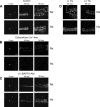
Ca2+ mobilization between epithelial cells in response to neuronal wound media. HCLE cells were cultured under normoxia or hypoxia for 24 h, washed, and incubated with fluo 3-AM. Conditioned (CM) or neuronal wound media were collected from cultures incubated under normoxia (Nx) or hypoxia (Hx). Epithelial response to the media was monitored for >600 frames at 0.789 s/frame. Data were exported into MATLAB software written for analysis. A: percent change in average fluorescence of HCLE cells in response to neuronal wound media. B: percentage of cells activated over time in response to neuronal wound media. C: percentage of cells participating in cluster formation in response to neuronal wound media. D: number of clusters per 7.89 s (10 frames) in response to neuronal wound media. E: N-methyl-d-aspartate (NMDA) receptor 1 localization is altered under hypoxic conditions in HCLE cells. Unwounded HCLE cells were incubated under normoxia or hypoxia for 24 or 48 h and stained for NMDA receptor 1 (green) and counterstained with DAPI (blue) and rhodamine phalloidin (red). Tiled confocal images were collected, and representative images are shown. NC, negative control. Scale bar, 50 μm. Traces in A–D are representative of ≥22 independent experiments; images in E are representative of 3 independent experiments.
Fig. 4.
Fig. 4.
Hypoxia alters Ca2+ mobilization. Cells were cultured under hypoxia or normoxia for 24 h and incubated with fluo 3-AM, and the response to injury was monitored. A series of images were taken over 0–90 s at 0.789 s/frame under various conditions, and distance of Ca2+ mobilization after injury was determined. Images are representative of ≥3 independent experiments with 13 individual runs/condition. Bracket indicates distance in mobilization from wound origin (∗). A: time series of Ca2+ mobilization after injury. B: time series of Ca2+ mobilization after preincubation in Ca2+-free HEPES buffer for 20 min followed by injury. C: time series of Ca2+ mobilization after preincubation in BAPTA-AM (25 μM) for 20 min followed by injury. D: Ca2+ mobilization at 0 and 90 s after injury of cultures preincubated for 20 min in the presence (+) or absence (−) of 1 μM thapsigargin (TG). Scale bars, 200 μm.
Fig. 5.
Fig. 5.
Role of mitochondrial uncoupling. A: initial fluorescence intensity of epithelial cells loaded with 7.5 nM tetramethylrhodamine ethyl ester (TMRE) after 24 h under hypoxic or normoxic conditions. Values are means ± SE of 7 experiments. B: mitochondrial depolarization in response to 20 μM carbonyl cyanide 4-(trifluoromethoxy)phenylhydrazone (FCCP) added at time indicated by arrow under normoxic and hypoxic conditions. Decrease in TMRE fluorescence indicates loss of mitochondrial membrane potential. Cells were incubated in normoxic (⧫) or hypoxic (◇) conditions for 24 h and imaged every 789 ms. FCCP (20 μM) was added at time indicated by arrow. Traces are representative of 7 independent experiments. C: ATP-induced Ca2+ mobilization after mitochondrial depolarization. Epithelial cells were stimulated repeatedly with 100 μM ATP in the presence (◇) or absence (⧫) of 20 μM FCCP. Traces are averages of 5 independent experiments.
Fig. 6.
Fig. 6.
Hypoxia alters ATP released with injury. A: injury releases less ATP under hypoxic conditions. Epithelia were cultured under normoxic or hypoxic conditions for 24 or 48 h. Media from unwounded (C) or wounded (W) cell cultures were harvested, and ATP released into the medium was immediately determined using a bioluminescence assay. B: trigeminal neuronal cells were cultured and incubated under normoxic or hypoxic conditions for 4 h. Media from cell cultures were harvested, and ATP released into the medium was determined immediately using a bioluminescence assay. C: total ATP in cells is decreased under hypoxia. After incubation under hypoxic or normoxic conditions, epithelia were immersed in boiling water, and whole cell ATP was determined. Values are means ± SE from ≥3 independent experiments. *P < 0.05 (by Student's t-test).
Fig. 7.
Fig. 7.
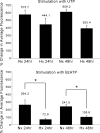
Hypoxia alters nucleotide-induced Ca2+ mobilization. Epithelial cells incubated for 24 or 48 h in normoxia or hypoxia were loaded with fluo 3-AM and stimulated with 100 μM UTP or 2′,3′-O-(4-benzoylbenzoyl)-ATP (BzATP) using a flow-through system, and images were collected at 0.789 s/frame. Values (means ± SE of ≥3 independent experiments) are presented as calculated maximum percent change in average fluorescence. *P < 0.05 (by Student's t-test).
Fig. 8.
Fig. 8.
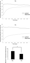
Hypoxia decreases cell communication. Epithelia were incubated under normoxic or hypoxic conditions for 24 h and loaded with 5-carboxyfluorescein diacetate acetoxymethyl ester (5-CFDA-AM). Cells were selected using the region of interest tool and photobleached, while control unbleached cells were also monitored over time. Fluorescence recovery of bleached cells over time is shown as percent change in average fluorescence and compared with control. Maximal fluorescence recovery at the end point (400 frames) is also shown. Data represent ≥7 independent experiments. *P < 0.05 (by Student's t-test).
Fig. 9.
Fig. 9.
Hypoxia diminishes activation of paxillin during migration. A: phosphorylation of paxillin at Y118 is reduced under hypoxia. Epithelial cells were incubated for 24 h under normoxic or hypoxic conditions and wounded, and respective wound media were collected immediately. Normoxic media were added immediately to normoxic cells in culture, and hypoxic media were added immediately to hypoxic cells. Protein lysates were collected at designated time points. Phosphorylation of paxillin at Y118 was normalized to total paxillin. Data are presented as ratio of phosphorylated to total paxillin [arbitrary units (AU)]. Values are means ± SE of 3 independent experiments. *P < 0.05 (by Student's t-test). B and C: recruitment of paxillin to the wound edge and presence of lamellipodial extensions are impaired under hypoxia. Epithelia were incubated under normoxia or hypoxia for 24 h prior to scratch wounding. After 1 and 4 h, cells were fixed and stained for paxillin (green) and counterstained with DAPI (blue) and rhodamine phalloidin (red), and images were obtained using a Zeiss Axiovert LSM 700 confocal microscope. Scale bar, 25 μm. Images represent ≥3 independent experiments. B: cells washed in the absence of cytoskeleton buffer (−). Arrows denote leading edge. C: cells washed in cytoskeleton buffer (+). Arrowheads denote paxillin.
Fig. 10.
Fig. 10.
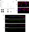
Hypoxia decreases the rate of corneal epithelial wound healing in organ culture. A: corneal epithelial abrasions on rat organ cultures heal more rapidly in normoxic conditions. Epithelial abrasions (3 mm diameter) were made on the central cornea prior to incubation under normoxic or hypoxic conditions. Black circles in schematic under the graph represent wounds. Wound closure was monitored at 0, 2, 12, 18, and 24 h using a digital camera mounted on a dissecting microscope to determine the size of the remaining abrasion. Values are means ± SE of ≥3 different eyes at each time point and condition. B and C: images obtained using a Zeiss Axiovert LSM 700 confocal microscope. B: hypoxia alters cellular morphology at the wound edge. After 12 h of incubation in normoxia or hypoxia, rat cornea organ cultures were fixed and counterstained with rhodamine phalloidin (red) and DAPI (blue). Scale bar, 50 μm. Arrows denote wound edge. Images represent ≥3 independent experiments. C: hypoxia alters migrating epithelium. Cross sections of wounded corneas were stained for paxillin (green) and counterstained with DAPI (blue) and rhodamine phalloidin (red). Tiled images (1 × 4) of the wound margin are presented. Note decreased paxillin and altered stratification of superficial cells at the leading edge of corneas cultured under hypoxic conditions compared with normoxia. Scale bar, 25 μm. Images represent ≥3 independent experiments.
Fig. 11.
Fig. 11.
Hypoxia results in decreased fibronectin along the basal lamina. A: air-lifted organ-cultured corneas were incubated in normoxia or hypoxia. Control (0-h) image shows cornea that was neither wounded nor incubated. All other corneas were wounded at 0 h, cultured for 2, 24, and 48 h, and then stained for fibronectin (green) and counterstained with DAPI (blue) and rhodamine phalloidin (red). Images were taken at ×40 magnification using a Zeiss Axiovert LSM 700 confocal microscope. Scale bar, 100 μm. B: fibronectin (FN) along the basal lamina and in the stroma was quantified using ImageJ. W(+), wounded; W(−), not wounded. Values are means ± SE of ≥3 independent experiments for each time point (0, 2, 24, and 48 h). Data were analyzed by Student's t-test (P < 0.05).

References

    1. Anumolu SS, DeSantis AS, Menjoge AR, Hahn RA, Beloni JA, Gordon MK, Sinko PJ. Doxycycline loaded poly(ethylene glycol) hydrogels for healing vesicant-induced ocular wounds. Biomaterials 31: 964–974, 2010 - PMC - PubMed
    1. Araki K, Ohashi Y, Kinoshita S, Hayashi K, Kuwayama Y, Tano Y. Epithelial wound healing in the denervated cornea. Curr Eye Res 13: 203–211, 1994 - PubMed
    1. Belmonte C, Gallar J, Pozo MA, Rebollo I. Excitation by irritant chemical substances of sensory afferent units in the cat's cornea. J Physiol 437: 709–725, 1991 - PMC - PubMed
    1. Berridge MJ, Lipp P, Bootman MD. Signal transduction. The calcium entry pas de deux. Science 287: 1604–1605, 2000 - PubMed
    1. Besshoh S, Chen S, Brown IR, Gurd JW. Developmental changes in the association of NMDA receptors with lipid rafts. J Neurosci Res 85: 1876–1883, 2007 - PubMed

Publication types

MeSH terms