. 2014 Mar 26;9(3):e93181.
doi: 10.1371/journal.pone.0093181. eCollection 2014.

Beta-actin deficiency with oxidative posttranslational modifications in Rett syndrome erythrocytes: insights into an altered cytoskeletal organization

Affiliations

Beta-actin deficiency with oxidative posttranslational modifications in Rett syndrome erythrocytes: insights into an altered cytoskeletal organization

Alessio Cortelazzo et al. PLoS One. .

Abstract

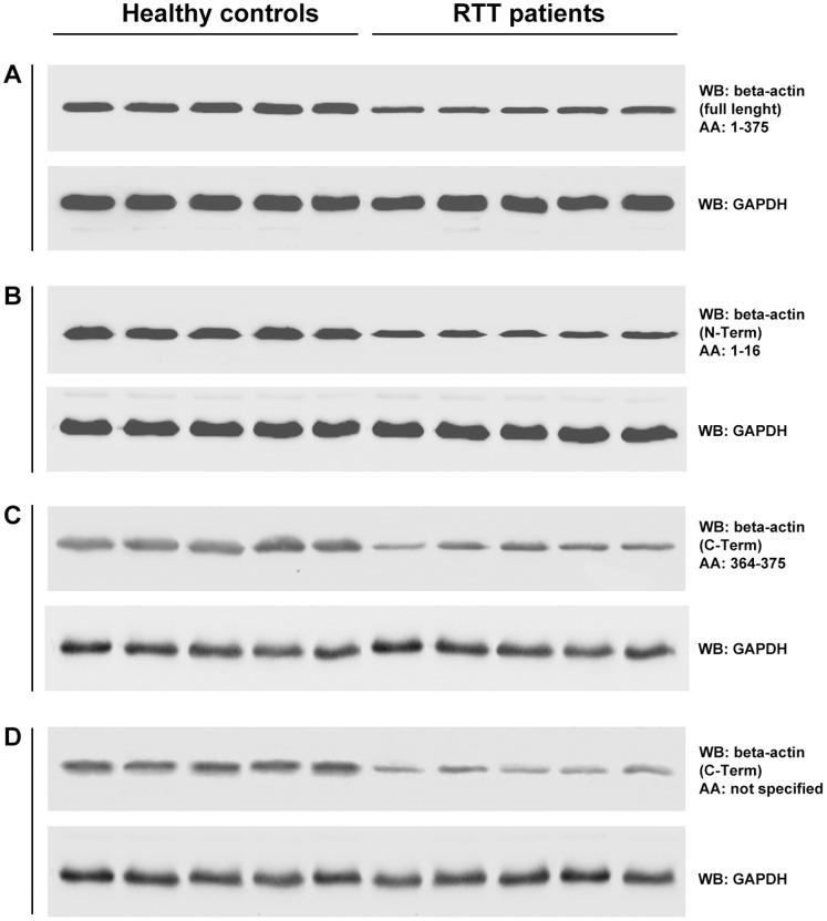
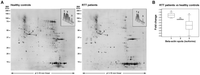
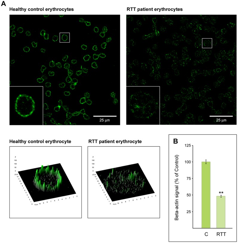
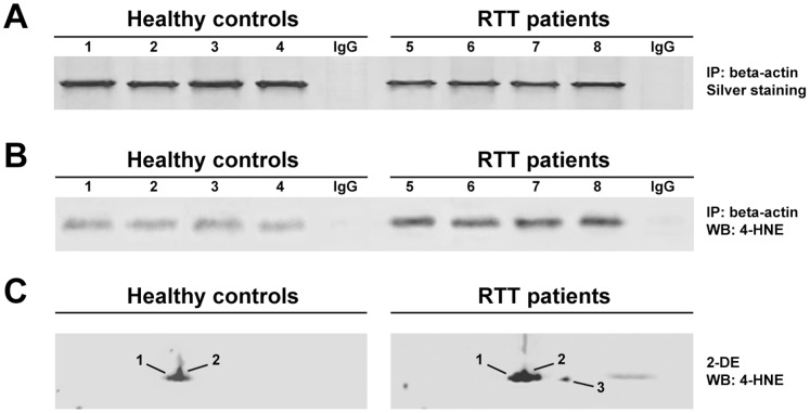
Beta-actin, a critical player in cellular functions ranging from cell motility and the maintenance of cell shape to transcription regulation, was evaluated in the erythrocyte membranes from patients with typical Rett syndrome (RTT) and methyl CpG binding protein 2 (MECP2) gene mutations. RTT, affecting almost exclusively females with an average frequency of 1∶10,000 female live births, is considered the second commonest cause of severe cognitive impairment in the female gender. Evaluation of beta-actin was carried out in a comparative cohort study on red blood cells (RBCs), drawn from healthy control subjects and RTT patients using mass spectrometry-based quantitative analysis. We observed a decreased expression of the beta-actin isoforms (relative fold changes for spots 1, 2 and 3: -1.82±0.15, -2.15±0.06, and -2.59±0.48, respectively) in pathological RBCs. The results were validated by western blotting and immunofluorescence microscopy. In addition, beta-actin from RTT patients also showed a dramatic increase in oxidative posttranslational modifications (PTMs) as the result of its binding with the lipid peroxidation product 4-hydroxy-2-nonenal (4-HNE). Our findings demonstrate, for the first time, a beta-actin down-regulation and oxidative PTMs for RBCs of RTT patients, thus indicating an altered cytoskeletal organization.


Conflict of interest statement

Competing Interests: The authors have declared that no competing interests exist.

Figures

Figure 1
Figure 1. Beta-actin expression in RBC membranes.
SDS-PAGE comparative analysis of RBC ghosts from healthy control subjects and RTT patients. Visible reduction of intensity for beta-actin (band 6) is present in RTT patients.
Figure 2
Figure 2. Western blot of beta-actin.
Comparative analysis of RBC ghosts from healthy controls and RTT patients using four antibodies, which recognize different beta-actin amino acidic sequences (A: full length, B: N-terminal, C: C-terminal and D: C-terminal, amino acidic sequence not specified), confirm a beta-actin decrease in RTT patients.
Figure 3
Figure 3. 2-DE analysis of beta-actin isoforms.
(A) Comparative analysis between RTT patients (right panel) and healthy controls (left panel). Arrows indicate the identified beta-actin isoforms which appear to be decrease in RTT patients. The top right panels represent the beta-actin differentially expressed spots on 3D view. (B) Quantitative analysis of the identified beta-actin spots changes in RTT patients as compared to control expression levels. Molecular mass and pI markers are indicated along the gels.
Figure 4
Figure 4. Confocal microscopy analysis of beta-actin distribution in RBCs.
(A) Comparative analysis shows beta-actin expression and distribution differences between RBCs from RTT patients (right panel) and healthy control subjects (left panel) at confocal microscopy. Small panels represent, with flat and 3D views, two typical erythrocytes in which beta-actin signal is differentially distributed. Scale bar 25 μm. (B) Quantitative analysis of beta-actin signal differences in RBCs from healthy control (C) and RTT patients (RTT).
Figure 5
Figure 5. 4-HNE/beta-actin adducts in RBC membranes.
(A) SDS-PAGE (silver staining) of immunoprecipitated beta-actin from RBC ghosts of healthy control subjects (1–4) and RTT patients (5–8). (B) Western blot analysis of 4-HNE on immunoprecipitated beta-actin from RBC ghosts of healthy control subjects (1–4) and RTT patients (5–8). Immunoprecipitation (IP) with normal rabbit IgG served as a negative control. (C) 2-DE/Western blot analysis of 4-HNE on beta-actin from RBC ghosts proteome.
Figure 6
Figure 6. Potential binding sites for 4-HNE in the beta-actin amino acid sequence.
The potential binding sites for 4-HNE are highlighted. Red color for 6 cysteine (C), yellow color for 8 hystidine (H) and green color for 19 lysine (K) residues. Blue double lines indicate sub-domain 1; orange lines indicate sub-domain 2; black lines indicate sub-domain 3 and purple lines indicate sub-domain 4 (primary sequence extracted from the ExPASy: SIB Bioinformatics Resource Portal, http://www.expasy.org/).

Similar articles

Cited by

References

    1. Dominguez R, Holmes KC (2011) Actin structure and function. Annu Rev Biophys 40: 169–86. - PMC - PubMed
    1. Herman IM (1993) Actin isoforms. Curr Opin Cell Biol 5: 48–55. - PubMed
    1. Kaufmann WE, Moser HW (2000) Dendritic anomalies in disorders associated with mental retardation. Cereb Cortex 10: 981–91. - PubMed
    1. Kunze D, Rüstow B (1993) Pathobiochemical aspects of cytoskeleton components. Eur J Clin Chem Clin Biochem 31: 477–89. - PubMed
    1. Kaufmann WE, MacDonald SM, Altamura CR (2000) Dendritic cytoskeletal protein expression in mental retardation: an immunohistochemical study of the neocortex in Rett Syndrome. Cereb Cortex 10: 992–1004. - PubMed

Publication types